EP2048228A2 - Human liver progenitors - Google Patents
Human liver progenitors Download PDFInfo
- Publication number
- EP2048228A2 EP2048228A2 EP08019659A EP08019659A EP2048228A2 EP 2048228 A2 EP2048228 A2 EP 2048228A2 EP 08019659 A EP08019659 A EP 08019659A EP 08019659 A EP08019659 A EP 08019659A EP 2048228 A2 EP2048228 A2 EP 2048228A2
- Authority
- EP
- European Patent Office
- Prior art keywords
- cells
- liver
- cell
- progenitors
- hepatic
- Prior art date
- Legal status (The legal status is an assumption and is not a legal conclusion. Google has not performed a legal analysis and makes no representation as to the accuracy of the status listed.)
- Granted
Links
Images


Classifications
-
- C—CHEMISTRY; METALLURGY
- C12—BIOCHEMISTRY; BEER; SPIRITS; WINE; VINEGAR; MICROBIOLOGY; ENZYMOLOGY; MUTATION OR GENETIC ENGINEERING
- C12N—MICROORGANISMS OR ENZYMES; COMPOSITIONS THEREOF; PROPAGATING, PRESERVING, OR MAINTAINING MICROORGANISMS; MUTATION OR GENETIC ENGINEERING; CULTURE MEDIA
- C12N5/00—Undifferentiated human, animal or plant cells, e.g. cell lines; Tissues; Cultivation or maintenance thereof; Culture media therefor
- C12N5/06—Animal cells or tissues; Human cells or tissues
- C12N5/0602—Vertebrate cells
- C12N5/067—Hepatocytes
- C12N5/0672—Stem cells; Progenitor cells; Precursor cells; Oval cells
-
- A—HUMAN NECESSITIES
- A61—MEDICAL OR VETERINARY SCIENCE; HYGIENE
- A61P—SPECIFIC THERAPEUTIC ACTIVITY OF CHEMICAL COMPOUNDS OR MEDICINAL PREPARATIONS
- A61P1/00—Drugs for disorders of the alimentary tract or the digestive system
-
- A—HUMAN NECESSITIES
- A61—MEDICAL OR VETERINARY SCIENCE; HYGIENE
- A61P—SPECIFIC THERAPEUTIC ACTIVITY OF CHEMICAL COMPOUNDS OR MEDICINAL PREPARATIONS
- A61P1/00—Drugs for disorders of the alimentary tract or the digestive system
- A61P1/16—Drugs for disorders of the alimentary tract or the digestive system for liver or gallbladder disorders, e.g. hepatoprotective agents, cholagogues, litholytics
-
- A—HUMAN NECESSITIES
- A61—MEDICAL OR VETERINARY SCIENCE; HYGIENE
- A61P—SPECIFIC THERAPEUTIC ACTIVITY OF CHEMICAL COMPOUNDS OR MEDICINAL PREPARATIONS
- A61P35/00—Antineoplastic agents
-
- A—HUMAN NECESSITIES
- A61—MEDICAL OR VETERINARY SCIENCE; HYGIENE
- A61P—SPECIFIC THERAPEUTIC ACTIVITY OF CHEMICAL COMPOUNDS OR MEDICINAL PREPARATIONS
- A61P7/00—Drugs for disorders of the blood or the extracellular fluid
-
- A—HUMAN NECESSITIES
- A61—MEDICAL OR VETERINARY SCIENCE; HYGIENE
- A61K—PREPARATIONS FOR MEDICAL, DENTAL OR TOILETRY PURPOSES
- A61K39/00—Medicinal preparations containing antigens or antibodies
- A61K2039/555—Medicinal preparations containing antigens or antibodies characterised by a specific combination antigen/adjuvant
- A61K2039/55588—Adjuvants of undefined constitution
- A61K2039/55594—Adjuvants of undefined constitution from bacteria
-
- A—HUMAN NECESSITIES
- A61—MEDICAL OR VETERINARY SCIENCE; HYGIENE
- A61K—PREPARATIONS FOR MEDICAL, DENTAL OR TOILETRY PURPOSES
- A61K35/00—Medicinal preparations containing materials or reaction products thereof with undetermined constitution
- A61K35/12—Materials from mammals; Compositions comprising non-specified tissues or cells; Compositions comprising non-embryonic stem cells; Genetically modified cells
-
- C—CHEMISTRY; METALLURGY
- C12—BIOCHEMISTRY; BEER; SPIRITS; WINE; VINEGAR; MICROBIOLOGY; ENZYMOLOGY; MUTATION OR GENETIC ENGINEERING
- C12N—MICROORGANISMS OR ENZYMES; COMPOSITIONS THEREOF; PROPAGATING, PRESERVING, OR MAINTAINING MICROORGANISMS; MUTATION OR GENETIC ENGINEERING; CULTURE MEDIA
- C12N2500/00—Specific components of cell culture medium
- C12N2500/05—Inorganic components
- C12N2500/10—Metals; Metal chelators
- C12N2500/20—Transition metals
- C12N2500/22—Zinc; Zn chelators
-
- C—CHEMISTRY; METALLURGY
- C12—BIOCHEMISTRY; BEER; SPIRITS; WINE; VINEGAR; MICROBIOLOGY; ENZYMOLOGY; MUTATION OR GENETIC ENGINEERING
- C12N—MICROORGANISMS OR ENZYMES; COMPOSITIONS THEREOF; PROPAGATING, PRESERVING, OR MAINTAINING MICROORGANISMS; MUTATION OR GENETIC ENGINEERING; CULTURE MEDIA
- C12N2500/00—Specific components of cell culture medium
- C12N2500/05—Inorganic components
- C12N2500/10—Metals; Metal chelators
- C12N2500/20—Transition metals
- C12N2500/24—Iron; Fe chelators; Transferrin
- C12N2500/25—Insulin-transferrin; Insulin-transferrin-selenium
-
- C—CHEMISTRY; METALLURGY
- C12—BIOCHEMISTRY; BEER; SPIRITS; WINE; VINEGAR; MICROBIOLOGY; ENZYMOLOGY; MUTATION OR GENETIC ENGINEERING
- C12N—MICROORGANISMS OR ENZYMES; COMPOSITIONS THEREOF; PROPAGATING, PRESERVING, OR MAINTAINING MICROORGANISMS; MUTATION OR GENETIC ENGINEERING; CULTURE MEDIA
- C12N2500/00—Specific components of cell culture medium
- C12N2500/30—Organic components
- C12N2500/36—Lipids
-
- C—CHEMISTRY; METALLURGY
- C12—BIOCHEMISTRY; BEER; SPIRITS; WINE; VINEGAR; MICROBIOLOGY; ENZYMOLOGY; MUTATION OR GENETIC ENGINEERING
- C12N—MICROORGANISMS OR ENZYMES; COMPOSITIONS THEREOF; PROPAGATING, PRESERVING, OR MAINTAINING MICROORGANISMS; MUTATION OR GENETIC ENGINEERING; CULTURE MEDIA
- C12N2503/00—Use of cells in diagnostics
- C12N2503/02—Drug screening
Definitions
- the present invention relates to human hepatic stem cells, pluripotent cells that give rise to hepatocytes and biliary cells, and other liver progenitor cell subpopulations that have the capacity to expand and differentiate into one or more liver cell lineages including hemopoietic, mesenchymal or hepatic cell lineages.
- the invention relates to markers and properties used to identify human liver progenitors, methods of their purification and cryopreservation, novel approaches that enable one to distinguish hepatic from hemopoietic subpopulations, and evidence proving that hepatic progenitors exist in livers from fetal to adult human livers.
- the inventions constitute the basis for cell and gene therapies and for the establishment of bioartificial organs.
- the primary structural and functional unit of the mature liver is the acinus, which in cross section is organized like a wheel around two distinct vascular beds: 3-7 sets of portal triads (each with a portal venule, hepatic arteriole, and a bile duct) for the periphery, and with the central vein at the hub.
- the liver cells are organized as cell plates lined on both sides by fenestrated endothelia, defining a series of sinusoids that are contiguous with the portal and central vasculature.
- hepatocytes have two basal domains, each of which faces a sinusoid, and an apical domain which is defined by the region of contact between adjacent hepatocytes.
- the basal domains contact the blood, and are involved in the absorption and secretion of plasma components, while the apical domains form bile canaliculi, specialized in the secretion of bile salts, and are associated through an interconnecting network with bile ducts.
- zone 1 the periportal region
- zone 2 the midacinar region
- zone 3 the pericentral region.
- Proliferative potential, morphological criteria, ploidy, and most liver-specific genes are correlated with zonal location ( Gebhardt, R., et al. 1988. FEBS Lett. 241:89-93 ; Gumucio, J. J. 1989, Vol. 19. Springer International, Madrid ; Traber, P. et al. 1988. Gastroenterology. 95:1130-43 ).
- hepatocytes In addition to hepatocytes, bile duct epithelial cells (cholangiocytes), and endothelial cells, the region between the portal and central tracts contains other cell types, such as Ito cells and Kupffer cells. These play prominent roles in pathogenic conditions of the liver, especially in inflammation and fibrosis, but their direct contribution to the main homeostatic functions of the normal organ are apparently small.
- the liver develops as a result of the convergence of a diverticulum formed from the caudal foregut and the septum transversum, part of the splanchnic mesenchyme.
- the formation of the hepatic cells begins after the endodermal epithelium interacts with the cardiogenic mesoderm, probably via fibroblast growth factors.
- the specified hepatic cells then proliferate and penetrate into the mesenchyme of the septum transversum with a cord like fashion, forming the liver strom.
- the direct epithelial-mesenchymal interaction is critical in these early developmental stages of the liver and dictates which cells will become hepatocytes or cholangiocytes, and the fenestrated endothelia, respectively.
- liver development consists of clusters of primitive hepatocytes bounded by a continuous endothelium lacking a basement membrane and abundant hemopoietic cells. As the endothelium is transformed to become a discontinuous, fenestrated endothelium, the vasculature, especially the portal vasculature, becomes more developed with the production of basement membranes.
- the portal interstitium may provide the trigger for the development of bile ducts, and as it surrounds the portal venules, hepatic arterioles, and bile ducts, portal triads are formed. Immature hepatocytes rapidly proliferate and parenchymal plates are formed, probably in response to changes in the amount and distribution of such tissue-organizing molecules as C-CAM 105, Agp 110, E-cadherin, and connexins, coincident with the relocation of most, but not all, of the hemopoietic cells to the bone marrow.
- tissue-organizing molecules as C-CAM 105, Agp 110, E-cadherin, and connexins
- hemopoietic progenitors persist in the adult quiescent rodent liver, and hemopoietic stem cells have been isolated from both adult human and murine liver ( Crosbie, O. M. et al. 1999. Hepatology. 29:1193-8 ).
- the mature physical organization is achieved within the first weeks after birth in rodents, and in humans, within the first few years. Metabolic zonation is established according to somewhat different schedules for different enzymes, but becomes evident in the period following birth.
- Stem cells have been defined as primitive cells that self-replicate, that are pluripotent, i.e. produce daughter cells with more than one fate, that can expand extensively and can reconstitute a tissue or tissues. Most of the literature on stem cells derives either from the literature on embryos or that on hemopoietic, epidermal, or intestinal tissues.
- Embryonic stem cells also called "ES" cells, consist of permanent cell populations derived from totipotent, normal cells in blastocysts, that were first reported in the early 1980s.
- ES cell lines can be cultured in vitro with maintenance of totipotency. ES cells are tumorigenic if introduced into immunocompromised hosts in any site other than in utero, forming teratocarcinomas.
- ES cell lines have been established from many species (mouse, rat, pig, etc.), only the mouse system has been used routinely to generate animals with novel phenotypes (knockouts, transgenics) by merging modified ES cells from culture to blastocysts and then implanting the blastocysts into pseudopregnant hosts.
- Embryonic germ (EG) cell lines which show many of the characteristics of ES cells, can be isolated directly in vitro from the primordial germ cell population. As with ES cells, the EG cells form teratocarcinomas when injected into immunocompromised mice and contributed to chimeras, including the germ line, when injected into blastocysts.
- Determined stem cells are pluripotent cells that have restricted their genetic potential to that for a limited number of cell types and have extensive growth potential. Increasing evidence such as that from the telomerase field suggest that determined stem cells do not self-replicate, that is their progeny can have less growth potential than the parent. Determined stem cells give rise to daughter cells that lose pluripotency by restricting their genetic potential to a single fate, e.g. hepatocytes, and are referred to as committed progenitors . In the hepatic lineage there are committed hepatocytic progenitors and committed biliary progenitors.
- liver cells are stem cells ( Kennedy, S. et al. 1995. Hepatology. 22:160-8 ; Michalopoulos, G. K. et al. 1997, Science. 276:60-6 .). These investigators believe that all parenchymal cells are co-equal, are highly plastic and with gene expression dictated only by the microenvironment. Under appropriate oncogenic conditions, the mature parenchymal cells are hypothesized to become stem cells that can subsequently convert to tumor cells.
- the silent stem cell model is based on the studies of Wilson and Leduc ( Wilson, J. W. et al. 1958. J. Pathol. Bacteriol. 76:441-449 .). As in the hemopoietic field, this concept gained the most credibility from extensive studies of liver carcinogenesis ( Marceau, N. 1994. Gut. 35:294-6 .). These investigators believe that progenitor cells, including bipotential progenitor cells, can persist in adult tissue but propose that they are rare holdovers or remnants of cell populations from embryonic development. They assume that progenitors play no role in normal or regenerative liver functioning but only in disease states ( Overturf K, et al. 1999. American Journal of Pathology. 155:2135-2143 .).
- silent stem cells similar to the satellite cells in muscle. These cells have been described as “oval cells” on account of the distinctive shape of the cell nuclei. They are small ( ⁇ 9 um) and express a characteristic antigenic profile on the cell surface. All mature liver cells are assumed to be co-equal with respect to growth and gene expression and that all aspects of heterogeneity of gene expression is dictated only by the cellular microenvironment. The proponents of the silent stem cell model strongly reject any idea of movement of parenchymal cells from periportal to pericentral locations. The importance of stem cells and other hepatic progenitors is thought to be relevant to disease states only, especially carcinogenesis.
- liver is a stem cell and maturational lineage system
- stem cells are organized as maturational lineages fed, like a spring, by stem cells or early progenitor cell populations ( Brill, S. et al. 1993. Proceedings of the Society for Experimental Biology & Medicine. 204:261-9 .).
- the tissue is defined as going from "young, to middle age, to old cells”.
- the maturational process is accompanied by lineage-position-dependent changes in cell size, morphology, antigenic profiles, growth potential and gene expression.
- microenvironment comprises the nutrients, gas exchange (oxygen, CO 2 ), pH, hormones, cell-cell interactions and extracellular matrix chemistry.
- Zones 1 2 3 Ploidy Diploid cells Tetraploid cells Mix of tetraploid and octaploid cells Average Size 7-20 ⁇ 20-30 ⁇ 30-50 ⁇ Growth Maximum Intermediate Negligible Extracellular Matrix A gradient in the matrix chemistry located in the space of Disse and consisting of type IV collagen mixed with laminin and heparan sulfate proteoglycans in the periportal area and converting to fibrillar collagens, fibronectins and heparin proteoglycans in the pericentral area.
- the stem cell and maturational lineage model contradicts other liver cell development models in suggesting that liver malignancy is most often an indirect, rather than a direct, result of an oncogenic insult.
- Oncogenic insults are proposed to kill most cells of the liver, specially the mature cells in the lineage, resulting in a dramatic induction of a regenerative response.
- the resultant expansion of the progenitors increases the risk of secondary mutational events in the rapidly growing cells, the progenitors, that can result in malignancy.
- the older hypotheses that cancer is blocked differentiation or that cancers are due to oncogenic insults targeting stem cells are accepted as correct but with the modification presented above.
- U.S. Patent No 5,559,022 to Naughton discloses isolation of cells from liver and further purification by the use of gradient centrifugation. However, the cell population isolated is the "acidophilic parenchymal cell population" which is not the liver progenitors of this invention as claimed.
- liver transplantation is a strong clinical and commercial interest in isolating and identifying immature progenitor cells from liver because of the impact that such cell population may have in treating liver diseases.
- Liver transplants are curative for some forms of liver failure, and approximately 4100 transplants are performed a year in United States.
- One of the limiting factors in liver transplantation is the availability of donor livers especially given the constraint that donor livers for organ transplantation must originate from patients having undergone brain death but not heart arrest. Livers from cadaveric donors have not been successful, although recent efforts to use such donors have supported the possibility of using them if the liver is obtained within an hour of death.
- liver transplantation into the liver is an attractive alternative therapy for most liver diseases.
- the surgical procedures for cell transplantation are minor relative to those needed for whole organ transplantation and, therefore, can be used for patients with various surgical risks such as age or infirmity.
- the use of human liver cells is superior to liver cells derived from other mammals because the potential pathogens, if any, are of human origin and could be better tolerated by patients and could be easily screened before use.
- liver cell transplantation Attempts to perform liver cell transplantation have made use of unfractionated mature liver cells and have shown some measure of efficacy ( Fox, I. J. et al. 1998. New England Journal of Medicine. 338:1422-1426 .).
- the successes require injection of large numbers of cells (10-20 billion), since the cells do not grow in vivo.
- the introduction of substantial numbers of large mature liver cells (average cell diameter 30-50 ⁇ ) is complicated by their tendency to form large aggregates upon injection, resulting in potentially fatal emboli.
- these cells elicit a marked immunological rejection response forcing patients to be maintained on immunosuppressive drugs for the remainder of their lives.
- mature liver cells have not been successfully cryopreserved and complicated logistics are required to coordinate the availability of suitable liver tissue, the preparation of cell suspensions and the immediate delivery of the cells for clinical therapies.
- liver progenitors from liver are known to be an extremely challenging task due to the shortage of markers that positively select for liver cells.
- the only available antibodies for candidates of hepatic progenitors are those monoclonal antibodies that are prepared against subpopulations of hepatic progenitors (oval cells) induced to proliferate after exposure to oncogenic insults. These antibodies however cross-react with antigens present in hemopoietic cells.
- liver progenitors from adult human liver is novel and unexpected partly due to the controversy regarding the mere presence of liver progenitors in the adult in which human hepatic progenitors have been assumed either not to be present or to be a physiologically silent remant from embryogenesis. Therefore, there have not been attempts to isolate them or study them except in disease states.
- alpha-fetoprotein AFP
- albumin cytoplasmic proteins alpha-fetoprotein
- AFP alpha-fetoprotein
- albumin albumin
- these cells are capable of producing offspring that enter both biliary and hepatocyte lineages. If these daughter cells commit to the biliary lineage alpha-fetoprotein expression ceases.
- alpha-fetoprotein expression persists in the hepatocyte lineage until the perinatal period when it is suppressed, leaving albumin expression as one of the principal characteristics of the adult hepatocyte.
- alpha-fetoprotein is an intracellular protein and can only be visualized after fixation and permeabilization of the cell, it is unsuitable as a marker for the identification of viable hepatic progenitor cells.
- the invention relates to a method of providing a composition comprising a mixture of cells derived from human liver tissue, which mixture comprises an enriched population of human hepatic progenitors, the method comprising: providing a substantially single cell suspension of human liver tissue comprising a mixture of cells of varying sizes, including immature cells and mature cells; and debulking the suspension under conditions that permit the removal of mature cells and those of relatively large size, while retaining immature cells and those of relatively small size, to provide a mixture of cells comprised of an enriched population of human hepatic progenitors which human hepatic progenitors themselves, their progeny, or more mature forms thereof exhibit one or more markers indicative of expression of alpha-fetoprotein, albumin, or both.
- the alpha-fetoprotein and albumin can be full-length or a variant.
- the debulking process can comprise a separation by cell size, buoyant density, or both.
- the debulking can also be based on sedimentation velocity, hydrodynamic radius, and sedimentation to equilibrium density.
- the separation can be by relative adherence of surface markers to binding components, for example antibodies or lectins.
- the isolated progenitors can be diploid and can be less than about 15 microns in diameter.
- the progenitors or their progeny can synthesize macromolecules characteristic of progenitors, including, but not limited to alpha-fetoprotein and albumin.
- the alpha-fetoprotein includes the exon1(aFP)-encoded peptide sequence.
- the alpha-fetoprotein is transcribed from an mRNA greater than 2Kb in size, a full-length aFP mRNA.
- the albumin preferably includes the exon1 (ALB)-encode peptide sequence.
- ALB exon1
- the present invention relates to a method of isolation, cryopreservation, and use of progenitors from human liver which includes processing human liver tissue to provide a substantially single cell suspension including progenitors and non-progenitors of one or more cell lineages found in human liver; subjecting the suspension to a debulking step, which reduces substantially the number of non-progenitors in the suspension, to provide a debulked suspension enriched in progenitors exhibiting one or more markers associated with at least one of the cell lineages; optionally selecting from the debulked suspension those cells, which themselves, their progeny, or more mature forms thereof express at least one marker associated with at least one liver cell lineage; optionally, suspending the cells under conditions optimal for cryopreservation; and optionally use for production of growth factors and for therapy in patients.
- liver progenitors expressing cytoplasmic proteins such as alpha-fetoprotein are selected.
- Processing or debulking steps of this invention preferably include a density gradient centrifugation or centrifugal elutriation of the liver cell suspension to separate the cells according to their buoyant density and/or size, which are associated with one or more gradient fractions having a lower buoyant density and/or smaller size.
- the density gradient method can include zonal centrifugation and continuous-flow centrifugation.
- One embodiment of the invention is negative selection of non-progenitors including mature hepatic, hemopoietic, and mesenchymal cells by the use of markers associated with mature hepatic cells, such as connexin, markers associated with hemopoietic cells, such as glycophorin A and CD45, and/or markers associated with mature mesenchymal cells, such as retinoids, and von Willebrand Factor.
- markers associated with mature hepatic cells such as connexin
- markers associated with hemopoietic cells such as glycophorin A and CD45
- markers associated with mature mesenchymal cells such as retinoids, and von Willebrand Factor.
- hepatic progenitors can overcome many of the shortcomings associated with use of mature liver cells, making them ideal cells for use in cell and gene therapies and for bioartifical organs.
- the cells are small (7-15 ⁇ ), therefore minimizing the formation of large emboli.
- the cells have extensive growth potential meaning that fewer cells are needed for reconstitution of liver tissue in a patient.
- the progenitors have minimal antigenic markers that might elicit immunological rejection providing hope that little or no immunosuppressive drugs might be needed.
- Therapy with liver cells involves either extracorporeal treatment or transplantation of liver cells.
- the cells preferably including progenitor cells, are supplied in any of various ways, including parenterally and intraperitoneally. An effective amount of cells is necessary, preferably between 10 3 and 10 10 cells. More preferably between 10 5 and 10 8 cells are transplanted, optimally about 10 6 cells.
- liver progenitors are extremely useful for production of growth factors and other proteins. These factors are associated with their own growth or that of other progenitors in the liver (e.g. hemopoietic or mesenchymal progenitors) and factors associated with early steps in the dedication of hepatic progenitor cells to a particular lineage. These novel growth factors can be used to treat liver disease or to control those cancers that are transformants of the liver progenitors. Furthermore, liver progenitors are important targets for gene therapy, wherein the inserted genetically transformed or normal hepatic progenitors promote the health of the individual into whom such hepatic progenitors are transplanted.
- factors are associated with their own growth or that of other progenitors in the liver (e.g. hemopoietic or mesenchymal progenitors) and factors associated with early steps in the dedication of hepatic progenitor cells to a particular lineage. These novel growth factors can be used to treat liver disease or to control those cancers that are transformants
- Another aspect of this invention is the determination of unique antigenic profiles on the cell surface that correlate with the expression of alpha-fetoprotein within the cell. Characterization of alpha-fetoprotein-containing cells in this way allows the subsequent enrichment of viable hepatic progenitor cells by flow cytometric methodology from living single cell suspensions prepared from whole livers or liver lobes. Moreover, the isolation and identification of human hepatic progenitors as described herein were obtained through application of a combination of unique methods, markers and parameters which the present inventors used for the first time to achieve the unique cell population of this invention.
- a further aspect of this invention provides for liver cell progenitors of hepatic, hematopoietic, or mesenchymal origin. These cell lineages, their progenies or their more mature forms are selected by antigenic markers selected from the group consisting of CD14, CD34, CD38, CD45, CD117, ICAM, glycophorin A, and/or cytoplasmic markers such as alpha-fetoprotein-like immunoreactivity, albumin-like immunoreactivity, or both.
- Alpha-fetoprotein can derive from a full-length mRNA (greater than 2 Kb, the form usually expressed in hepatic progenitors) or from a variant form (less than 2 Kb, i.e.
- liver progenitors of this invention can be isolated from the liver of a fetus, a neonate, an infant, a child, a juvenile, or an adult.
- isolated human liver progenitors are isolated in a highly enriched to substantially pure form.
- Such liver progenitors contain hepatic, hemopoietic and mesenchymal progenitors.
- the hepatic progenitors have the capacity to develop into hepatocytes, biliary cells, or a combination thereof;
- the hematopoietic progenitors have the capacity to develop into macrophages, neutrophils, granulocytes, lymphocytes, platelets, neutrophils eosinophils, basophils, or a combination thereof.
- the mesenchymal progenitors have the capacity to develop into endothelial cells, stromal cells, hepatic stellate cells (Ito cells), cartilage cells, bone cells or combinations thereof.
- the method of this invention can be used to select mesenchymal progenitors expressing alpha-fetoprotein-like immunoreactivity, CD45, albumin-like reactivity, CD34, osteopontin, bone sialoprotein, collagen (types I, II, III, or IV), or a combination thereof.
- a still further aspect of this invention provides for liver progenitors that harbor exogenous nucleic acid.
- exogenous nucleic acid can encode at least one polypeptide of interest, or can promote the expression of at least one polypeptide of interest.
- a method of alleviating the negative effects of one or more human disorders or dysfunctions by administering to an individual suffering from such negative effects an effective amount of isolated human liver progenitors.
- the progenitors can be administered either intraperitoneally, or parenterally via a vascular vessel, or administered directly into the liver.
- the direct administration may be effected surgically via portal vein, mesenteric vein, hepatic artery, hepatic bile duct, or combinations thereof.
- the liver progenitors can be administered into an ectopic site of the individual, such as spleen or peritoneum.
- the human disorders or dysfunctions that can be alleviated by the method of this invention include: hepatocholangitis, hepatomalacia, hepatomegalia, cirrhosis, fibrosis, hepatitis, acute liver failure, chronic liver failure, or inborn errors of metabolism, and liver cancer such as hepatocarcinoma, or hepatoblastoma.
- liver cancer such as hepatocarcinoma, or hepatoblastoma.
- the cancer of the liver can be a primary site of cancer or one that has metastasized into the liver.
- the metastatic tumor could be derived from any number of primary sites including, intestine, prostate, breast, kidney, pancreas, skin, brain, lung or a combination thereof.
- a bioreactor which includes biological material comprising isolated progenitors from human liver, their progeny, their maturing or differentiated descendants, or combinations thereof; and culture media, such as basal media; one or more compartments holding the biological material or the components comprising the biological material; and optionally one or more connecting ports.
- the bioreactor can, optionally, also include: extracellular matrix; hormones, growth factors, nutrients, or combinations thereof; and a biological fluid such as serum, plasma, or lymph.
- the bioreactor is adapted for sustaining said progenitors in a viable, functional state, and can sustain liver progenitors for a period ranging from about one week to about 55 weeks.
- the bioreactor is adapted for use as an artificial liver, for product manufacturing, toxicological studies, or metabolic studies, including studies involving the activity of cytochrome P450, or other types of drug metabolism.
- a composition of isolated human liver progenitors, or a suspension enriched in progenitors obtained from human liver is provided.
- the cell suspension is provided in a pharmaceutically acceptable carrier or diluent and is administered to a subject in need of treatment.
- the composition of this invention includes liver progenitors that exhibit one or more markers associated with at least one of one or more cell lineages found in human liver and are substantially free of mature cells.
- isolated liver progenitors are derived from one or more liver cell lineages including hepatic, hemopoietic, or mesenchymal cell lineages and themselves, their progeny, or more mature forms of the progenitors thereof express at least one or more of antigenic markers CD14, CD34, CD38, CD90, or CD117, CD45, glycophorin A, and cytoplasmic markers of alpha-fetoprotein-like immunoreactivity, albumin-like immunoreactivity, or both.
- the immature cells, their progeny, or more mature forms express osteopontin, bone sialoprotein, collagen I, collagen III, collagen IV, or a combination thereof.
- a cell culture system of liver progenitors which includes isolated progenitors from human liver, their progeny, their maturing or differentiated descendants, or combinations thereof.
- the cell culture system additionally includes extracellular matrix comprising one or more collagens, one or more adhesion proteins (laminins, fibronectins), and other components such as proteoglycans (such as heparan sulfate proteoglycans); or an individual matrix component.
- the matrix component includes fragments of matrix components, matrix mimetics that can be synthetic and biodegradable materials (i.e . microspheres) coated with one or more of the factors from one of the classes of extracellular matrices.
- the cell culture system additionally can include basal or enriched media and other nutrients; hormones, growth factors, and, optionally, a biological fluid such as serum, plasma or lymph. Additionally, the cell culture system can have one or more compartments that holds the biological material such as a culture dish, plate, flask, roller bottle, transwell or other such container.
- the cultures or bioreactors of this invention can be used in one or more metabolic studies including studies involving the activity of phase I or II biotransformation enzyme systems, one or more transport studies including studies involving the expression, regulation and activity of hepatic sinusoidal and canalicular transport systems, facets of drug metabolism, and the activity of cytochrome P450 among others.
- a method of cryopreservation of adherent cells comprises (a) providing adherent cells and a matrix or a viscosity enhancer; (b) suspending the cells in a cryopreservation mixture comprising culture medium, an ice-crystal inhibitor, a carbohydrate regulating factor, an iron donator, a lipoprotein, and a lipid; and (c) cooling the suspension to below the freezing point of the cells.
- the freezing point here means the temperature at which the cells become a solid mass, whether that is a supercooled liquid or glass, a microcrystalline or macrocrystalline mass.
- a mixture for cryopreservation comprises a culture medium, an ice-crystal inhibitor, a carbohydrate regulating factor, an iron donator, a lipoprotein, and a lipid.
- the cryopreservation mixture can also include an antioxidant, such as ascorbic acid, glycerol (10% v/v) or dimethylsulfoxide (DMSO, 10% v/v), the latter two agents which can act as inhibitors of ice crystal formation.
- the carbohydrate-regulating factor can be insulin or insulin-like growth factor.
- the iron donator, lipoprotein, and lipid can be transferrin, high density lipoprotein, and free fatty acids, respectively. The free fatty acids are optionally complexed with albumin.
- the cryopreservation mixture can include collagen, a collagen-like substance, agarose, methylcellulose, or gelatin, where the collagen can be collagen I, collagen, III, or collagen IV.
- the components of the cryopreservation mixture can be prepared in Viaspan or University of Wisconsin cryopreservation solution.
- a further embodiment of the invention is a collection, cell bank, catalog or biologic repository having a plurality of cryopreserved hepatic progenitors and/or their progeny.
- the progenitors can be isolated by the method described above and can also be hepatic progenitors isolated by any acceptable method that provides hepatic progenitors that express full-length alpha-fetoprotein, albumin, or both. Similarly, the progenitors can express markers indicative of expression of full-length alpha-fetoprotein, albumin, or both.
- the repository can include a system of indexing of cell markers. Upon thawing, the cells of the repository can be used to inoculate bioreactors, to initiate cell cultures, or for therapy of patients.
- a yet further embodiment of the invention comprises a variant alpha-fetoprotein which is the gene product of a gene or mRNA missing exon1, defined below.
- the variant alpha-fetoprotein is often associated with hemopoietic progenitors and their progeny and not associated with hepatic progenitors.
- a still further embodiment of the invention comprises a three to ten amino acid peptide taken from the alpha-fetoprotein exon 1-encoded sequence.
- Another embodiment of the invention comprises a conjugate of a macromolecule and a peptide comprising between three and ten amino acids from the alpha-fetoprotein exon 1-encoded sequence and suitable for use as an antigen.
- the macromolecule can be albumin, hemocyanin, casein, ovalbumin, polylysine, e.g. poly -L-lysine or poly-D-lysine, and any other suitable macromolecule known in the art.
- the antigen can be used generate antibodies specific for the alpha-fetoprotein whose expression is indicative of hepatic progenitors and not indicative of hemopoietic progenitors or their progeny.
- the antibodies can be produced by immunizing an animal with the antigen in the absence or presence of adjuvant, or by exposing spleen cells to the antigen followed by fusion of the spleen cells to form hybridomas, as is known in the art.
- a method for isolating progenitors from human liver comprising processing human liver tissue to provide a substantially single cell suspension comprising progenitors and non-progenitors of one or more cell lineages found in human liver, subjecting the suspension to a debulking step, which reduces substantially the number of non-progenitors in the suspension to provide a debulked suspension enriched in progenitors exhibiting one or more markers associated with at least one of the one or more cell lineages, and selecting from the debulked suspension those cells, which themselves, their progeny, or more mature forms thereof express one or more markers associated with at least one of the one or more cell lineages.
- Alpha-fetoprotein-like immunoreactivity Any immune reactions caused by alpha-fetoprotein.
- Alpha-fetoprotein can be full-length or truncated, including isomers and splice variants of alpha-fetoprotein.
- Committed progenitors Immature cells that have a single fate such as hepatocytic committed progenitors (giving rise to hepatocytes)) or biliary committed progenitors (giving rise to bile ducts).
- the commitment process is not understood on a molecular level. Rather, it is recognized to have occurred only empirically when the fates of cells have narrowed from that of a predecessor.
- Hepatic cells A subpopulation of liver cells which includes hepatocytes and biliary cells.
- Liver cells As used herein, the term “liver cells” refers to all type of cells present in normal liver, regardless of their origin or fate.
- Stem cells refers to immature cells that can give rise to daughter cells with more than one fate, that is they are pluripotent.
- Totipotent stem cells such as embryonic stem cells (ES cells) or embryonic cells up to the 8 cell stage of a mammalian embryo, have self-renewal (self-maintaining) capacity in which the stem cell produces a daughter cell identical to itself.
- ES cells embryonic stem cells
- determined stem cells such as hemopoietic, neuronal, skin or hepatic stem cells, are pluripotent and have extensive growth capacity but have questionable self-renewal capacity.
- Hepatic progenitors These cells give rise to hepatocytes and biliary cells.
- the hepatic progenitors include three subpopulations: "hepatic stem cells”, “committed hepatocytic progenitors”, and committed biliary progenitors, the last two being immature cells that are descendants of the hepatic stem cell and that have a single fate, either hepatocytes or biliary cells, but not both.
- Hepatic stem cells A subpopulation of hepatic progenitors.
- Liver progenitors A cell population from liver, including hepatic progenitors, hemopoietic progenitors and mesenchymal progenitors.
- Hemopoiesis yielding blood cells with cell fates of lymphocytes (B and T), platelets, macrophages, neutrophils, and granulocytes.
- Mesengenesis yielding mesenchymal derivatives with cell fates of endothelia, fat cells, stromal cells, cartilage, and even bone (the last two occurring in the liver only under disease conditions).
- Cell Therapy refers to the in vivo or ex vivo transfer of defined cell populations used as an autologous or allogenic material and transplanted to, or in the vicinity of, a specific target cells of a patient.
- Cells may be transplanted in any suitable media, carrier or diluents, or any type of drug delivery systems including, microcarriers, beads, microsomes, microspheres, vesicles and so on.
- Gene Therapy refers to the in vivo or ex vivo transfer of defined genetic material to specific target cells of a patient, thereby altering the genotype and, in most-situations, altering the phenotype of those target cells for the ultimate purpose of preventing or altering a particular disease state. This can include modifying the target cell ex vivo and introducing the cells into the patient. Alternatively, a vector can be targeted to liver progenitor cells in vivo to deliver the exogenous genetic material and transfect the progenitors. Furthermore, genetically engineered progenitor cells can be used in a bioreactor as a therapy for patients or as source of biological products. As this definition states, the underlying premise is that these therapeutic genetic procedures are designed to ultimately prevent, treat, or alter an overt or covert pathological condition. In most situations, the ultimate therapeutic goal of gene therapy procedures is to alter the phenotype of specific target cell population.
- CD “Cluster of differentiation” or “common determinant” as used herein refers to cell surface molecules recognized by monoclonal antibodies. Expression of some CDs are specific for cells of a particular lineage or maturational pathway, and the expression of others varies according to the state of activation, position, or differentiation of the same cells.
- Alpha-fetoprotein and albumin as diagnostic markers for hepatic lineages.
- Alpha-fetoprotein (AFP) and albumin are especially reliable markers for hepatic lineages.
- the expression of these proteins is the foundation for identification of the hepatic subpopulations from other cell types in the liver.
- AFP Human leukemia cell lines and normal T lymphocytes after in vitro stimulation can also express AFP.
- the data do not address whether the AFP mRNA's in the leukemia cell lines and activated T lymphocytes are an identical form to the authentic AFP mRNA in hepatic cells. It has to be determined whether or not the expression of AFP or albumin mRNA's can be measured by routine protein assays, such as immunofluorescence, western blots, etc, because RT-PCR is the most sensitive technique known for identifying particular RNA templates.
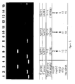
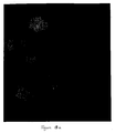
- Figure 1 illustrates the analysis of liver and non-liver cells by polymerase chain reaction (PCR) with primers to several exons of alpha-fetoprotein mRNA.
- PCR Analysis reveals truncated AFP in hemopoietic cells.
- the complete AFP species is observed in lanes 3 and 6.
- the inventors have designed nine PCR primers in order to characterize variant forms of hAFP mRNA, as exemplified in Example 1.
- the coding sequence of AFP extends from exon 1 to exon 14. All primer combinations other than the one for exon 1 of AFP mRNA amplify the portion of the AFP mRNA in a human erythroleukemia cell line, K562, whereas all combinations detected AFP mRNA in human hepatic cell lines HepG2 and Hep3B. This demonstrates that variant forms of AFP mRNA contain from exon 2 to exon 14, as expressed in K562, but do not cover the entire coding sequence of AFP.
- the only useful primers for identifying hepatic cells are those that detect the portion of exon 1 of AFP, the expression of which is more provably restricted in a tissue-specific manner.
- exon 1 is unique to hepatic progenitor subpopulations enables one to use it as a probe for identifying hepatic versus hemopoietic progenitor cell types.
- albumin is also analyzed in both hepatic and hemopoietic cells. Primers for albumin are developed in a fashion analogous to that for AFP (see above) and used to assess albumin expression in hepatic versus hemopoietic cell lines. As for AFP, a truncated form is found in K562, the hemopoietic cell line, and a transcript that is detected by the primer for exon 12-14.
- This invention discloses the design and preparation of specific primers of RT-PCR to determine the expression pattern of variant forms of AFP and albumin mRNA in hepatic versus hemopoietic cell populations.
- the invention as disclosed herein demonstrates that variant forms of both AFP and albumin mRNA can be found in hemopoietic progenitors. It means that when such sensitive assays are used, additional criteria, such as the use of an exon 1 probe for AFP, must be used to define hepatic from hemopoietic cell populations.
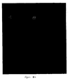
- Figure 2 illustrates the analysis of liver and non-liver cells by PCR to several exons of albumin. Since a truncated form of AFP mRNA is found in some subpopulations of hemopoietic cells, albumin is also analyzed in both hepatic and hemopoietic cells. Primers for albumin are developed in a fashion analogous to that for AFP (see above) and used to assess albumin expression in hepatic versus hemopoietic cell lines. As for AFP, a truncated form is found in K562, the hemopoietic cell line, and a transcript is detected by the primer for exon 12-14.
- liver progenitor cells of this invention demonstrate that fetal liver is both a hepatopoietic and hematopoietic organ during intrauterine development.
- the fetal liver contains large numbers of hematopoietic cells, especially of the erythroid lineage.
- this interrelationship includes the joint expression of AFP and albumin, or perhaps isotypes of this protein.
- exon 1 of AFP is unique to hepatic progenitor subpopulations enables one to identify specific subpopulations of liver progenitor cells of this invention.
- AFP and albumin are critical guides in the identification of hepatic cells, AFP is especially diagnostic of the hepatic progenitor cells after their purification by flow cytometry because of its intense expression in the hepatic progenitors. AFP is adopted also to estimate the purity of hepatic progenitors after any kind of fractionation strategy.
- the inventors have established methods that optimally yield dissociated human liver progenitors from fetal or adult livers.
- the isolation of mature liver cells usually involves enzymatic and mechanical dissociation of the tissue into single cell suspensions followed by fractionation with density gradient centrifugation, centrifugal elutriation, differential enzymatic digestion protocols (i.e. hepatic stellate cells), and/or with selection using cell culture (reviewed in Freshney, "Culture of Animal Cells, A Manual of Basic Technique" 1983, Alan R Liss, Inc. NY ). Density gradient centrifugation is used routinely by most investigators to eliminate what they assume to be debris and dead cells by discarding all fractions and retaining only the final pellet.
- the protocol disclosed herein is unique in that it makes use of the upper fractions of a density gradient and excludes the pellet.
- the novel variation to the density gradient centrifugation, as disclosed herein, is that the pellet is discarded and cells with a lower buoyant density (i.e., cells collecting at or near the top of the gradient) are retained.
- the inventors have found that younger (i.e. diploid) and cells more robust upon cryopreservation are present at the top of or within the Percoll density gradient, rather than in the pellet.
- Debulking is a process for enrichment of liver progenitors.
- the progenitors may be any of several lineages, including hepatic, hemopoietic, and mesenchymal.
- As the liver has a variety of mature cells, which can be tetraploid or polyploid, it is useful to remove some, or all, mature cells to prepare an enriched population of progenitors. It is advantageous but not essential to carry out the debulking step at 4 °C.
- the cells After preparation of a single cell suspension of liver cells, the cells are separated into multiple fractions according to cell size, buoyant density, or a combination of both.
- the liver progenitor cells are less than 15 microns in diameter. Any separation method that separates such small cells from larger cells and from cell debris is suitable, including sedimentation velocity in culture medium (which can be basal medium or enriched medium), gradient sedimentation, chromatography using large pore size separation beads, among others.
- the gradient material can be polyvinylpyrrolidone-coated silica (Percoll), cross-linked sucrose (Ficoll), dextran or any known to those in the art, and prepared to be isotonic to prevent cell lysis, in, for example, phosphate-buffer saline or Eagle's basal medium (BME).
- BME Eagle's basal medium
- the suspension of dissociated cells is typically applied to the top of a layer of the gradient material and subjected to a centrifugal field, while kept at 4°C.
- the cell suspension may be applied to an apheresis unit, such as is used for isolation of blood components, i.e. plasmapheresis.
- the debulking step can comprise centrifugal elutriation, panning based on cell surface adherence proteins, affinity chromatography or batch processing, tagging with fluorescent labels, zonal centrifugation, continuous-flow centrifugation, magnetic sorting after incubation with magnetic beads, e.g. magnetic beads complexed to antibodies, or combinations of these methods.
- the density gradient centrifugation can be a discontinuous gradient or a continuous gradient.
- the Percoll fraction is suitable for immediate use, cryopreservation, establishment in culture, or further enrichment. Further enrichment can be accomplished by panning, affinity selection, FACS sorting or any of the techniques known in the art and described above.
- Negative selection is accomplished by removal of cells expressing markers for CD45, glycophorin A, or other markers as mentioned below.
- Positive selection is accomplished by selection of cells expressing CD14, CD34, CD 38, ICAM or other markers indicative of expression of full-length alpha-fetoprotein, albumin, or both.
- non-progenitors are selectively removed by selective lysis.
- Red cells are lysed by brief exposure of the cell suspension to an isotonic solution of ammonium chloride, followed by dilution with culture medium and centrifugation to remove red cell "ghosts" and free hemoglobin.
- non-progenitors are selectively and hydrolytically lysed by freezing using the cryopreservation mixture described below.
- the various methods of debulking remove polyploid cells, cells that express markers associated with mature hemopoietic cells, cells that express markers associated with mature hepatic cells, cells that express markers associated with mature mesenchymal cells, and combinations of these cells.
- Cryopreservation methodologies of this invention are unique and distinct from the methods used in the prior art. Major distinctions are the use of different buffers and cryopreservation of a hepatic progenitor population which is low in density and, thus, buoyant in gradient centrifugation. The hepatic progenitors are small is size and diploid.
- successful cryopreservation of mature human liver cells is highly desired but has never been achieved in the art.
- successful cryopreservation is defined as the ability to freeze the cells at liquid nitrogen temperatures (-160-180°C) and then to thaw them, observe viabilities of>75% and with the ability to attach onto culture dishes.
- mature hepatocytes of rodent or human origin have viabilities of 30-40% with no ability to attach after freezing under the above conditions (for example see Toledo-Pereya, et al.,U.S. Patent No. 4,242,883 ; Fahy et al., U.S. Patent No. 5,217,860 ; Mullon et al., U.S. Patent No.
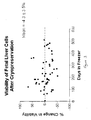
- Figure 3 illustrates the excellent viability of liver cells cryogenically stored accordingly to the method of the invention. Data are expressed as the percent change in viability measured at the time of processing versus the time of thawing. These data indicate that the cryopreservation did not affect significantly the viability of the cells. There was no significant change in viability over a period extending to 550 days in storage.
- the special cryopreservation methodology of this invention includes the use of a novel buffer, a novel cell population, and optionally embedding the cells in forms of extracellular matrix. This methodology for the first time achieves a viability upon thawing that is not different from the viability measured prior to freezing, immediately after cell dispersion.
- the invention teaches a method of isolating progenitors from human liver comprising providing a substantially single cell suspension of human liver tissue, and subjection the suspension to a positive or negative immunoselection.
- the method of immunoselection can comprise selecting from the suspension those cells, which themselves, their progeny, or more mature forms thereof express at least one marker associated with at least one of the cell lineages. These cell lineages can be hemopoietic, mesenchymal, hepatic, or some combination of these cell lineages.
- the cell selection step can include removing cells that express glycophorin A, CD45, an adult-liver-cell-specific marker, connexin 32, or combinations of these.
- the selection method can include removing polyploid cells, cells that express markers associated with mature hemopoietic cells, cells that express markers associated with mature hepatic cells, cells that express markers associated with mature mesenchymal cells, or combinations thereof.
- the selection of cells can comprise selecting cells that express CD 14, CD34, CD38, ICAM, or combinations thereof.
- the method can identify and select mature hemopoietic cells that express glycophorin A, CD45, or a combination of these.
- the selection method can select mature mesenchymal cells that express retinoids, von Willebrand Factor, Factor VIII, or combinations thereof.
- the immunoselection method can be carried out in conjunction with debulking based on cell size, buoyant density, or a combination thereof.
- the selection method can select cells that express at least one marker associated with at least one cell lineage, which may be hemopoietic, hepatic, or mesenchymal.
- the selection of cells, their progeny, or more mature forms thereof can express at least one marker associated with at least one hepatic cell lineage. That lineage can be parechymal cells or hepatocytes, or biliary cells.
- the markers expressed by the cells can be CD14, CD34, CD38, CD117, ICAM, or combinations thereof.
- liver progenitors as immature cell populations that express alpha-fetoprotein with or without expression of albumin, we have assessed markers that will select specifically for these cells using immunoselection technologies.
- markers i.e. CD34
- CD34 markers that are classical ones for hemopoietic progenitors, also identify hepatic progenitor subpopulations.
- single color sorts for CD34 resulted in significant enrichment (at least 9-fold) for cells that express AFP.
- not all of these AFP-positive cells can be verified to be hepatic progenitors.
- This invention uses a unique flow cytometric sorting strategy. Using the combination of AFP and albumin expression as two uniquely defining features of hepatic progenitors, we have identified antigenic markers and other flow cytometric parameters that define the hepatic progenitor cells.
- the sorting strategies to date involve sorts for small cells ( ⁇ 15 ⁇ by measures of forward scatter), that are diploid (using fluorescence from Hoechst dye 33342), are agranular by side scatter, are negative for certain hemopoietic antigens (i.e. glycophorin A, the red blood cell antigen and CD45) followed by positive markers shared between hepatic cell subpopulations and hemopoietic cell subpopulations (i.e. CD14 and/or CD38.)
- hemopoietic antigens i.e. glycophorin A, the red blood cell antigen and CD45
- the inventors identify hepatic progenitor cells by sorting for those cells that strongly express alpha-fetoprotein , weakly express albumin, and express CD14, CD34, CD38, CD117, or a combination thereof. Also, described herein is the evidence that hemopoietic cells also express AFP, albeit a truncated form.
- the inventors describe a novel cell population and process of isolation, identification, culture, and a method of using such cell population. The success in the isolation, identification, and culture of the particular cell population of the invention is achieved partly through advanced methods of isolation, affinity debulking, high-speed fluorescence-activated cell sorting, greater speed and accuracy, and modified cryopreservation and culture techniques.
- Applicants demonstrate flow cytometric sorting strategies and methods to purify liver progenitors from freshly isolated cell suspensions and/or from thawed cryopreserved liver cells. These methods involve 1) staining of the cells with several fluoroprobe-labeled antibodies to specific cell surface markers and 2) using a combination of negative and positive sorting strategies in multiparametric flow cytometric technologies.
- the methods for purification of specific lineage stages from human hepatic cell populations can be used with livers from any age donor, since the markers appear to be lineage-position specific.
- Figure 4 illustrates a univariant FACS sort.
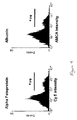
- the cell suspension is prepared for immunofluorescence analysis of alpha-fetoprotein (AFP) using antibodies conjugated to the red dye, Cy5, and for albumin using antibodies conjugated to the blue dye (AMCA).
- AFP alpha-fetoprotein
- ACA blue dye
- Thirty thousand cells are screened for red (AFP) and blue (albumin) fluorescence.
- the results show a clear group of cells positive for each protein. Further analysis shows that about 80% of the positive populations for each protein are represented by the same cells (i.e. co-expression of the two proteins).
- the expression of AFP and albumin like immunoreactivity is well defined in the cell suspensions, with a clear group of cells showing a clear differentiation from the background signal.
- Alpha-fetoprotein is expressed in 6.9 ⁇ 0.86% of cells in unfractionated cell suspensions and albumin is present in 7.7 ⁇ 1.1%. Among AFP positive cells 75.6 ⁇ 4.9% co-expressed albumin while 80 ⁇ 5.5 % of albumin positive cells also expressed AFP. Thus, approximately 25% of cells expressing alpha-fetoprotein do not express albumin and 20% of cells expressing albumin do not express alpha-fetoprotein.
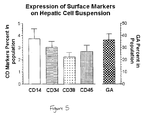
- Figure 5 illustrates the percent of cells expressing surface markers CD14, CD34, CD38, CD45, and Glycophorin A (GA) in unfractionated liver cell suspensions. Note that the GA data is plotted on the right axis to preserve scale.
- Figure 6 illustrates the percentage of cells in the original cell suspension expressing alpha-fetoprotein and other antigenic markers. Mean ⁇ SEM for percent of cells positive for alpha-fetoprotein (AFP) and specific cell surface markers (CD14, 34, 38, 45 and glycophorin A).
- AFP alpha-fetoprotein
- CD14, 34, 38, 45 and glycophorin A specific cell surface markers
- glycophorin A (GA) positive cells i.e. erythroid cells
- Figure 7 illustrates the co-expression of alpha-fetoprotein and albumin.
- the percent of AFP positive cells co-expressing albumin is also increased to 80.5 ⁇ 8.2% and the proportion of albumin-positive cells co-expressing AFP increased to 89 ⁇ 3.1%, though neither change is statistically significant.
- Figure 7 illustrates the effect of debulking by Percoll fractionation on alpha-fetoprotein and albumin co-expression.
- the proportion of cells expressing both alpha-fetoprotein and albumin expressed as a percentage of AFP or albumin positive cells.
- Data for cells with and without red cell depletion are shown using Percoll fractionation.
- Percoll fractionation When cell suspensions are depleted of red cells by Percoll fractionation the proportion of cells expressing AFP is increased significantly to 12.9 ⁇ 1.9% and those expressing albumin to 12.1 ⁇ 2.3%.
- Table 3 Percentage of CD Positive Populations in Liver Cell Suspension after Depletion of Red Cells and percentage of these that are positive for AFP CD14 CD34 CD38 CD45 GA Red cell depleted % in population 7.4 ⁇ 1.3 3.4 ⁇ 0.5 4.8 ⁇ 0.9 8.2 ⁇ 0.3 27.5 ⁇ 4.7 % AFP positive 89.8 ⁇ 1.3 77.1 ⁇ 2.9 53.5+7.2 32.5 ⁇ 1.3 1.8 ⁇ 0.9
- Figure 8 illustrates a FACS analysis of fetal liver cell suspension for co-expression of CD14, CD38 and AFP.
- the bivariate scattergram shows the distribution of TriColor staining for CD14 (ordinate) versus FITC staining for CD38 (abscissa). Gates are created to select specific cell groupings according to the CD14 and CD38 signals. These are then used to display the intensity of AFP staining in each of these subgroups.
- the AFP results show that a high level of enrichment for AFP is produced by selecting cells positive for either CD38 or CD14.
- the AFP signal generated from the entire cell suspension (30,000 cells) is shown at the lower left.
- AFP in the subgroups selected by cell surface marker is distributed continuously with a clear preponderance of cells showing staining intensities in the positive range.
- CD38 positive cells with respect to co-expression of AFP was unique.
- CD38-positive cells a bimodal distribution for AFP co-expression is apparent in which two distinct groups of cells are apparent, one group positive for AFP, the other negative.
- alpha-fetoprotein is present in 7% of the cells in single cell suspensions of fetal liver tissue (i.e. in the original cell suspension).
- the antibody to glycophorin A an antigen on red blood cells, erythrocytes
- erythrocytes an antigen on red blood cells, erythrocytes
- the CD38 antigen identifies a population of cells that shows significant enhancement in the proportion of AFP positive cells ( i . e ., greater than 7 times the proportion in unfractionated samples.
- Both antigens show a number of isoforms, depending on whether or not there are sections of the molecule, encoded by splicing variants, present. Antibodies are available that identify the various isoforms.
- CD34 hemopoietic progenitor cells
- the classic marker for hemopoietic progenitor cells, CD34 is found to be present on many cells that also express AFP.
- the sorting of cells positive for CD34 results in enrichment of AFP-positive cells at least 9 fold over that found in the original cell suspension (67%, in the CD34-positive cells vs 7% in the original cell suspension).
- CD14 the most effective single antibody for enrichment of AFP positive cells is CD14, which produces a greater than 11 fold increase in the proportion of these cells compared to the original population (81% versus 7%).
- the yield of AFP positive cells could be improved by using a combination of surface markers.
- the extent of co-expression of AFP with selected combinations of surface markers is determined to establish the extent to which the selection the intracellular marker can be increased.
- the data are expressed as the proportion of AFP positive cells expressing surface markers (termed the “yield” of AFP positive cells) and as the proportion of all AFP positive cells that appear in the population defined by the surface marker (termed the "enrichment” factor for AFP positive cells).
- Results for combinations of CD 14, CD34 and CD38 are shown in Table 4 together with the results from individual markers for comparison. Table 4.
- Figure 9 illustrates how selection for CD14 and CD38 enriches for AFP positive cells.
- the proportion of AFP-positive cells in cell suspensions prepared from fetal liver is enhanced dramatically by selecting cells with positive surface labeling for the markers CD38 and CD14.
- the combination of the two markers produces a significantly better enrichment of AFP-containing cells than that obtained with either marker alone.
- Figure 10 illustrates fluorescence microscopy of human hepatic progenitor cells.
- the morphology of cells staining positive for AFP is variable and encompassed the entire range of cell size and shape in the cell suspension from fetal livers but not adult liver.
- the largest of the AFP-positive cells approximately 12-15 ⁇ , is much smaller than mature hepatocytes, ranging in size from 20-50 ⁇ ).
- Figure 11 illustrates representative cells selected by expression of AFP.
- the cells with positive staining for CD14 (right side) are characteristic of hepatoblasts.
- the cells with negative staining for surface markers are smaller and consistent in size and morphology with early hepatic progenitors. In all cases a certain proportion of AFP positive cells show no expression of any surface antibodies used in this study.
- the appearance of these AFP-positive "null" cells is illustrated in Figure 11 where they can be compared with the appearance CD14 positive/AFP positive cells sorted from the same suspension. It is clear that while both cell types are positive for AFP, the cells staining negative for surface antigens are consistently smaller and less complex than the CD14 positive cells.
- the probable markers for sorting hepatic progenitors are: Glycophorin A - , CD45 - , ICAM + , and one or more CD14 + , CD34, CD38 + , CD117, diploid, agranular (by side scatter), less than 15 ⁇ (by forward scatter).
- the phenotype of these sorted cells is small cells ( ⁇ 15 ⁇ ), with little cytoplasm (high nucleus/cytoplasm ratio), albumin + and/or AFP +++
- Confocal microscopy has been used to obtain the images from human fetal and adult cells that express alpha-fetoprotein. This methodology enables one to observe the morphology and size of these cells and to show directly the location of intracellular proteins, such as AFP and ALB, and that of membrane surface markers such as CD34 and CD38.
- Figure 12 illustrates confocal miscroscopy of alpha-fetoprotein expressing cells, that is, hepatic progenitors in adult human livers.
- the figure shows three view of one field, and that there are two AFP-positive cells in this field.
- the overlay of panel (A) and panel (B) is shown in panel (C) and indicates the morphology of AFP positive cells (colored pink, in the original) in a group of liver cells.
- Figure 13 illustrates cells that are labeled with calcein (A) to show all cell types.
- Fig. 13(B) consist of the same cells co-expressing AFP and showing that only two cells are AFP-positive. Cell size is not a factor for AFP positivity.
- AFP-expressing cells are found in both fetal and adult livers. Fetal livers, as expected, have the highest percentage (6-7%), whereas adult livers have a small percentage ( ⁇ 1%) and with the numbers declining with age of the donor. The few hepatic progenitors found in adult livers can be enriched significantly through the Percoll fractionation process to yield up to 2% of the cells in Percoll fractions 1 and 2 from the adult livers (Table 5). No AFP-expressing cells are found in a liver from donors older than 71 years of age.
- Table 5 shows the cell size and viability from Percoll-isolated fractions of adult liver cells. Smaller cells (fractions 1-3) have higher viability than larger cells (fraction 4) after being cryopreserved under the same cryopreservation condition. Percoll Fraction Viability(%) Cell Size ( ⁇ m) % AFP+ cells Fraction 1 82 > 12 0.5-1 % Fraction 2 84 10-15 2 % Fraction 3 85 15-25 ⁇ 0.2 % Fraction 4 56 25-50 ⁇ 0.01%
- liver cell therapies as well as for organ transplantation will consist of those from of young donors (up to about 45 years of age.
- livers from geriatric patients >65 years of age will be inappropriate donors for cell therapies and perhaps also for whole organ transplants, especially for children, since they will have little if any regenerative capacity from hepatic progenitors and only the intermediate or minimal regenerative capacity known to be available from the mature cells.
- adult liver contains a hepatic progenitor cell population capable of growth and differentiation into hepatocytes and biliary cells under both normal and disease conditions.
- This invention stands for the proposition that every position in the liver lineage is a distinct maturational stage, and that there are multiple stem cell populations in the liver.
- the embryonic liver of the present invention yields progenitor cells for 3 separate maturational lineages: hepatopoiesis, with cell fates of hepatocytes and biliary cells (bile duct); hemopoiesis, with cell fates of lymphocytes (B and T), platelets, macrophages, neutrophils, and granulocytes; and mesengenesis, with cell fates of endothelia, fat cells, stromal cells, cartilage, and even bone (the last two occurring in the liver only under disease conditions).
- stem cells are immature cells that can give rise to daughter cells with more than one fate.
- the stem cells produce daughter cells, some of which are identical to the parent and some of which "commit” to a specific fate.
- the commitment process is not understood on a molecular level. Rather, it is recognized to have occurred only empirically when the fates of cells have narrowed from that of a predecessor.
- “Committed progenitors” are defined as immature cells that have a single fate such as hepatocytic committed progenitors (giving rise to hepatocytes) or biliary committed progenitors (giving rise to bile ducts).
- the transitions from the stem cell to the adult cells occur in a step-wise process yielding a maturational lineage in which cell size, morphology, growth potential and gene expression is tied to the lineage.
- the metaphor of aging is useful in defining the process.
- the "young" cells have early gene expression and the greatest growth potential; the cells late in the lineage have “late” gene expression and usually are limited in their growth or do not grow at all.
- the late cells can be considered “old” or in biological terms, apoptotic, and ultimately are sloughed off.
- the maturational lineage process results in a natural turnover for the tissue and allows for regeneration after injuries. Tissues differ in the kinetics of the maturational process.
- the maturational lineage of the gut is quite rapid with a complete cycle occurring in less than a week; that of the liver is slow occurring, and in the rat liver is about a year.
- the rat liver forms in embryonic life at about day 10, referred to as "embryonic day 10" or E10, with the invagination of the cardiac mesenchyme by endoderm located in the midgut region of the embryo( Zaret, K. 1998. Current Opinion in Genetics & Development. 8:526-31 .). Earliest recognition of liver cells in the embryos has been by achieved using in situ hybridization studies for mRNA encoding alpha-fetoprotein (AFP) (( Zaret, K. 1998. Current Opinion in Genetics & Development. 8:526-31 ; Zaret, K. 1999 Developmental Biology (Orlando). 209:1-10 ).
- AFP alpha-fetoprotein
- AFP-expressing cells are observed in the midgut region of the embryo near the mesenchyme that produces the heart on day 9-10 in all rat and mouse livers assayed.
- the liver becomes macroscopically visible by E12 and is about 1 mm in diameter by E13.
- hemopoiesis occurs with the first identifiable hemopoietic cells appearing by E15-E16 (in rodents) and by the 3 rd to 4 th month (in humans) and with the peak of erythropoiesis (formation of erythroid cells or red blood cells) occurring by E18 (in rodents) and by the 5 th -6 th month (in humans).
- E15-E16 in rodents
- E18 in rodents
- 5 th -6 th month in humans
- the end of the gestational period is on day 21 in rodents and 9 months in humans.
- the hemopoietic progenitors prefer relatively anaerobic conditions and flee to the bone marrow (which is relatively anaerobic) with the elevated oxygen levels in the liver with the activation of the lungs; and second, the loss of the pregnancy hormones are the cause of the migration.
- Postnatally the loss of the hemopoietic progenitors in the liver is associated with a dramatic reduction in the numbers of hepatic progenitors and a parallel increase in the numbers and maturity of the hepatocytes and biliary cells.
- Full maturity of the liver is completed by 2-3 weeks postnatal life (in rodents) and within a few months (humans). By then the remaining hepatic progenitor cells are localized to the regions of the portal triads in the periphery of each liver acinus.
- each acinus being defined peripherally by six sets of portal triads, each one having a bile duct, an hepatic artery and an hepatic vein, and in the center a central vein that connects to the vena cava.
- Plates of liver cells like spokes in a wheel, extend from the periphery to the center.
- the plates are divided into three zones: Zone 1 is near the portal triads; zone 2 is midacinar; and zone 3 is near the central veins.
- Zone 1 is near the portal triads; zone 2 is midacinar; and zone 3 is near the central veins.
- the only diploid cells of the liver are in zone 1; tetraploid cells are in zone 2; and tetraploid, octaploid and multinucleated cells are in zone 3.
- the pattern is highly suggestive of a maturational lineage that ends in an apoptotic process (( Sigal, S. H., S. et al. 1995.
- the in vitro and in vivo growth and differentiation characteristics of the cell population of this invention is in agreement with the concept and implications of a lineage -position lineage model in liver.
- a lineage -position lineage model in liver For example, in an in vitro parenchymal culture, the ability of the parenchymal cells to divide and the number of cell divisions are predicted to be strictly lineage-position dependent. Therefore, periportal parenchymal cells should have greater division potential than pericentral ones. This explains the long-standing mystery of why primary cultures of liver, the most renowned regenerative organ in the body, show such limited cell division in culture.
- hepatomas Stem cells and their transformed counterparts, hepatomas, are predicted to express early genes such as alpha-fetoprotein and insulin-like growth factor II, but not genes expressed later in the lineage. In the maturity-lineage model no hepatoma should express late genes, because full progression through the lineage requires undisturbed regulation of differentiation, growth, and cell cycling. This indeed has been observed in the cell population of the invention.
- Molecular biological studies comparing liver-specific gene expression in embryonic versus adult tissues define several classes of genes: those diagnostic of the compartments (stem cell, amplification, differentiation); those expressed zonally and potentially crossing compartmental boundaries; and those expressed early, middle, or late in the lineage but discretely in one few cells.
- Various morphological and gene expression patterns of primary liver tumors may be understood by studying the cell population of the invention. If tumors represent the proliferation of transformed stem cells with varying capacities of differentiation, the common expression of alpha-fetoprotein in hepatomas is not an induced tumor marker but an indicator of an expanded immature cell population that normally expresses alpha-fetoprotein.
- the isolated cell population of this invention has a great impact on the success of liver-directed cell and/or gene therapy.
- This invention as described in the Examples, has identified key conditions in which nonhuman primate and human hepatic progenitors can be successfully cryopreserved.
- the cell population of this invention can be used as a "punch biopsy material" to provide the cell seed for ex vivo expansion. This would eliminate the necessity for major invasive surgical resection of the patient's liver.
- gene transfer is performed. This can be accomplished with a number of different gene delivery vector systems.
- An important consideration at this point is that successful gene transfer requires a rapidly growing culture, and since human hepatic progenitors of the invention significantly divide under normal physiological conditions, these cells are ideal candidates for gene transfer to liver. Also, the growing characteristics of the cell population of this invention permits the use in an ex vivo gene transfer using certain gene delivery vectors ( i.e. , retroviral vectors) which will require cell proliferation for efficient gene insertion and expression.
- An alternative approach for gene therapy is to design vectors that target the progenitors specifically and then to inject the vector, coupled with the gene of interest, directly into the patient.
- the vectors would target and modify the endogenous progenitor cell population.
- the progenitor cell population of this invention can be used in an autologous or allogeneic liver-directed cell or gene therapy.
- autologous hepatic progenitors will eliminate a significant concern regarding rejection of the transplanted cells.
- the cell population of this invention is particularly attractive for allogenic cell transfer, because their antigenic profile suggests minimal immunological rejection phenomena.
- other cellular elements such as blood cells, endothelial cells, Kupffer cells, that are known to be highly immunogenic are substantially eliminated through the purification process.
- the autologous or allogenic hepatic progenitors are isolated purified and cultured, they can be genetically modified or remain intact, expanded in vitro, and then transplanted back into the host. If genetic modification is desired, after genetic modification and before transplant, those genetically modified cells may be expanded and/or selected based on the incorporation and expression of a dominate selectable marker. Transplant can be back into the hepatic compartment or an ectopic or heterotopic site. For transplant into the Hepatic compartment, portal vein infusion or intrasplenic injection could be used. Intrasplenic injection may be the administration route of choice because hepatic progenitors transplanted via an intrasplenic injection move into the hepatic compartment.
- Additional medical procedures may assist in the efficacy of hepatic engraftment of the transplanted hepatic progenitors.
- Animal models have demonstrated that in partial hepatectomy, administration of angiogenesis factors, and other growth factors aide in the engraftment and viability of the transplanted hepatocytes.
- An alternative approach is to transplant the genetically modified hepatocytes to an ectopic site.
- the cell therapy approaches with respect to liver have shown little efficacy. This may be due to the fact that the donor cells being used are predominantly adult liver cells and are short-lived after isolation and reinjection. In addition, the use of adult cells results in strong immunological rejection.
- the hepatic progenitor cells of the instant invention offer greater efficacy because of their limited capacity to elicit immunological rejection phenomena and because of their extensive regenerative potential.
- the improved methodologies enabled the inventors to more closely study and characterize hepatic progenitors. These studies revealed a specially close relationship between hepatic progenitors and hemopoietic progenitors suggesting a close relationship between these two lineages. Indeed, these studies show that the progenitor cells of the hepatic and hemopoietic lineages share numerous antigenic markers (CD14, CD34, CD38, CD117 or ckit, oval cell antigens), share biochemical properties (i.e. transferrins, glutathione-S-transferases, and a truncated isoform of alpha-fetoprotein), and have extensive overlap in the culture requirements (forms of extracellular matrix and specific hormonal requirements) for expansion ex vivo.
- antigenic markers CD14, CD34, CD38, CD117 or ckit, oval cell antigens
- biochemical properties i.e. transferrins, glutathione-S-transferases, and a truncated isoform of al
- progenitor cells of both lineages are located in the same sites within the liver acinus.
- paracrine signaling is present throughout the cells of the two maturational lineages; that is signals produced by each of the lineages regulates cells in the other lineage. Indeed, it may be concluded that there may be a common lineage or at the very least interdependent lineages between the hepatic and hemopoietic cells.
- the cell populations disclosed herein are purified and utilized to yield either myelo-hemopoietic cells or hepatic derivatives depending on the conditions under which the cells are isolated and cultured. Therefore, bioreactor systems inoculated with cell populations sorted for a set of antigens that defines both hepatic and hemopoietic progenitors (e.g. CD38 + , ckit + , CD45 + ) can result in cell populations with multiple fates. The fate depends on how the cells are reintroduced in vivo or under what culture conditions the cells are placed.
- CD34 positive cells of bone marrow has been used as a convenient positive selection marker for hemopoietic stem cells.
- CD34 antigenic marker for hemopoietic stem cells Nakauchi H. Nature Medicine 4:1009-1010 (1998 )
- Experimental evidence demonstrates the existence of cells in the CD34 negative population of human bone marrow and cord blood that can repopulate the bone marrow of immunodeficient mice.
- This invention discloses ways to purify both the hemopoietic and the hepatic progenitor cell populations which are used subsequently in the clinical and pre-clinical programs, utilizing the close relationships between the hepatic and hemopoietic cells.
- human hepatic progenitors are many and diverse. They include: 1) research on human cells; 2) production of vaccines or antivirals; 3) toxicological studies; 4) drug development; 5) protein manufacturing (using the cells as hosts for production of various human-specific factors); 6) liver cell therapies; 7) liver gene therapies; 8) bioartificial livers that can be used in research, toxicological and antimicrobial studies, protein manufacturing, or clinically as a liver assist system.
- the same cells can be used both for hepatic or hemopoietic fates depending upon the microenvironment in which they are placed.
- the availability of highly purified human hepatic progenitor cells will enable much more extensive research on human cells, will facilitate the development of successful forms of liver cell and gene therapy, and should enable the development of human bioartificial livers for use both in research and as clinical assist devices.
- the limited supply of healthy human tissues precludes clinical programs in liver cell therapy or in human bioartificial livers.
- the progenitor cell populations should have sufficient expansion potential to overcome, or at least greatly alleviate, that limited supply.
- Cell lines Two human hepatomas, Hep3B and HepG2, are maintained in Eagle's MEM supplemented with 1 mM sodium pyruvate, 2mM L-glutamine, 50 U/ml penicillin, 50 ⁇ g/ml streptomycin, 0.1 mM MEM non-essential amino acid solution, 5 ⁇ g/ml insulin and 10% FBS.
- a human erythroleukemia cell line, K562 and a mouse embryonic fibroblast cell line, STO are maintained in DMEM/F12 supplemented with 2 mM L-glutamine, 50 U/ml penicillin, 50 ⁇ g/ml streptomycin, 5 x 10 -5 M 2-ME and 10% FBS.
- RNAs are extracted from Hep3B, HepG2, and STO by the method of Chomcznski and Sacchi N. Anal. Biochem 162:156-159 (1987 ).
- the cDNAs are synthesized by oligo-dT priming and subjected to PCR amplification using primer sets designed by the inventors and prepared for human AFP or albumin.
- the primer sequences are as follows, For AFP: SEQ ID 1 1 hAFP1: 5'-ACCATGAAGTGGGTGGAATC-3', SEQ ID 2 hAFP2: 5'-CCTGAAGACTGTTCATCTCC-3', SEQ ID 3 hAFP3: 5'-TAAACCCTGGTGTTGGCCAG-3', SEQ ID 4 hAFP4: 5'-ATTTAAACTCCCAAAGCAGCAC-3', SEQ ID 5 hAFPexon2: 5'-CTTCCATATTGGATTCTTACCAATG-3'.
- PCR is performed in a total volume of 50 ⁇ l consisting of1 ⁇ M each primer, 200 ⁇ M each dNTP, 50mM KCI, 1.5mM MgCI2, 10mM Tris HCl, pH 8.3, and 1.25U Amplitaq polymerase (Cetus Corp). Samples are heated to 94°C for 3 min followed by amplification for 30 cycles of 2 min at 94°C, 2 min 62°C, and 3 min at 72°C. After the last cycle, a final extension step is performed at 72°C for 7 min. Then 5 ⁇ l of each PCR reaction is run on 2% agarose gel containing 5 ⁇ g/ml ethidium bromide in Tris-acetate-EDTA buffer.
- RT-PCR for AFP Human AFP gene consists of 15 exons ( Gibbs et al., Biochemistry, 26: 1332-1343 ). To distinguish truncated transcripts from functional complete AFP mRNA, two different portions of AFP cDNA sequence are selected as target molecules of RT-PCR. The primer combination of hAFP1 and hAFP2 is used for the amplification of exon 1 containing the initiation MET to exon 3, whereas that of hAFP3 and hAFP4 amplify exon 12 to exon 14 containing the stop codon. The results of the PCR are shown in Figure 1 .
- the PCR of Hep3B and HepG2 cDNA shows the single remarkable band of 1.8 Kb (lanes 3 and 6), whereas there is no band in K562 (lane 9).
- the controls are samples with no RNA and a sample derived from the mouse embryonic fibroblast cell line (STO). Neither shows any detectable band.
- RT-PCR for albumin Human albumin gene consists of 15 exons also ( Minghetti et al., J. Biol. Chem, 261: 6747-6757 ).
- AFP the primer combination of hALB1 and hALB2 is used for the amplification of exon 1 containing the initiation MET to exon 4, whereas that of hALB3 and hALB4 amplify exon 12 to exon 14 containing the stop codon.
- the results of the PCR are shown in Figure 17. Both combinations of the primers result in strongly detected amplification bands in the RNA from Hep3B and HepG2 (lanes 1, 2, 4, and 5).
- Suppliers for reagents include:
- Fetal Livers The fetal livers come from multiple clinics affiliated with Advanced Biosciences Research (ABR), all in California, or from the Anatomical Gift Foundation (AGF) with clinics in the South (i.e., Georgia, Virginia), Northeast (Pennsylvania) or Midwest (Kansas, Colorado).
- ABR Advanced Biosciences Research
- AGF Anatomical Gift Foundation
- the fetuses are collected from clinics; the tissues dissected free from the fetuses and placed into RPMI 1640 (Gibco) supplemented with insulin (Sigma, 5 ⁇ g/ml), transferrin (Sigma, 5 ⁇ g/ml), selenium (10 -9 M, and 5% fetal bovine serum (Gibco).
- the samples are then put on ice and shipped by courier to our lab, a process that can take 10-16 hours. Thus, we receive the samples approximately 24 hours after surgery.
- the samples are assigned a number with the prefix REN, given in chronological order of being received (REN 1, 2, 3, etc), where REN is an abbreviation for Renaissance.
- the adult livers come from the Anatomical Gift Foundation or from local surgeons (UNC) and consist of rejected liver tissue, explants from transplant recipients, or livers donated for organ transplantation but then rejected for reasons other than pathogens.
- the patients providing explant tissue or rejected donor tissue are screened for an array of diseases and only those found safe by these tests are used for cell professing
- the livers are put into University of Wisconsin solution (also called Viaspan) and shipped on ice to the lab.
- the time interval between organ removal from a brain-dead patient (“clamp time”) and its arrival in the lab is extremely variable. The specimens arrive within less than 24 hours of "clamp time", the time at which the liver is removed from the donor.
- Cadaveric Livers Livers obtained postmortem within at least 30 hours of death are obtained through local organ procurement associations (e.g. Carolina Organ Procurement Association or COPA). The livers are processed as for the adult livers.
- organ procurement associations e.g. Carolina Organ Procurement Association or COPA.
- COPA Carolina Organ Procurement Association
- HIV I and II The list of elements checked for investigator's safety is: HIV I and II, HTLV I and II, hepatitis B and C; tuberculosis.
- the list for clinical usage is: HIV I and II, HTLV I and II; hepatitis A, B, C, and G; EBV, CMV; tuberculosis, syphilis and mycoplasma.
- Fetal and adult livers are processed using a combination of enzymatic digestion and mechanical dissociation, fetal livers are prepared primarily by mechanical dissociation, whereas the adult livers are dissociated primarily by enzymatic digestion. A description of each is given below. Both fetal and adult livers are digested for varying lengths of time in an enzyme buffer that serves to dissolve the extracellular matrices that bind the cells together in a tissue.
- the collagenase enzyme mix used for isolation of liver cells is a high purity "Liberase” enzyme preparation manufactured by Boehringer-Mannheim, consisting of a mixture of purified collagenase and elastase. This enzyme mix can be used at much lower concentrations and with fewer deleterious "side effects.”
- Enzyme solution collagenase solution--60-70 mg/100 mls of buffer (Sigma's type IV collagenase, catalog #C5138 or Worthington's type B, catalog #LS005273; both being bacterial preparations enriched in collagenase but with many enzymatic impurities) or Liberase -- (purified collagenase/elastase preparation by Boehringer-Mannheim, catalog 1814184) prepared in P2 buffer (see below) and used at 0.23 mgs/ml
- Cell Wash Solution RPMI 1640 (Gibco) supplemented with insulin (5 ⁇ g/ml), transferrin (5 ⁇ g/ml), free fatty acid mixture (see below) bound 1:1 molar ratio to purified bovine or human serum albumin.
- Free Fatty Acid Mixture Immature cell populations, and damaged older liver cells, require lipids to maintain and to synthesize their membranes. Although fully mature hepatocytes can synthesize their membranes from a single fatty acid source (linoleic acid) younger parenchymal cells cannot and thus require a mixture of many different fatty acids to handle their lipid requirements.
- a complex mixture that is then bound in a 1:1 molar ratio with a highly purified albumin. A detailed description of the method for preparation of that fatty acid preparation is given below:
- the stock solutions are prepared as follows, for a combined total of 100 mM free fatty acids: Palmitic 31.0 mM Oleic 13.4 mM Palmitoleic 2.8 mM Linoleic 35.6 mM Stearic 11.6 mM Linolenic 5.6 mM
- Each individual component is dissolved in 100% EtOH as follows: Palmitic 1 M stock, soluble in hot EtOH Palmitoleic 1 M stock, readily soluble in EtOH Stearic 151 mM stock, soluble in heated EtOH at 1 g / 21ml Oleic 1 M stock, readily soluble in EtOH Linoleic 1 M stock, readily soluble in EtOH Linolenic 1 M stock, readily soluble in EtOH
- P1 Perfusion buffer calcium and magnesium free perfusion buffer (pH 7.2) with final concentrations as specified for each of the following components: 118 mM NaCl, 4.7 mM KC1, 1.2 mM KPO 4 pH 7.4, 2.5 mM NaHCO 3 , 0.5 mM EDTA, 5.5 mM glucose, 0.5% bovine or human serum albumin (BSA), Ascorbic acid (50 ⁇ g/ml), insulin (4 ⁇ g/ml), dexamethasone (1 ⁇ M).
- BSA bovine or human serum albumin
- Ascorbic acid 50 ⁇ g/ml
- insulin 4 ⁇ g/ml
- dexamethasone (1 ⁇ M).
- P2 Perfusion buffer Dulbecco's modified Eagle's medium or RPMI 1640 supplemented with 0.5% BSA, ascorbic acid (50 ⁇ g/ml), insulin (4 ⁇ g/ml) and dexamethasone (1 ⁇ M).
- DMEM Dulbecco's Modified Eagle's medium (Gibco) with glucose, sodium pyruvate and L-glutamine and further supplemented with 5% fetal bovine serum, insulin (4 ⁇ g/ml) and dexamethasone (1 ⁇ M).
- Percoll (Pharmacia, catalog #17089102) is diluted 9:1 with 10X Dulbecco's phosphate buffered saline.
- the fetal livers arrive in the transport buffer (described above) and on ice. They are rinsed with a "cell washing buffer” consisting of RPMI 1640 (Gibco) supplemented with insulin (Sigma; 5 ⁇ g/ml), transferrin (Sigma; 5 ⁇ g/ml selenium (Johnson Matthey's mass spec trace elements; 10 -9 M), and a free fatty acid mixture bound to bovine serum albumin in a 1:1 molar ratio.
- the fetal livers are then put into a collagenase buffer for 15-20 minutes and then gently pressed through a "cellector” (Sigma) with an 800 mesh grid to yield small aggregates of cells; the "cell wash buffer” is used to facilitate the dissociation process.
- the aggregates of cells are then fully dissociated by pressing them through a 70 Micron filter (Falcon cell strainer, 70 ⁇ m nylon, catalog #2350) using the "cell wash buffer" to facilitate the process.
- the cells that pass through the 70 micron filter are kept separate from those that do not. Both samples are cryopreserved and checked for percentage viability using the Trypan blue dye exclusion assay.
- the livers are catheterized by the portal vein, vena cava, or by both, perfused with buffers to eliminate blood; and then perfused with buffers containing collagenases/proteases to enzymatically dissociate the cells.
- the tissue is pressed through cheesecloth or a nylon filter or raked with a comb to mechanically complete the cell dissociation process.
- the dissociated cells are rinsed with a buffer containing serum to inactivate the collagenase and other enzymes used in the perfusion process.
- the perfusion buffers, P1 and P2 are placed in a water bath at 37°C.
- the perfusion is carried out in a Miller type perfusion box, which is maintained at 37°C throughout the perfusion.
- the buffers are oxygenated during the perfusion. All tubing in the box is rinsed with 70% ethanol, followed by distilled water and then with PI to ensure that the air has been removed from the system.
- the liver is cannulated using a Teflon cannula from a 16-gauge needle attached to 60 ml syringe to flush ice-cold PI buffer through the liver using various blood vessels available on the cut surface of the liver for large pieces of liver (100-300 gms).
- the remnants of the vena cava can be cannulated.
- the various blood vessels in chunks of liver are tested to learn which will offer optimal perfusion of the tissue. This procedure also removes any excess blood from the liver.
- the chosen blood vessel is cannulated and sealed into place using medical grade adhesive (medical grade "superglue"). All other large vessels and surface openings are sealed using the medical grade adhesive, and, if required, using Q-tips with the adhesive to help seal the openings.
- the liver specimen is placed on a nylon mesh within an appropriate size glass bowl.
- the P1 buffer is added to the bowl, and the liver submerged in the buffer.
- the bowl containing the liver is placed inside the perfusion box, and the outlet tubing of the cannula is attached.
- the P1 buffer is recirculated for 15 minutes starting at a low speed of about 24 mls/min and then slowly increased to between 58 mls/min and 90 mls/minute to optimize a flow rate with an acceptable back pressure. One must check that there are no excessive leaks of the perfusate from the liver.
- the P1 buffer is removed from the bowl and replaced with the P2 buffer containing the collagenase.
- the P2 buffer is recirculated until the liver is sufficiently digested (evaluated by color-conversion of liver from dark reddish brown to pale brown and by acquisition of mushy texture to liver).
- the P2 buffer is recirculated for no longer than 20-25 minutes. Once the perfusion has ended, the P2 buffer is drained from the bowl and the liver transferred in the bowl to a biological hood.
- the cell culture medium (DMEM) is added to the bowl, and the cannula and the adhesive is removed along with any undigested regions of the liver.
- the capsule of the liver (Glisson's capsule) is broken using tissue forceps and scissors. This allows the release of the digested tissue into the medium leaving behind the connective tissue and any undigested material.
- the digested material is put into the DMEM and then filtered through a series of different size filters. The filters are placed inside a large funnel to aid the filtration.
- the digested material is filtered first with a single layer of cheesecloth, followed by a 400 ⁇ m nylon filter, and finally through a 70 ⁇ m Teflon filter.
- the filtrate is divided equally into centrifuge tubes and centrifuged at 70 g for 4 minutes.
- Fraction 1 After centrifugation, prior to the addition of Percoll, the supernatant is referred to as the Fraction 1 (F1).
- DMEM and isotonic Percoll are added to give a final ratio of 3:1 respectively.
- Fraction 2 The sample is centrifuged at 100 g for 5 minutes.
- the supernatant is obtained: the top layer is referred to as Fraction 2 (F2).
- Fraction 3 The middle layer of the Percoll is referred to as Fraction 3 (F3).
- the pellet of cells that remains is Fraction 4 (F.).
- the cells of the different fractions are suspended and assessed for viability using the Trypan blue dye exclusion assay.
- the viabilities of these different fractions are presented in Table 3, along with their viabilities after cryopreservation.
- Percoll fractionation is used routinely by most investigators to eliminate what they assume to be debris and dead cells; only the final pellet is preserved.
- the novel variation to the perfusion routine, as disclosed herein, is that the pellet was discarded and cells with a lowest buoyant density (i.e., cells collecting at the top of the gradient) are being retained and used for further studies. These cells are younger parenchymal cells and have a much greater ease of freezing (see section on cryopreservation).
- the livers used for cryopreservation methodologies have derived from donors as young as fetal livers (gestational ages 12 weeks to 25 weeks) and as old as 77 years of age.
- the variation in the composition is to combine the key nutrients, lipids, hormones and growth factors that were identified as part of serum-free hormonally defined media tailored for liver cells.
- the novel buffer results in viabilities of the liver cells for the F4 fractions that are as low as 50% (from very poor samples) to as high as 80% (for good samples).
- the viabilities of the FI-F3 fractions are consistently above 80%, a fact that we suspect is because these fractions have younger cells with ploidy states and metabolic activity more conducive to synthesis of extracellular matrix components and/or other cellular factors needed for viability and growth; thus, they are likely to be easier to freeze.
- the use of a superoxide dismutase mimetic in the buffer increased the viability of the cells by 5-10%.
- Fetal liver cells processed as described above, are suspended in the cryopreservation buffer (described above), aliquoted into 3 ml cryovials at 5-10 X 10 6 cells/ml and maintained under that condition for 1-2 hours.
- the cells are then frozen to liquid nitrogen temperatures of -100°C to -180°C, preferably -160°C using a computerized control rate freezer (Forma Cryomed) and then stored in a large vapor phase, liquid nitrogen (-160°C) storage tank. Cells survive the process well and no significant loss of viability occurs over storage periods ranging from 50-270 days (see Fig 3 ).
- F1 contains debris, red blood cells, hepatic stellate cells, and small hepatic cells ( ⁇ 10 ⁇ ) that are probable progenitor cell populations (of either hepatic or hemopoietic lineages);
- the F2 fraction the top of the Percoll solution, contains larger hepatic cells (10-15 ⁇ ) that are diploid, small parenchymal cells;
- the F3 fraction at the bottom of the Percoll contains yet larger parenchymal cells (15-25 ⁇ ) consisting of a mixture of diploid and tetraploid cells;
- the F4 fraction (the one used by all other investigators) consisting of the largest of the parenchymal cells (25-50 ⁇ ) and that are entirely polyploid (tetraploid and octaploid).
- the parenchymal cells in the F1-F3 fraction have a viability after freezing of 85-95%; the parenchymal cells in the F4 fraction have a 50-80% viability after freezing (depending upon the conditions of the liver upon arrival).
- the identified variables influencing viability of the parenchymal cells in the F4 fraction are: 1) age of the donor (the older the age of the donor, the worse the prognosis for the cells); 2) the time between "clamp time" and delivery to the lab (the shorter the better); 3) health status of the liver tissue prior to removal (i.e., severe ischemic condition confers a bad prognosis).
- the extreme range of viabilities of the F4 fractions both after processing and after freezing are due to the varying lengths of time between "clamp time” and receiving the samples in the lab and also to the varying conditions of the liver (fibrotic, ischemic, etc.).
- the F4 fraction is the most sensitive to the vagaries of treatment of the livers and the general health of the tissue.
- the F2 and F3 fractions were routinely viable and readily cryopreserved even when obtained from poor liver specimens.
- the F1 fractions were more variable, containing a large amount of debris, fat droplets as well as numerous small cells that included both small parenchymal cells (assumed to include hepatic progenitors) and various hemopoietic subpopulations ( i . e ., erythrocytes). Table 6.
- the cells are passed in single file through a flow cell where they are exposed to laser light.
- the approximate volume of each cell is determined by "forward scatter", or the amount of light that is refracted as the beam is intersected.
- Scattered light, "side scatter” from internal cellular structures such as the nucleus, endoplasmic reticulum Golgi bodies, vesicles, etc., are used to determine the amount of internal complexity (i.e. an active cell and a more mature cells win contain more internal components than a quiescent one or a younger one). More selective information on cell characteristics is obtained by binding highly specific, characteristic antigens to protein complexes on the cell surface.
- These antibodies can be covalently bonded to fluorescent molecules such as Fluorescein Isothiocyanate (FITC), Phycoerythrin (PE), and tandem conjugates of PE and Cytochrome which are excited by the laser beams, generating emitted light at specific wavelengths for each fluorophore.
- fluorescent molecules such as Fluorescein Isothiocyanate (FITC), Phycoerythrin (PE), and tandem conjugates of PE and Cytochrome which are excited by the laser beams, generating emitted light at specific wavelengths for each fluorophore.
- Cells are analyzed based on their parameters input.
- a variety of collection devices are used to collect the desired cells, including Eppendorf and conical tubes, and any size multi-well plate at the speed of up to 40,000 events per second or higher.
- Antibody Supplier Cat #, Lot # Goat anti-human AFP Chemicon, AB635, C4P168 Monoclonal mouse X human Thy Chemicon, MAB1294,293CCD Monoclonal mouse antihuman AFP-PE conjugate Chromaprobe, P41020, A45P7 Biotinylated Rabbit anti-Goat Vector Laboratories, BA-5000,J0313 Biotinylated Rabbit anti-Goat, Jackson Immunochemicals 200-152-096,25985 Streptavidin/AMCA conjugate, Jackson Immunochemicals, 016-150-084,40001 Donkey anti-sheepAMCA conjugate, Jackson Immunochemicals, 713-156-4732202 Donkey anti-Goat CY5 conjugate, Jackson Immunochemicals,705-156-147,38756 Goat IgG, Jackson Immunochemicals, 005-000-002, 38837 Sheep IgG Jackson Immunochemicals, 013-000-002, 39945 Sheep anti-human Albumin
- Each cryovial of liver (each containing about 3 mL of buffer containing 5-10 X 10 6 cells/mL) is brought up to 10 mL at a rate of 1 mL per min. on ice with HC-DMEM.
- the sample is then centrifuged at 1200 RPM for 5 min at 4°C.
- the supernatant is discarded, and the pellet of cells resuspended in 5 mL of HC-DMEM.
- the washing of the cells is repeated until the supernatant becomes clear.
- the cells are counted and the viabilities assessed with a hemocytometer using the trypan blue dye exclusion assay.
- the cells are split into fractions according to the experimental protocol.
- Standard tubes are prepared for control data containing between 1 and 2 X 10 6 cells, usually achieved by taking 200 ⁇ L for each from a cell suspension of 5-10 X 10 6 /mL. The following standard tubes are needed:
- Sort tubes are prepared for the acquisition of selected cell populations expressing particular combinations of CD markers. Normally these tubes contain 50-70 X 10 6 cells. Cells are resuspended in 1 mL of staining buffer comprised of HC_DMEM + 1% BSA + 500 pM 7AAD (5 ⁇ L of 100 ⁇ M stock). Between 15 and 25 ⁇ L each of CD 34 FITC, CD38 PE, or CD 45 PE are added to the staining buffer according to cell numbers (normally 3 ⁇ L of Pharmingen antibody per 10 X 10 6 cells). Antibody to c-Kit is added at a 1:60 dilution, glycophorin A is used at a 1:500 dilution. Stain for 40 min on ice in the dark. After staining wash cells twice with HBSS-mod and fix with 2% paraformaldehyde in PBS for 30 min on ice.
- HBSS_ mod a mixture of saponin (Sigma S4521) 0.05% in HBSS_ mod for 10 min on ice.
- Cells are then blocked in a mixture of HBSS_ mod containing 1% teleostean fish gel and 0.8% BS and 0.005% saponin for 20 min, followed by incubation with goat anti-human AFP and sheep anti human albumin (both 1:800 in blocking buffer) for 90 min at room temperature in the dark.
- HBSS_mod containing 0.01% saponin followed by incubation with Cy5-conjugated donkey anti-goat IgG and AMCA-conjugated donkey anti sheep IgG for 90 min.
- cells are incubated with biotinylated rabbit anti goat IgG (1: 500 in blocking buffer containing 2% human serum and 0.01% saponin for 90 min at room temp in dark). This is followed by 2 washes with HBSS_mod containing 0.01% saponin and then incubation with 9 ⁇ g/mL streptavidin/Cy5 conjugate in 0.01% saponin/ HBSS-mod for 90 minutes at room temperature in dark. Finally, cells are washed 2 times with HBSS-mod and resuspended in HBSS-mod, filtered though a 50 ⁇ m sieve to remove clumps of cells for analysis and sorting on the flow cytometer.
- the immunoselection includes removing cells that are polyploid and/or express markers associated with mature hemopoietic cells from the liver such as glycophorin A on red blood cells. Additionally cells exhibiting CD45, which is expressed on all mature hemopoietic cells; cells exhibiting markers associated with mature hepatic cells such as connexin 32, which is found on all hepatocytes and biliary cells; and cells expressing markers associated with mature mesenchymal cells, such as retinoids in hepatic stellate cells or von Willebrand Factor or Factor 8 in endothelia, are all removed.
- Cells are stained for alpha-fetoprotein after analysis and sorting by the flow cytometer.
- the sorted cell fractions are collected in 0.3% HBSS-mod containing 1% BSA.
- the volume of collected samples is adjusted to provide 0.5 X10 6 cells/mL and 200 ⁇ L aliquots are spun onto microscope slides with a Shandon Cytospin apparatus.
- the cytospun slide preparations are air dried and stored for later staining for alpha-fetoprotein and/or albumin.
- the attached cell "disk" of the microscope slide are ringed with a rubber dam to produce a "well” for application of immunohistochemical reagents.
- Slides are soaked in tris buffer ("low salt” 10 mM tris with 0.9% NaCl at pH 7.4) containing 0.3% Triton X for 10 min, followed by 10 min in low salt Tris alone.
- Cells are then blocked in 10% rabbit serum contained in a teleostean gel blocking solution described above for 90 min at room temperature. After two washes in low salt Tris, cells are incubated overnight at 4 °C with goat anti-human AFP antibody diluted to 1:100 in blocking buffer containing 2% rabbit serum. Two washes in Tris buffer are then followed by a 90 min incubation with biotinylated rabbit anti goat IgG (1:200) in blocking buffer at room temp. Final incubation with streptavidin/AMCA complex (9 ⁇ g/mL in low salt Tris buffer) is used to locate AFP-like immunoreactivity through binding of the AMCA fluorochrome with the biotinylated rabbit antibody.
- Control slides are prepared by omission of the primary or the secondary antibody to demonstrate no AMCA labeling of cells in the absence of either the anti alpha protein antibody or the biotinylated secondary antibody. Slides are inspected with epifluorescence microscopy using UV excitation of the AMCA dye which emits light in the blue (450 nm) region.
- Ad-RSV-uPA human urokinase plasminogen activator
- the 1.326 kb Hindlil/Asp718 uPA fragment that contains the protein coding sequence is insetted into the Hindill/Asp718 sites of pXCJL.1 under the transcriptional control of the Rous Sarcoma Virus LTR (RSV) promoter, and upstream of the bovine growth hormone polyadenylation signal.
- the virus is prepared after co-transfection with pJMI7 and the vector designated Ad-RSV-uPA.
- the screening for Ad-RSV-uPA is carried out by amplification of individual plaques in 293 cells.
- the supernatant is tested for immunological reactive uPA by ELISA and fibrinolytic activity by fibrin plaque assay demonstrating the catalytic activity of uPA produced upon Ad-RSVuPA infection.
- the purified virus is stored in aliquots at-80°C and freshly diluted with HGDMEM media prior to injection.
- the viral titers are determined by OD measurements and standard plaque assay.
- the construction of the vectors is essentially carried out as described in the U. S. Pat. No. 5,980,886 .
- the viruses are titered on 208F cells.
- mice C57BL/6 female mice aged 5 to 6 weeks (Jackson Laboratories, Bar Harbor, ME) are housed in a specific pathogen free environment. Ischemic liver samples at various time periods are obtained from euthanased mice and liver progenitors are isolated as disclosed supra.
- recipient mice are anesthetized by an intraperitoneal administration of 0.5 ml of 20 mg/ml 2,2,2-Tribromoethanol. A midline abdominal incision is made and the skin is separated from the peritoneum to create a subcutaneous pocket. The peritoneum is opened and the portal vein is exposed.
- a silicone tube (0.02" I.D., 0.037" O.D., S/P Medical Grade, Baxter, 111.) is inserted in the portal vein and perfused with heparinized saline. Thereafter the cannula is tunneled through the peritoneum and secured with a 4.0 silk suture. The 3cm long cannula is tied off at the distal end and placed subcutaneously in the previously created pocket. The mice are given the virus-infected progenitor cells no earlier than 24 hrs later. In some mice the portal vein cannulation is performed together with a 2/3 hepatectomy. The partial hepatectomy is then carried out.
- mice are anesthetized, the skin is opened at the proximal site of the already existing abdominal incision.
- the cannula is exposed and connected to a syringe pump.
- the preps of adenovirus in DMEM are injected over 5 to 10 min into the portal vein through the cannula.
- the ELISA assay for uPA is based on two different monoclonal antibodies directed against the catalytic and receptor-binding domain of uPA.
- One of the monoclonal antibodies is labelled with peroxidase.
- Serum total protein and albumin are analyzed by routine automated methods in the clinical pathology laboratories. Infusion of adenovirus into the portal vein of C57BL/6 mice is known to result in transduction of 100% of hepatocytes with more than 1 copy of adenoviral DNA per cell.
- Ad-RSV-uPA results in 90% mortality that at least in part was related to hemorrhage.
- the mortality rate is less than 5% and this dose is selected for the liver regeneration experiments.
- the infusion of Ad-RSV-uPA results in transient elevations of serum urokinase reaching a peak value of about 350 ng/mi (70 to 100 times greater than endogenous levels) four days later before failing to background concentrations by day 12.
- the rise in uPA is also associated with an increase in the serum SGPT concentrations.
- mice are infused with 3H-thymidine, and the amount of radioactivity incorporated into liver DNA is determined as a means to quantitate cell proliferation.
- the animals treated with Ad-RSV-uPA had an increased period of thymidine uptake that began on day 3 and persisted for 8 days.
- the period of hepatic 3H-thymidine uptake with Ad-RSV-uPA/oval cells treatment is much greater than that obtained with partial hepatectomy.
- the recipients of the negative control adenovirus show peak of hepatic 3H-thymidine uptake on day 4 that returned to baseline levels 24 h later and a minimal rise in 3H-thymidine uptake on day 11.
- Hepatic damage as measured by SGPT levels and high rates of 3H-thymidine uptake is attributed to intrahepatic urokinase production indicating that significant liver biosynthetic regeneration occurs.
- Hepatic progenitor cells infused without uPA are better than adenovirus without uPA insert.
- mice Microscopic histological findings from animals treated with recombinant adenovirus/progenitors derived from non-heart beating cadaver donors indicate that by day 3 treated mice had a moderate inflammatory infiltrate that contained macrophages and neutrophils.
- Degenerative changes in hepatocytes included vacuolization, pyknotic and few mitotic nuclei.
- Eight to 10 days after Ad-RSV-uPA/oval cell administration there is evidence of hepatic recovery including the presence of multifocal regeneration, heterogenous size of nuclei, and a much decreased inflammatory reaction with few degenerating hepatocytes. By three to four weeks, the infiltrate resolved and the liver appears normal.
- liver progenitors in including liver stem cells, uncommitted progenitors, and committed progenitors. Variations of these techniques are known to those skilled in the art and are equally suitable as long as they are agreeable with the goal of debulking liver cell suspensions to provide an enriched population of progenitors.
- a substantially single cell suspension of liver cells in culture medium e.g. the basal medium of Eagle (BME)
- BME basal medium of Eagle
- the gradients are centrifuged at 600 to 1200 rpm, preferably 750 to 1000 rpm for 10 min.
- the supernatant is collected and centrifuged again, but at 1200 to 2000 rpm, preferably about 1500 rpm.
- the supernatant fraction is enriched in progenitors and the pellet (F3 fraction) contains cells capable of at least one cell cycle.
- the supernatant cells are collected separately and centrifuged again, at 2000 to 3000 rpm, preferably about 2500 rpm. In this latter centrifugation, progenitor cells frequently sediment into the upper regions of the Percoll, leaving cell debris at the upper levels, and the pellet has cells capable of several cycles of mitosis.
- the Percoll fraction is suitable for immediate use, cryopreservation, establishment in culture, or further enrichment. Further enrichment can be accomplished by panning, affinity selection, FACS sorting or any of the techniques known in the art and described above. Negative selection is accomplished by removal of cells expressing markers for CD45, glycophorin A, or other markers as mentioned below. Positive selection is accomplished by selection of cells expressing CD14, CD34, CD 38, ICAM or other marker indicative of expression of full-length alpha-fetoprotein, albumin, or both.
- This example provides steps for an isolation of committed and uncommitted liver progenitor cells. While various techniques are known in the art, one of the preferred embodiments is disclosed in detail with understanding that other preparation techniques are equally suitable as long as they are agreeable with desired goals. For examples of preferred, non-limiting techniques see for example U. S. Pat. Nos. 5,807,686 , 5,916,743 , 5,672,346 , 5,681,559 , 5,665,557 , 5,672,346 , and 5,663,051 as incorporated herein by way of reference.
- Pluripotent or committed hepatic small liver cells can be preliminary isolated using either Percoll, or other suitable density gradients such as Histopaque, and after centrifugation, washed twice with media and resuspended in 10 ml of elutriation media.
- the washed small mononuclear cells are injected via a sampling site coupler into the inlet stream of a Beckman J6M/E centrifuge equipped with a JE-5 rotor and standard chamber.
- any of a number of commercial continuous flow centrifuges and elutriators that preferably employ disposable plastic insets including chamber means for facilitating density based separation can be used, such as the "Fenwal Models CS 3000" and “Autopheresis C” sold by Baxter International Inc, of Deerfield, IL; or Spectra Apherisis v 7/6, sold by Cobe manufacturing of Lakewood, CO.
- the choice of instruments is up to one skilled in the art.
- a peristaltic pump (Cole Palmer Instruments, Chicago, IL) provides continuous flow of elutriation medium, which is 0.9% normal saline solution with 100 mg/dl D-glucose, 0.3 Mm disodium ethylenediaminetetraacetic acid (EDTA) and 50 mg/dl bovine serum albumin with pH adjusted to 7.2.
- the medium is sterilized prior to use.
- Cells are delivered at a total flow rate of 15 ml/min, rotor speed of 900g and at room temperature. After 100 ml of eluate are collected, the flow rate is increased to 25 ml/min.
- the flow rates are sequentially increased to 29 ml/min, 33 ml/min, and 37 ml/min, collecting 200 ml with each increment.
- the cells that remain in the chamber are captured by turning the rotor off and flushing the chamber with 100 ml of elutriation media.
- Each cell fraction is washed and centrifuged at 300g for 10 minutes. Suitable fractions are collected, viability is determined by trypan blue dye exclusion and cell recoveries are determined with cell counter (Coulter Electronics, Hialeah, FL).
- liver cells are not separated by density gradient separation and are suspended in phosphate buffered saline (PBS), pH 7.4, containing 5% fetal calf serum, 0.01% EDTA wt/vol., and 1.0 g/l D-glucose, and injected into a Beckman counterflow centrifugal elutriation system at 10°C at a rotor speed of 1,950 rpm using a JA-17 rotor and standard separation chamber (Beckman Instruments) and samples are eluted at flow rates between 12 and 14 ml/min.
- PBS phosphate buffered saline
- pH 7.4 containing 5% fetal calf serum, 0.01% EDTA wt/vol., and 1.0 g/l D-glucose
- the cells obtained in the suitable fractions generally have cell diameters in a range of 5 to 15 microns, preferably 8.0 to 9.4 microns; the majority of the cells had diameters that fell within a range of 8.3 to 9.2 microns. These diameters are measured according to techniques known in the art. If necessary, further selection either positive or negative, based on cell markers is carried out.
- liver progenitor markers A variety of other antibodies known to those of skill in the art may be used alone or in combination with liver progenitor markers. The choice will depend upon the cell type desired to be isolated or enriched and include, but are not limited to, antibodies specific to hematopoietic and lymphoid antigens such as, anti-CD2, anti-CD2R, anti-CD3, anti-CD4, anti-CD5 and anti-CD8 specific for T cells; anti-CD6 specific for T-cell subset and B-cell subset; anti-CD7 specific for major T-cell subset; anti-CD12, anti-CD19 and anti-CD20, anti-CD72, anti-CDw78, specific for B cells; anti-CD13 and anti-CD14 specific for monocytes; anti-CD16 and anti-CD56 specific for natural killer cells; anti-CD41 for platelets; anti-CD1a, CD1b and CD1c specific for cortical thymocytes and Langerhans cells; anti-CD9 specific for pre-B-cells
- IL1-IL13 EGF, IGF I and II, TGF-.alpha. and beta., TNF-.alpha. and .beta., FGF, NGF, CIF, IFN-.alpha. and beta., CSF's
- viral antigens e.g. Hepatitis B virus envelope proteins or HIV envelope proteins
- hormones e.g. Hepatitis B virus envelope proteins or HIV envelope proteins
- adhesion molecules esion molecules
- hemostasis molecules endothelial cells.
- endothelial cells endothelial cells.
- Other markers and enrichment procedures are equally suitable such as disclosed in U.S. Pat. No. 5,840,502 incorporated by reference.
- a high performance bioreactor is employed to cultivate human hepatocyte progenitors and their progeny. This process will provide a large number of cells useful for further medical purposes or the bioreactor by itself serves as a production unit for biologically useful cell-secreted proteins and factors that may include, but are not limited to hepatocyte growth factor (HGF), insulin-like growth factor-I and II (IGF-I and II), epidermal growth factor (EGF), type a and type b transforming growth factor (TGF-a and TGF-beta), nerve growth factor (NGF), fibroblast growth factor (FGF), platelet-derived growth factor (PDGF), sarcoma growth factor (SGF), granulocyte macrophage colony stimulating growth factor (GM-CSF), vascular endothelial growth factor (VEGF), prolactin and growth hormone releasing factor (GHRF) and various hemopoietic growth factors such as interleukins (IL) IL-1, IL-2, IL-3, IL-4,
- these cellular factors refer to a secreted protein which is selected from the group consisting of a cytokine, a lymphokine, an interleukine, a colony-stimulating factor, a hormone, a chemotactic factor, an anti-chemotactic factor, a coagulation factor, a thrombolytic protein, a complement protein, an enzyme, an immunoglobulin, and an antigen.
- Factor VIII Factor IX
- Factor VII erythropoietin
- alpha-1-antitrypsin calcitonin
- growth hormone insulin, low density lipoprotein, apolipoprotein E, IL-2 receptor and its antagonists
- IFN alpha, beta, or gamma interferons
- nerve growth factors glucocerebrosidase
- colony stimulating factor interleukins 1 to 15, granulocyte colony stimulating factor (G-CSF), granulocyte, macrophage-colony stimulating factor (GM-CSF), macrophage-colony stimulating factor (M-CSF), fibroblast growth factor (FGF), platelet-derived growth factor (PDGF), adenosine deaminase, insulin-like growth factors (IGF-1 and IGF-2), megakaryocyte promoting ligand (M
- the instant device contains 450 10 kD cellulose fibers 540 polypropylene fibers and details on other parameters are found for example in U. S. Pat. No. 5,622,857 as incorporated herein by way of reference.
- Cells are isolated as disclosed above. All necessary materials are obtained from either Sigma Chemical Co. or Life Technologies.
- Attachment media for long-term culture media is as follows: RPMI 1640 (500 mL); 50 mL (10%) FBS; 4 mM L-glutamine; 1x Penicillin/streptomycin; Gentamicin; 15 mM HEPES; 10 mU/mL Insulin; 10 mU/mL transferrin; selenium.
- the HPBR system is flushed with media for one day before attachment media is applied.
- the HPBR is equally suitable in the cultivation and genetic transformation of cells (e.g., HGF gene expression).
- the following is a genetic non-viral protocol for anchorage dependent cells (e.g., SW 480 P3; ATCC #CCL228), that can be appropriately modified and optimized from published procedures using culture wells and dishes, by those skilled in the art.
- Media fiber with 10 kD properties are preferred in the HPBR.
- the bioreactor is operated in much the same manner as described supra.
- Cytodex 1 microcarrier (Pharmacia, sold by Sigma Chemical Co.) are widely use for culturing anchorage dependent cells.
- a broad range of cell densities can be inoculated into the ECS of the HPBR, ranging from: 1x10 4 to 1x10 15 cells or higher as desired.
- the recommended cell-to-microcarrier inoculum ratio is in the range of about 10, although one skilled in the art can modify this as desired.
- the device is gently rotated throughout the experiment at about 10 cpm (or greater). After culturing the cells for about one day (or more, depending on the specific cell), optimal confluence is attained to obtain efficient transfection.
- the cell-to-microcarrier inoculation ratio is adjustable to positively impact this time frame for therapeutic and economic efficiency.
- prepare the DNA plasmid solution e.g., pCMV
- cationic lipid solution e.g., LIPOFECTIN Reagent, Life Technologies.
- Xenotransplantation (the transplantation of organs between species) may help alleviate the shortage of donor livers by using animal organs.
- a potential danger of transplanting animal organs into humans is that viruses that infect the donor animals may infect the recipients.
- the organ transplant recipients would be taking drugs to surpress the immune system and prevent organ rejection, they may be unable to fight off the infecting animal virus.
- the animal virus may mutate in the infected host into a form that can infect human contacts with normal immune systems. As a result, a new pathogenic human virus may arise.
- a favorite animal species for human organ transplantation is the pig and also primates. Nevertheless it is clear that if human cell-based artificial liver is available, it would be preferable to animal livers.
- hepatocytes and/or biliary cells derived from a population enriched in liver progenitors are obtained. Routinely 2 to 5 billion cells of high (over 80%) viability are obtained.
- the culture medium used is the hormone-supplemented Waymouth medium.
- the bioreactor is scaled up to two containment vessels, each with an internal diameter of 40 mm and a height of 100 mm. In this particular situation glass beads of approximately 2 mm in diameter and a total volume of 250 ml per containment vessel are used. Medium is supplied at a recycle rate of 360 ml/min. The high viability of the hepatocytes is evidenced by the stable oxygen consumption rate.
- the bioreactor is then attached to an ahepatic human recipient whose liver is removed by surgery due to total hepatic failure. Similarly, the bioreactor is attached to a human subject with a dysfunctional liver.
- a skilled artisan will know the procedures for attaching the bioreactor as an extracorporeal hepatic support system or will know alternative means known in the art such as disclosed for example in the U. S. Pat. Nos. 6,008,049 ; 5,981,211 ; 5,976,870 ; 5,891,713 ; 5,827,729 ; 5,643,794 ; 5,622,857 ; 5,605,835 ; and 5,270,192 incorporated herein by way of reference.
- liver cell from pigs or primates are equally suitable for human use. It is equally evident that the methods and compositions of the instant invention permit preparation of human liver cells for use in cell therapy or extracorporeal liver therapy, with all the advantages attendant thereto.
- Blood from the left femoral artery is directed into a Minntech hemoconcentrator.
- a 12 fringe elecath canula is inserted into the femoral artery and connected to a 1/4" PVC tubing to the hemoconcentrator.
- the hemoconcentrator separated the blood into a cell free ultrafiltrate fraction, and a blood cell fraction.
- the blood cell fraction is returned to the femoral vein via a similar tubing.
- the ultrafiltrate exited the hemoconcentrator via a 1/4" PVC tubing and entered the hepatocyte bioreactor system with the flow rate adjusted to 40 ml/min. using a roller pump. After perfusion through the bioreactor, the ultrafiltrate is returned to the patient via the left jugular vein.
- Short peptides corresponding to the exon 1 of alpha-fetoprotein are used to unambiguously distinguish alpha-fetoprotein in various cell lineages by evaluating expression with specific antibodies.
- the exon 1-encoded peptide sequence is: SEQ. ID 14 MKWVESIFLIFLLNFTESRTLHRNEYGI These amino acids can be also represented by an alphabetical string such as ABCDEFGHIJKLMNOPRSTUVWXYZ such that letter A from this string starts from position M, K, W, V, E, S, I, F, L, I, F, L, L, or N of the peptide.
- Peptides of the exon 1-encoded sequence and between four and twelve amino acid residues in length are conjugated to a macromolecule to produce an antigen.
- the peptide is optionally linked to the macromolecule by a spacer of from two to eight carbon atoms in length.
- the macromolecule is albumin, hemocyanin, casein, ovalbumin, or polylysine.
- Suitable peptides include the peptides in the table and analogs with at least 80% homology or standard substitute amino acids. The following is the example one skilled in the art construes to obtain desired peptide sequence and length according to specific needs:
Abstract
Description
- The present invention relates to human hepatic stem cells, pluripotent cells that give rise to hepatocytes and biliary cells, and other liver progenitor cell subpopulations that have the capacity to expand and differentiate into one or more liver cell lineages including hemopoietic, mesenchymal or hepatic cell lineages. In particular, the invention relates to markers and properties used to identify human liver progenitors, methods of their purification and cryopreservation, novel approaches that enable one to distinguish hepatic from hemopoietic subpopulations, and evidence proving that hepatic progenitors exist in livers from fetal to adult human livers. The inventions constitute the basis for cell and gene therapies and for the establishment of bioartificial organs.
- The primary structural and functional unit of the mature liver is the acinus, which in cross section is organized like a wheel around two distinct vascular beds: 3-7 sets of portal triads (each with a portal venule, hepatic arteriole, and a bile duct) for the periphery, and with the central vein at the hub. The liver cells are organized as cell plates lined on both sides by fenestrated endothelia, defining a series of sinusoids that are contiguous with the portal and central vasculature. Recent data have indicated that the Canals of Hering, small ducts located around each of the portal triads, produce tiny ductules that extend and splice into the liver plates throughout
zone 1 forming a pattern similar to that of a bottle brush (Theise, N. 1999 Hepatology. 30:1425-1433). - A narrow space, the Space of Disse, separates the endothelia from hepatocytes all along the sinusoid. As a result of this organization, hepatocytes have two basal domains, each of which faces a sinusoid, and an apical domain which is defined by the region of contact between adjacent hepatocytes. The basal domains contact the blood, and are involved in the absorption and secretion of plasma components, while the apical domains form bile canaliculi, specialized in the secretion of bile salts, and are associated through an interconnecting network with bile ducts. Blood flows from the portal venules and hepatic arterioles through the sinusoids to the terminal hepatic venules and the central vein.
- Based on this microcirculatory pattern, the acinus is divided into three zones:
zone 1, the periportal region;zone 2, the midacinar region, andzone 3, the pericentral region. Proliferative potential, morphological criteria, ploidy, and most liver-specific genes are correlated with zonal location (Gebhardt, R., et al. 1988. FEBS Lett. 241:89-93; Gumucio, J. J. 1989, Vol. 19. Springer International, Madrid; Traber, P. et al. 1988. Gastroenterology. 95:1130-43). Gradients in the concentration of blood components, including oxygen, across the acinus, and following the direction of blood flow from the portal triads to the central vein, are responsible for some of this zonation, for example the reciprocal compartmentation of glycolysis and gluconeogenesis. However, the periportal zonation of the gap junction protein connexin 26 and the pericentral zonation of glutamine synthetase, to name only two, are insensitive to such gradients, are more representative of most tissue-specific genes and appear to be determined by factors intrinsic to the cells or to variables other than blood flow in the microenvironment. - In addition to hepatocytes, bile duct epithelial cells (cholangiocytes), and endothelial cells, the region between the portal and central tracts contains other cell types, such as Ito cells and Kupffer cells. These play prominent roles in pathogenic conditions of the liver, especially in inflammation and fibrosis, but their direct contribution to the main homeostatic functions of the normal organ are apparently small.
- The liver develops as a result of the convergence of a diverticulum formed from the caudal foregut and the septum transversum, part of the splanchnic mesenchyme. The formation of the hepatic cells begins after the endodermal epithelium interacts with the cardiogenic mesoderm, probably via fibroblast growth factors. The specified hepatic cells then proliferate and penetrate into the mesenchyme of the septum transversum with a cord like fashion, forming the liver anlage. The direct epithelial-mesenchymal interaction is critical in these early developmental stages of the liver and dictates which cells will become hepatocytes or cholangiocytes, and the fenestrated endothelia, respectively. Mutations in the mesenchyme-specific genes hlx and jumonji block liver development, illustrating the importance of contributions from this tissue. Early in its development, the liver consists of clusters of primitive hepatocytes bounded by a continuous endothelium lacking a basement membrane and abundant hemopoietic cells. As the endothelium is transformed to become a discontinuous, fenestrated endothelium, the vasculature, especially the portal vasculature, becomes more developed with the production of basement membranes. The portal interstitium may provide the trigger for the development of bile ducts, and as it surrounds the portal venules, hepatic arterioles, and bile ducts, portal triads are formed. Immature hepatocytes rapidly proliferate and parenchymal plates are formed, probably in response to changes in the amount and distribution of such tissue-organizing molecules as C-CAM 105, Agp 110, E-cadherin, and connexins, coincident with the relocation of most, but not all, of the hemopoietic cells to the bone marrow. Recent studies suggest that some hemopoietic progenitors persist in the adult quiescent rodent liver, and hemopoietic stem cells have been isolated from both adult human and murine liver (Crosbie, O. M. et al. 1999. Hepatology. 29:1193-8). The mature physical organization is achieved within the first weeks after birth in rodents, and in humans, within the first few years. Metabolic zonation is established according to somewhat different schedules for different enzymes, but becomes evident in the period following birth.
- Stem cells have been defined as primitive cells that self-replicate, that are pluripotent, i.e. produce daughter cells with more than one fate, that can expand extensively and can reconstitute a tissue or tissues. Most of the literature on stem cells derives either from the literature on embryos or that on hemopoietic, epidermal, or intestinal tissues.
- More recently, the definitions have been modified to recognize particular classes of stem cells. Those with the potential to participate in the development of all cell types including germ cells are referred to as totipotent stem cells and include the zygote and normal embryonic cells up to the 8 cell stage (the morula). Embryonic stem cells, also called "ES" cells, consist of permanent cell populations derived from totipotent, normal cells in blastocysts, that were first reported in the early 1980s. ES cell lines can be cultured in vitro with maintenance of totipotency. ES cells are tumorigenic if introduced into immunocompromised hosts in any site other than in utero, forming teratocarcinomas. However, when they are injected back into normal blastocysts, they are able to resume embryonic development and participate in the formation of a normal, but chimeric, mouse. Although ES cell lines have been established from many species (mouse, rat, pig, etc.), only the mouse system has been used routinely to generate animals with novel phenotypes (knockouts, transgenics) by merging modified ES cells from culture to blastocysts and then implanting the blastocysts into pseudopregnant hosts. Embryonic germ (EG) cell lines, which show many of the characteristics of ES cells, can be isolated directly in vitro from the primordial germ cell population. As with ES cells, the EG cells form teratocarcinomas when injected into immunocompromised mice and contributed to chimeras, including the germ line, when injected into blastocysts.
- Determined stem cells are pluripotent cells that have restricted their genetic potential to that for a limited number of cell types and have extensive growth potential. Increasing evidence such as that from the telomerase field suggest that determined stem cells do not self-replicate, that is their progeny can have less growth potential than the parent. Determined stem cells give rise to daughter cells that lose pluripotency by restricting their genetic potential to a single fate, e.g. hepatocytes, and are referred to as committed progenitors. In the hepatic lineage there are committed hepatocytic progenitors and committed biliary progenitors.
- Recent, highly publicized experiments have reported that human ES cell cultures can be established from human embryos. It has been suggested that these human ES cells may be injected into tissues in the hope that they will be able to reconstitute damaged organs and tissues. Given the findings that ES and EG cells form tumors when injected into sites other than in utero (see above), the plan to inoculate human ES cells into patients is unrealistic and with the grave possibility of creating tumors in the patients. To overcome this impasse, some groups are pursuing the plan of differentiating the ES cells under defined microenviron-mental conditions to become determined stem cells that can then be safely inoculated into patients. For example, there is some measure of success in generating hemopoietic progenitors. However, the concern remains that residual ES cells in the culture could pose the risk of tumorigenesis, if the cultures are inoculated into a patient. In summary, until research in developmental biology reveals the myriad controls dictating the fates of cells during embryogenesis, the ES cells will remain as an experimental tool with little hope for clinical programs in cell or gene therapies. The only realistic option for clinical programs in cell and gene therapies is to use determined stem cells in which the genetic potential is restricted to a limited number of cell types. By contrast, the ES cells may hold great promise for bioartificial organs for those tissue types (e.g. hemopoietic cells) that are produced by ES cells under known conditions.
- The presence of stem cells in adult normal liver is the subject of great controversy in the field of liver cell biology. Below are summarized the several prevailing models competing in the field. The italicized text indicates the key idea of the different models.
- It is believed by some experts in the field that hepatic stem cells exist only in embryonic tissue, that there are no stem cells in adult livers, and that all mature liver cells participate equally in liver regenerative processes (Farber, E. 1992. In The Role of Cell Types in Hepatocarcinogenesis. S. A. E, editor. Academic Press, New York.). The Farber model considers all mature parenchymal cells to be phenotypically co-equal and that the known heterogeneity of growth potential and gene expression in liver is due only to microenvironment. Farber proposes that under oncogenic conditions, adult parenchymal cells retro-differentiate and become tumor cells. This model dominated the liver carcinogenesis field for decades and still has impact in liver regeneration studies.
- Other experts believe that all liver cells are stem cells (Kennedy, S. et al. 1995. Hepatology. 22:160-8; Michalopoulos, G. K. et al. 1997, Science. 276:60-6.). These investigators believe that all parenchymal cells are co-equal, are highly plastic and with gene expression dictated only by the microenvironment. Under appropriate oncogenic conditions, the mature parenchymal cells are hypothesized to become stem cells that can subsequently convert to tumor cells.
- The silent stem cell model is based on the studies of Wilson and Leduc (Wilson, J. W. et al. 1958. J. Pathol. Bacteriol. 76:441-449.). As in the hemopoietic field, this concept gained the most credibility from extensive studies of liver carcinogenesis (Marceau, N. 1994. Gut. 35:294-6.). These investigators believe that progenitor cells, including bipotential progenitor cells, can persist in adult tissue but propose that they are rare holdovers or remnants of cell populations from embryonic development. They assume that progenitors play no role in normal or regenerative liver functioning but only in disease states (Overturf K, et al. 1999. American Journal of Pathology. 155:2135-2143.). That is, they are presumed to be "silent," similar to the satellite cells in muscle. These cells have been described as "oval cells" on account of the distinctive shape of the cell nuclei. They are small (∼9 um) and express a characteristic antigenic profile on the cell surface. All mature liver cells are assumed to be co-equal with respect to growth and gene expression and that all aspects of heterogeneity of gene expression is dictated only by the cellular microenvironment. The proponents of the silent stem cell model strongly reject any idea of movement of parenchymal cells from periportal to pericentral locations. The importance of stem cells and other hepatic progenitors is thought to be relevant to disease states only, especially carcinogenesis. Thus, these investigators have focused their efforts on candidate progenitors in animals treated with various oncogenic insults. These studies show that "oval cells" do not form a recognizable body of rapidly proliferating cells under regenerative conditions or under conditions of mild to moderate injuries. Significant numbers of proliferating oval cell populations are observed only after quite severe liver injuries, (Grisham, J. W. et al. 1997. In Stem Cells. C. S. Potter, editor. Academic Press, London. 233-282.).
- A model based on streaming of liver cells (Arber, N. et al. 1988. Liver. 8:80-7; Zajicek, G. et al. 1991. Liver. 11:347-51.) has been sharply criticized and largely ignored (Jirtle, R. L. 1995. Liver Regeneration and Carcinogenesis: Molecular and Cellular Mechanisms. Academic Press, New York.). This proposal postulates that a stem cell compartment at each of the portal triads yields adult parenchymal cells that "stream" towards the central vein. The streaming process brings the daughter cells into contact with distinct microenvironments resulting in changes in the phenotype of the cells. Again, the microenvironment is hypothesized to be the critical determinant of phenotype. A majority of investigators have argued against this model suggesting that it is inconsistent with studies showing no movement of marked donor cells reintroduced into liver (Kennedy, S. et al. 1995. Hepatology. 22:160-8.). However, even in studies that have provided the most definitive evidence countering the streaming model, it is unknown if the microenvironment or lineage position influences the expression of markers used in donor cells. Moreover, the streaming liver hypothesis is likely to be revisited after the recent findings by Thiese and his associates ( Theise, N. 1999. Hepatology. 30:1425-1433.) that the Canals of Herring, long suspected of being related to hepatic progenitors, extend ductules throughout the liver plate at least in
zone 1. - Reid and associates have advocated that the liver is a stem cell and maturational lineage system (Sigal, S. H. et al. 1992. Am J Physiol. 263:G139-48.). They propose that tissues are organized as maturational lineages fed, like a spring, by stem cells or early progenitor cell populations (Brill, S. et al. 1993. Proceedings of the Society for Experimental Biology & Medicine. 204:261-9.). The tissue is defined as going from "young, to middle age, to old cells". The maturational process is accompanied by lineage-position-dependent changes in cell size, morphology, antigenic profiles, growth potential and gene expression. These changes are hypothesized to be due to a combination of autonomous cellular changes, independent of microenvironment, and of microenvironmentally induced changes; the microenvironment comprises the nutrients, gas exchange (oxygen, CO2), pH, hormones, cell-cell interactions and extracellular matrix chemistry.
Table 1. Zones 1 2 3 Ploidy Diploid cells Tetraploid cells Mix of tetraploid and octaploid cells Average Size 7-20 µ 20-30 µ 30-50 µ Growth Maximum Intermediate Negligible Extracellular Matrix A gradient in the matrix chemistry located in the space of Disse and consisting of type IV collagen mixed with laminin and heparan sulfate proteoglycans in the periportal area and converting to fibrillar collagens, fibronectins and heparin proteoglycans in the pericentral area. Gene Expression Early Intermediate Late - Growth is hypothesized to be maximal in the stem cells and early progenitors and to wane with progression through the lineage. This model takes into account that the majority of the cells in the adult liver tissue are polyploid, mostly tetraploid or octaploid, less than a third of the cells are diploid. Recent data support the concept that the bulk of the regenerative potential in a tissue derives from the diploid cell population and that the older cells contribute to regeneration by increasing cell mass via hypertrophic responses associated with polyploidy. (Sigal, S. H. et al. 1999. American Journal of Physiology. 276:G1260-72.). Therefore, these researchers advocate that the best hopes for cell growth, whether in cell or gene therapies or in bioartificial organs, is with the diploid cell population of the tissue.
- The stem cell and maturational lineage model contradicts other liver cell development models in suggesting that liver malignancy is most often an indirect, rather than a direct, result of an oncogenic insult. Oncogenic insults are proposed to kill most cells of the liver, specially the mature cells in the lineage, resulting in a dramatic induction of a regenerative response. The resultant expansion of the progenitors increases the risk of secondary mutational events in the rapidly growing cells, the progenitors, that can result in malignancy. Thus, the older hypotheses that cancer is blocked differentiation or that cancers are due to oncogenic insults targeting stem cells are accepted as correct but with the modification presented above.
- Increasing acceptance of a maturational lineage model is now based on the data that liver is replete with features indicative of an apoptotic or terminal differentiation process (Sigal, S. H. 1995. Differentiation. 59:35-42.) and the findings that only certain subpopulations of liver cells present in adult livers are capable of extensive cell division ( Overturf K, et al. 1999. American Journal of Pathology. 155:2135-2143; Tateno, C et al. 2000. Hepatology. 31:65-74.). In this model the progenitors and a subpopulation of adult cells (presumed to be the diploid subpopulation) are capable of reconstituting liver tissue when re-injected in vivo, and are capable of extensive growth including clonal growth.
-
U.S. Patent No 5,559,022 to Naughton discloses isolation of cells from liver and further purification by the use of gradient centrifugation. However, the cell population isolated is the "acidophilic parenchymal cell population" which is not the liver progenitors of this invention as claimed. - There is a strong clinical and commercial interest in isolating and identifying immature progenitor cells from liver because of the impact that such cell population may have in treating liver diseases. Each year in the United States, there are about 250,000 people hospitalized for liver failure. Liver transplants are curative for some forms of liver failure, and approximately 4100 transplants are performed a year in United States. One of the limiting factors in liver transplantation is the availability of donor livers especially given the constraint that donor livers for organ transplantation must originate from patients having undergone brain death but not heart arrest. Livers from cadaveric donors have not been successful, although recent efforts to use such donors have supported the possibility of using them if the liver is obtained within an hour of death.
- Cell transplantation into the liver is an attractive alternative therapy for most liver diseases. The surgical procedures for cell transplantation are minor relative to those needed for whole organ transplantation and, therefore, can be used for patients with various surgical risks such as age or infirmity. The use of human liver cells is superior to liver cells derived from other mammals because the potential pathogens, if any, are of human origin and could be better tolerated by patients and could be easily screened before use.
- Attempts to perform liver cell transplantation have made use of unfractionated mature liver cells and have shown some measure of efficacy (Fox, I. J. et al. 1998. New England Journal of Medicine. 338:1422-1426.). However, the successes require injection of large numbers of cells (10-20 billion), since the cells do not grow in vivo. Furthermore, the introduction of substantial numbers of large mature liver cells (average cell diameter 30-50 µ) is complicated by their tendency to form large aggregates upon injection, resulting in potentially fatal emboli. Moreover, these cells elicit a marked immunological rejection response forcing patients to be maintained on immunosuppressive drugs for the remainder of their lives. Finally, mature liver cells have not been successfully cryopreserved and complicated logistics are required to coordinate the availability of suitable liver tissue, the preparation of cell suspensions and the immediate delivery of the cells for clinical therapies.
- Isolation of liver progenitors from liver is known to be an extremely challenging task due to the shortage of markers that positively select for liver cells. The only available antibodies for candidates of hepatic progenitors are those monoclonal antibodies that are prepared against subpopulations of hepatic progenitors (oval cells) induced to proliferate after exposure to oncogenic insults. These antibodies however cross-react with antigens present in hemopoietic cells.
- Attempts have been made in the past to obtain the hepatic progenitor cell population, suggested to be the most versatile population for cell and gene therapy of the liver.
U.S. Pat. Nos 5,576,207 ;5,789,246 to Reid et al . utilize cell surface markers and side scatter flow cytometry to provide a defined subpopulation in the liver. Subpopulations of rat hepatic cells have been isolated by removal of lineage-committed cells followed by selection for immature hepatic precursors which were detected as being agranular cells bearing OC.3-positive (oval cell antigenic marker), AFP-positive, albumin-positive, and CK19-negative (cytokeratin 19) cell markers. The foregoing rat liver subpopulations demonstrate particular characteristics important in isolation and identification of enriched hepatic progenitors from rodent liver. - Isolation of liver progenitors from adult human liver, as disclosed herein, is novel and unexpected partly due to the controversy regarding the mere presence of liver progenitors in the adult in which human hepatic progenitors have been assumed either not to be present or to be a physiologically silent remant from embryogenesis. Therefore, there have not been attempts to isolate them or study them except in disease states.
- By way of contrast, within the developing liver the presence of the cytoplasmic proteins alpha-fetoprotein (AFP) and albumin is recognized as a strong positive indicator of progenitor cells. In the earliest stages of liver development these cells are capable of producing offspring that enter both biliary and hepatocyte lineages. If these daughter cells commit to the biliary lineage alpha-fetoprotein expression ceases. However, alpha-fetoprotein expression persists in the hepatocyte lineage until the perinatal period when it is suppressed, leaving albumin expression as one of the principal characteristics of the adult hepatocyte.
- However, since alpha-fetoprotein is an intracellular protein and can only be visualized after fixation and permeabilization of the cell, it is unsuitable as a marker for the identification of viable hepatic progenitor cells.
- The invention relates to a method of providing a composition comprising a mixture of cells derived from human liver tissue, which mixture comprises an enriched population of human hepatic progenitors, the method comprising: providing a substantially single cell suspension of human liver tissue comprising a mixture of cells of varying sizes, including immature cells and mature cells; and debulking the suspension under conditions that permit the removal of mature cells and those of relatively large size, while retaining immature cells and those of relatively small size, to provide a mixture of cells comprised of an enriched population of human hepatic progenitors which human hepatic progenitors themselves, their progeny, or more mature forms thereof exhibit one or more markers indicative of expression of alpha-fetoprotein, albumin, or both. The alpha-fetoprotein and albumin can be full-length or a variant. The debulking process can comprise a separation by cell size, buoyant density, or both. The debulking can also be based on sedimentation velocity, hydrodynamic radius, and sedimentation to equilibrium density. Alternatively, the separation can be by relative adherence of surface markers to binding components, for example antibodies or lectins. The isolated progenitors can be diploid and can be less than about 15 microns in diameter. Furthermore, the progenitors or their progeny can synthesize macromolecules characteristic of progenitors, including, but not limited to alpha-fetoprotein and albumin. Preferably, the alpha-fetoprotein includes the exon1(aFP)-encoded peptide sequence. Thus the alpha-fetoprotein is transcribed from an mRNA greater than 2Kb in size, a full-length aFP mRNA. Likewise, the albumin preferably includes the exon1 (ALB)-encode peptide sequence. Thus the albumin is transcribed from a full-length mRNA.
- In another embodiment, the present invention relates to a method of isolation, cryopreservation, and use of progenitors from human liver which includes processing human liver tissue to provide a substantially single cell suspension including progenitors and non-progenitors of one or more cell lineages found in human liver; subjecting the suspension to a debulking step, which reduces substantially the number of non-progenitors in the suspension, to provide a debulked suspension enriched in progenitors exhibiting one or more markers associated with at least one of the cell lineages; optionally selecting from the debulked suspension those cells, which themselves, their progeny, or more mature forms thereof express at least one marker associated with at least one liver cell lineage; optionally, suspending the cells under conditions optimal for cryopreservation; and optionally use for production of growth factors and for therapy in patients. Preferably liver progenitors expressing cytoplasmic proteins such as alpha-fetoprotein are selected. Processing or debulking steps of this invention preferably include a density gradient centrifugation or centrifugal elutriation of the liver cell suspension to separate the cells according to their buoyant density and/or size, which are associated with one or more gradient fractions having a lower buoyant density and/or smaller size. The density gradient method can include zonal centrifugation and continuous-flow centrifugation.
- One embodiment of the invention is negative selection of non-progenitors including mature hepatic, hemopoietic, and mesenchymal cells by the use of markers associated with mature hepatic cells, such as connexin, markers associated with hemopoietic cells, such as glycophorin A and CD45, and/or markers associated with mature mesenchymal cells, such as retinoids, and von Willebrand Factor.
- The inventors have found that use of hepatic progenitors can overcome many of the shortcomings associated with use of mature liver cells, making them ideal cells for use in cell and gene therapies and for bioartifical organs. The cells are small (7-15 µ), therefore minimizing the formation of large emboli. Also, the cells have extensive growth potential meaning that fewer cells are needed for reconstitution of liver tissue in a patient. Finally, the progenitors have minimal antigenic markers that might elicit immunological rejection providing hope that little or no immunosuppressive drugs might be needed. Therapy with liver cells involves either extracorporeal treatment or transplantation of liver cells. The cells, preferably including progenitor cells, are supplied in any of various ways, including parenterally and intraperitoneally. An effective amount of cells is necessary, preferably between 103 and 1010 cells. More preferably between 105 and 108 cells are transplanted, optimally about 106 cells.
- In another embodiment of the invention, liver progenitors are extremely useful for production of growth factors and other proteins. These factors are associated with their own growth or that of other progenitors in the liver (e.g. hemopoietic or mesenchymal progenitors) and factors associated with early steps in the dedication of hepatic progenitor cells to a particular lineage. These novel growth factors can be used to treat liver disease or to control those cancers that are transformants of the liver progenitors. Furthermore, liver progenitors are important targets for gene therapy, wherein the inserted genetically transformed or normal hepatic progenitors promote the health of the individual into whom such hepatic progenitors are transplanted.
- Another aspect of this invention is the determination of unique antigenic profiles on the cell surface that correlate with the expression of alpha-fetoprotein within the cell. Characterization of alpha-fetoprotein-containing cells in this way allows the subsequent enrichment of viable hepatic progenitor cells by flow cytometric methodology from living single cell suspensions prepared from whole livers or liver lobes. Moreover, the isolation and identification of human hepatic progenitors as described herein were obtained through application of a combination of unique methods, markers and parameters which the present inventors used for the first time to achieve the unique cell population of this invention.
- A further aspect of this invention provides for liver cell progenitors of hepatic, hematopoietic, or mesenchymal origin. These cell lineages, their progenies or their more mature forms are selected by antigenic markers selected from the group consisting of CD14, CD34, CD38, CD45, CD117, ICAM, glycophorin A, and/or cytoplasmic markers such as alpha-fetoprotein-like immunoreactivity, albumin-like immunoreactivity, or both. Alpha-fetoprotein can derive from a full-length mRNA (greater than 2 Kb, the form usually expressed in hepatic progenitors) or from a variant form (less than 2 Kb, i.e. approximately 0.5, 0.8,1, 1.5, or 2 Kb, the form usually expressed in hemopoietic progenitors). The liver progenitors of this invention can be isolated from the liver of a fetus, a neonate, an infant, a child, a juvenile, or an adult.
- In accordance with yet a further aspect of this invention, isolated human liver progenitors are isolated in a highly enriched to substantially pure form. Such liver progenitors contain hepatic, hemopoietic and mesenchymal progenitors. The hepatic progenitors have the capacity to develop into hepatocytes, biliary cells, or a combination thereof; the hematopoietic progenitors have the capacity to develop into macrophages, neutrophils, granulocytes, lymphocytes, platelets, neutrophils eosinophils, basophils, or a combination thereof. The mesenchymal progenitors have the capacity to develop into endothelial cells, stromal cells, hepatic stellate cells (Ito cells), cartilage cells, bone cells or combinations thereof. The method of this invention can be used to select mesenchymal progenitors expressing alpha-fetoprotein-like immunoreactivity, CD45, albumin-like reactivity, CD34, osteopontin, bone sialoprotein, collagen (types I, II, III, or IV), or a combination thereof.
- A still further aspect of this invention provides for liver progenitors that harbor exogenous nucleic acid. Such exogenous nucleic acid can encode at least one polypeptide of interest, or can promote the expression of at least one polypeptide of interest.
- In accordance with yet a further aspect of this invention, there is provided a method of alleviating the negative effects of one or more human disorders or dysfunctions by administering to an individual suffering from such negative effects an effective amount of isolated human liver progenitors. The progenitors can be administered either intraperitoneally, or parenterally via a vascular vessel, or administered directly into the liver. The direct administration may be effected surgically via portal vein, mesenteric vein, hepatic artery, hepatic bile duct, or combinations thereof. Alternatively, the liver progenitors can be administered into an ectopic site of the individual, such as spleen or peritoneum.
- The human disorders or dysfunctions that can be alleviated by the method of this invention include: hepatocholangitis, hepatomalacia, hepatomegalia, cirrhosis, fibrosis, hepatitis, acute liver failure, chronic liver failure, or inborn errors of metabolism, and liver cancer such as hepatocarcinoma, or hepatoblastoma. The cancer of the liver can be a primary site of cancer or one that has metastasized into the liver. The metastatic tumor could be derived from any number of primary sites including, intestine, prostate, breast, kidney, pancreas, skin, brain, lung or a combination thereof.
- In accordance with yet a further aspect of the invention, a bioreactor is provided which includes biological material comprising isolated progenitors from human liver, their progeny, their maturing or differentiated descendants, or combinations thereof; and culture media, such as basal media; one or more compartments holding the biological material or the components comprising the biological material; and optionally one or more connecting ports. Furthermore the bioreactor can, optionally, also include: extracellular matrix; hormones, growth factors, nutrients, or combinations thereof; and a biological fluid such as serum, plasma, or lymph.
- The bioreactor is adapted for sustaining said progenitors in a viable, functional state, and can sustain liver progenitors for a period ranging from about one week to about 55 weeks. Specifically, the bioreactor is adapted for use as an artificial liver, for product manufacturing, toxicological studies, or metabolic studies, including studies involving the activity of cytochrome P450, or other types of drug metabolism.
- In accordance with yet another aspect of this invention, a composition of isolated human liver progenitors, or a suspension enriched in progenitors obtained from human liver is provided. The cell suspension is provided in a pharmaceutically acceptable carrier or diluent and is administered to a subject in need of treatment. The composition of this invention includes liver progenitors that exhibit one or more markers associated with at least one of one or more cell lineages found in human liver and are substantially free of mature cells. More particularly, isolated liver progenitors are derived from one or more liver cell lineages including hepatic, hemopoietic, or mesenchymal cell lineages and themselves, their progeny, or more mature forms of the progenitors thereof express at least one or more of antigenic markers CD14, CD34, CD38, CD90, or CD117, CD45, glycophorin A, and cytoplasmic markers of alpha-fetoprotein-like immunoreactivity, albumin-like immunoreactivity, or both. In a further embodiment, the immature cells, their progeny, or more mature forms express osteopontin, bone sialoprotein, collagen I, collagen III, collagen IV, or a combination thereof.
- In accordance with yet another embodiment of this invention, a cell culture system of liver progenitors is provided which includes isolated progenitors from human liver, their progeny, their maturing or differentiated descendants, or combinations thereof. The cell culture system additionally includes extracellular matrix comprising one or more collagens, one or more adhesion proteins (laminins, fibronectins), and other components such as proteoglycans (such as heparan sulfate proteoglycans); or an individual matrix component. The matrix component includes fragments of matrix components, matrix mimetics that can be synthetic and biodegradable materials (i.e. microspheres) coated with one or more of the factors from one of the classes of extracellular matrices. The cell culture system additionally can include basal or enriched media and other nutrients; hormones, growth factors, and, optionally, a biological fluid such as serum, plasma or lymph. Additionally, the cell culture system can have one or more compartments that holds the biological material such as a culture dish, plate, flask, roller bottle, transwell or other such container.
- The cultures or bioreactors of this invention can be used in one or more metabolic studies including studies involving the activity of phase I or II biotransformation enzyme systems, one or more transport studies including studies involving the expression, regulation and activity of hepatic sinusoidal and canalicular transport systems, facets of drug metabolism, and the activity of cytochrome P450 among others.
- In a yet further embodiment of the invention, a method of cryopreservation of adherent cells is provided. The method for cryopreservation of adherent cells comprises (a) providing adherent cells and a matrix or a viscosity enhancer; (b) suspending the cells in a cryopreservation mixture comprising culture medium, an ice-crystal inhibitor, a carbohydrate regulating factor, an iron donator, a lipoprotein, and a lipid; and (c) cooling the suspension to below the freezing point of the cells. The freezing point here means the temperature at which the cells become a solid mass, whether that is a supercooled liquid or glass, a microcrystalline or macrocrystalline mass. Moreover, a mixture for cryopreservation is disclosed that comprises a culture medium, an ice-crystal inhibitor, a carbohydrate regulating factor, an iron donator, a lipoprotein, and a lipid. The cryopreservation mixture can also include an antioxidant, such as ascorbic acid, glycerol (10% v/v) or dimethylsulfoxide (DMSO, 10% v/v), the latter two agents which can act as inhibitors of ice crystal formation. The carbohydrate-regulating factor can be insulin or insulin-like growth factor. The iron donator, lipoprotein, and lipid can be transferrin, high density lipoprotein, and free fatty acids, respectively. The free fatty acids are optionally complexed with albumin. The cryopreservation mixture can include collagen, a collagen-like substance, agarose, methylcellulose, or gelatin, where the collagen can be collagen I, collagen, III, or collagen IV. The components of the cryopreservation mixture can be prepared in Viaspan or University of Wisconsin cryopreservation solution.
- A further embodiment of the invention is a collection, cell bank, catalog or biologic repository having a plurality of cryopreserved hepatic progenitors and/or their progeny. The progenitors can be isolated by the method described above and can also be hepatic progenitors isolated by any acceptable method that provides hepatic progenitors that express full-length alpha-fetoprotein, albumin, or both. Similarly, the progenitors can express markers indicative of expression of full-length alpha-fetoprotein, albumin, or both. The repository can include a system of indexing of cell markers. Upon thawing, the cells of the repository can be used to inoculate bioreactors, to initiate cell cultures, or for therapy of patients.
- A yet further embodiment of the invention comprises a variant alpha-fetoprotein which is the gene product of a gene or mRNA missing exon1, defined below. As disclosed in this invention, the variant alpha-fetoprotein is often associated with hemopoietic progenitors and their progeny and not associated with hepatic progenitors. A still further embodiment of the invention comprises a three to ten amino acid peptide taken from the alpha-fetoprotein exon 1-encoded sequence.
- Another embodiment of the invention comprises a conjugate of a macromolecule and a peptide comprising between three and ten amino acids from the alpha-fetoprotein exon 1-encoded sequence and suitable for use as an antigen. The macromolecule can be albumin, hemocyanin, casein, ovalbumin, polylysine, e.g. poly -L-lysine or poly-D-lysine, and any other suitable macromolecule known in the art. The antigen can be used generate antibodies specific for the alpha-fetoprotein whose expression is indicative of hepatic progenitors and not indicative of hemopoietic progenitors or their progeny. The antibodies can be produced by immunizing an animal with the antigen in the absence or presence of adjuvant, or by exposing spleen cells to the antigen followed by fusion of the spleen cells to form hybridomas, as is known in the art.
- In another embodiment of the invention a method for isolating progenitors from human liver is disclosed, comprising processing human liver tissue to provide a substantially single cell suspension comprising progenitors and non-progenitors of one or more cell lineages found in human liver, subjecting the suspension to a debulking step, which reduces substantially the number of non-progenitors in the suspension to provide a debulked suspension enriched in progenitors exhibiting one or more markers associated with at least one of the one or more cell lineages, and selecting from the debulked suspension those cells, which themselves, their progeny, or more mature forms thereof express one or more markers associated with at least one of the one or more cell lineages.
-
- Figure 1.
- PCR Analysis of alpha-Fetoprotein mRNA
- Figure 2.
- PCR Analysis of Albumin mRNA
- Figure 3.
- Effect of Cryopreservation on Fetal Liver Cell Viability
- Figure 4.
- Left panel, Histogram of alpha-Fetoprotein Immunofluorescence by FACS Right panel, Histogram of Albumin Immunofluorescence by FACS
- Figure 5.
- Percent of cells Expressing Surface Markers CD14, CD34, CD38, CD45, and Glycophorin A (GA) in Unfractionated Liver Cell Suspensions.
- Figure 6.
- Coexpression of Cell Surface Markers and alpha-Fetoprotein by Fetal Liver Cells
- Figure 7.
- Top left, Percent of Cells Positive for alpha-Fetoprotein Top right, Percent of cells Positive for Albumin Bottom, Effect of Percoll Fractionation on alpha-Fetoprotein and Albumin Coexpression
- Figure 8.
- FACS Analysis of a Fetal Liver Cell Suspension for Co-Expression of CD14, CD38 and alpha-Fetoprotein
- Figure 9.
- Yield of alpha-Fetoprotein-positive cells using selection with
CD 14 and/orCD 38. - Figure 10.
- Four Representative Immunofluorescence views of Fetal Hepatic Progenitor Cells Stained for alpha-Fetoprotein.
- Figure 11.
- Effect of Selection for CD14 (right): Differential Interference Contrast (top) and Immunofluorescence Views (bottom).
- Figure 12A.
- A cluster of Liver Cells by Phase Contrast Microscopy.
- Figure 12B.
- The same cluster of Liver Cells by Immunofluorescence with antibody to alpha-Fetoprotein.
- Figure 12C.
- An overlay of A and B.
- Figure 13A.
- Liver Cells Stained with Calcein.
- Figure 13B.
- Liver Cells Stained with alpha-Fetoprotein, same view as panel A.
- In the description that follows, a number of terms are used extensively to describe the invention. In order to provide a clear and consistent understanding of the specification and claims, including the scope to be given such terms, the following definitions are provided.
- Alpha-fetoprotein-like immunoreactivity: Any immune reactions caused by alpha-fetoprotein. Alpha-fetoprotein can be full-length or truncated, including isomers and splice variants of alpha-fetoprotein.
- Committed progenitors: Immature cells that have a single fate such as hepatocytic committed progenitors (giving rise to hepatocytes)) or biliary committed progenitors (giving rise to bile ducts). The commitment process is not understood on a molecular level. Rather, it is recognized to have occurred only empirically when the fates of cells have narrowed from that of a predecessor.
- Hepatic cells: A subpopulation of liver cells which includes hepatocytes and biliary cells.
- Liver cells: As used herein, the term "liver cells" refers to all type of cells present in normal liver, regardless of their origin or fate.
- Stem cells: As used herein, the term "stem cells" refers to immature cells that can give rise to daughter cells with more than one fate, that is they are pluripotent. Totipotent stem cells, such as embryonic stem cells (ES cells) or embryonic cells up to the 8 cell stage of a mammalian embryo, have self-renewal (self-maintaining) capacity in which the stem cell produces a daughter cell identical to itself. By contrast, determined stem cells, such as hemopoietic, neuronal, skin or hepatic stem cells, are pluripotent and have extensive growth capacity but have questionable self-renewal capacity. In the case of totipotent stem cells, some daughter cells are identical to the parent, and some "commit" to specific fate(s) restricting their genetic potential to that which is less than the parent's. In the case of determined stem cells, some daughter cells retain pluripotency and some lose it, committing to a single, specific fate.
- Hepatic progenitors: These cells give rise to hepatocytes and biliary cells. The hepatic progenitors include three subpopulations: "hepatic stem cells", "committed hepatocytic progenitors", and committed biliary progenitors, the last two being immature cells that are descendants of the hepatic stem cell and that have a single fate, either hepatocytes or biliary cells, but not both.
- Hepatic stem cells: A subpopulation of hepatic progenitors.
- Liver progenitors: A cell population from liver, including hepatic progenitors, hemopoietic progenitors and mesenchymal progenitors.
- Hemopoiesis: yielding blood cells with cell fates of lymphocytes (B and T), platelets, macrophages, neutrophils, and granulocytes.
- Mesengenesis: yielding mesenchymal derivatives with cell fates of endothelia, fat cells, stromal cells, cartilage, and even bone (the last two occurring in the liver only under disease conditions).
- Cell Therapy: As used herein, the term "cell therapy" refers to the in vivo or ex vivo transfer of defined cell populations used as an autologous or allogenic material and transplanted to, or in the vicinity of, a specific target cells of a patient. Cells may be transplanted in any suitable media, carrier or diluents, or any type of drug delivery systems including, microcarriers, beads, microsomes, microspheres, vesicles and so on.
- Gene Therapy: As used herein, the term "gene therapy" refers to the in vivo or ex vivo transfer of defined genetic material to specific target cells of a patient, thereby altering the genotype and, in most-situations, altering the phenotype of those target cells for the ultimate purpose of preventing or altering a particular disease state. This can include modifying the target cell ex vivo and introducing the cells into the patient. Alternatively, a vector can be targeted to liver progenitor cells in vivo to deliver the exogenous genetic material and transfect the progenitors. Furthermore, genetically engineered progenitor cells can be used in a bioreactor as a therapy for patients or as source of biological products. As this definition states, the underlying premise is that these therapeutic genetic procedures are designed to ultimately prevent, treat, or alter an overt or covert pathological condition. In most situations, the ultimate therapeutic goal of gene therapy procedures is to alter the phenotype of specific target cell population.
- CD: "Cluster of differentiation" or "common determinant" as used herein refers to cell surface molecules recognized by monoclonal antibodies. Expression of some CDs are specific for cells of a particular lineage or maturational pathway, and the expression of others varies according to the state of activation, position, or differentiation of the same cells.
- When the terms "one," "a," or "an" are used in this disclosure, they mean "at least one" or "one or more," unless otherwise indicated.
- Alpha-fetoprotein (AFP) and albumin, both cytoplasmic proteins, are especially reliable markers for hepatic lineages. The expression of these proteins is the foundation for identification of the hepatic subpopulations from other cell types in the liver.
- Human leukemia cell lines and normal T lymphocytes after in vitro stimulation can also express AFP. The data, however, do not address whether the AFP mRNA's in the leukemia cell lines and activated T lymphocytes are an identical form to the authentic AFP mRNA in hepatic cells. It has to be determined whether or not the expression of AFP or albumin mRNA's can be measured by routine protein assays, such as immunofluorescence, western blots, etc, because RT-PCR is the most sensitive technique known for identifying particular RNA templates.
- Prior to the studies described herein, no one had ever investigated in detail the forms of AFP or albumin mRNA's in hemopoietic cells in human. This invention demonstrates the expression of the variant forms of AFP and albumin in hematopoietic cells.
-
Figure 1 illustrates the analysis of liver and non-liver cells by polymerase chain reaction (PCR) with primers to several exons of alpha-fetoprotein mRNA. PCR Analysis reveals truncated AFP in hemopoietic cells. RT-PCT using the primer combination of hAFP1, hAFP2, hAFP3, and hAFP4 was performed. M=molecular weight markers, lanes 1-3=Hep3B; lanes 10-12=STO fibroblasts; lanes 13-15=no RNA. Note, there is a shared band, a truncated AFP isoform, inlanes lanes lanes exon 1 toexon 14. All primer combinations other than the one forexon 1 of AFP mRNA amplify the portion of the AFP mRNA in a human erythroleukemia cell line, K562, whereas all combinations detected AFP mRNA in human hepatic cell lines HepG2 and Hep3B. This demonstrates that variant forms of AFP mRNA contain fromexon 2 toexon 14, as expressed in K562, but do not cover the entire coding sequence of AFP. The result suggests that the only useful primers for identifying hepatic cells are those that detect the portion ofexon 1 of AFP, the expression of which is more provably restricted in a tissue-specific manner. The fact thatexon 1 is unique to hepatic progenitor subpopulations enables one to use it as a probe for identifying hepatic versus hemopoietic progenitor cell types. - Since a truncated form of AFP is found in some subpopulations of hemopoietic cells, albumin is also analyzed in both hepatic and hemopoietic cells. Primers for albumin are developed in a fashion analogous to that for AFP (see above) and used to assess albumin expression in hepatic versus hemopoietic cell lines. As for AFP, a truncated form is found in K562, the hemopoietic cell line, and a transcript that is detected by the primer for exon 12-14.
- This invention discloses the design and preparation of specific primers of RT-PCR to determine the expression pattern of variant forms of AFP and albumin mRNA in hepatic versus hemopoietic cell populations. The invention as disclosed herein demonstrates that variant forms of both AFP and albumin mRNA can be found in hemopoietic progenitors. It means that when such sensitive assays are used, additional criteria, such as the use of an
exon 1 probe for AFP, must be used to define hepatic from hemopoietic cell populations. -
Figure 2 illustrates the analysis of liver and non-liver cells by PCR to several exons of albumin. Since a truncated form of AFP mRNA is found in some subpopulations of hemopoietic cells, albumin is also analyzed in both hepatic and hemopoietic cells. Primers for albumin are developed in a fashion analogous to that for AFP (see above) and used to assess albumin expression in hepatic versus hemopoietic cell lines. As for AFP, a truncated form is found in K562, the hemopoietic cell line, and a transcript is detected by the primer for exon 12-14. - Developmental studies of liver demonstrate that fetal liver is both a hepatopoietic and hematopoietic organ during intrauterine development. During various stages of liver development, the fetal liver contains large numbers of hematopoietic cells, especially of the erythroid lineage. Furthermore, there is an increasing awareness that hepatopoietic and hematopoietic systems are closely inter-related and the possibility exists that this interrelationship includes the joint expression of AFP and albumin, or perhaps isotypes of this protein. The fact that
exon 1 of AFP is unique to hepatic progenitor subpopulations enables one to identify specific subpopulations of liver progenitor cells of this invention. - Although the PCR analyses reveal that hemopoietic progenitors can express both AFP and albumin mRNA species, the mRNA expression levels are very small. Indeed, when AFP and albumin are measured by flow cytometric analysis, no detectable AFP or albumin could be found in K562. Although both AFP and albumin are critical guides in the identification of hepatic cells, AFP is especially diagnostic of the hepatic progenitor cells after their purification by flow cytometry because of its intense expression in the hepatic progenitors. AFP is adopted also to estimate the purity of hepatic progenitors after any kind of fractionation strategy.
- The inventors have established methods that optimally yield dissociated human liver progenitors from fetal or adult livers. The isolation of mature liver cells usually involves enzymatic and mechanical dissociation of the tissue into single cell suspensions followed by fractionation with density gradient centrifugation, centrifugal elutriation, differential enzymatic digestion protocols (i.e. hepatic stellate cells), and/or with selection using cell culture (reviewed in Freshney, "Culture of Animal Cells, A Manual of Basic Technique" 1983, Alan R Liss, Inc. NY). Density gradient centrifugation is used routinely by most investigators to eliminate what they assume to be debris and dead cells by discarding all fractions and retaining only the final pellet.
- Whereas all other investigators use the final pellet after density gradient fractionation, the protocol disclosed herein is unique in that it makes use of the upper fractions of a density gradient and excludes the pellet. The novel variation to the density gradient centrifugation, as disclosed herein, is that the pellet is discarded and cells with a lower buoyant density (i.e., cells collecting at or near the top of the gradient) are retained. The inventors have found that younger (i.e. diploid) and cells more robust upon cryopreservation are present at the top of or within the Percoll density gradient, rather than in the pellet.
- Debulking is a process for enrichment of liver progenitors. The progenitors may be any of several lineages, including hepatic, hemopoietic, and mesenchymal. As the liver has a variety of mature cells, which can be tetraploid or polyploid, it is useful to remove some, or all, mature cells to prepare an enriched population of progenitors. It is advantageous but not essential to carry out the debulking step at 4 °C.
- After preparation of a single cell suspension of liver cells, the cells are separated into multiple fractions according to cell size, buoyant density, or a combination of both. According to the invention the liver progenitor cells are less than 15 microns in diameter. Any separation method that separates such small cells from larger cells and from cell debris is suitable, including sedimentation velocity in culture medium (which can be basal medium or enriched medium), gradient sedimentation, chromatography using large pore size separation beads, among others. The gradient material can be polyvinylpyrrolidone-coated silica (Percoll), cross-linked sucrose (Ficoll), dextran or any known to those in the art, and prepared to be isotonic to prevent cell lysis, in, for example, phosphate-buffer saline or Eagle's basal medium (BME). The suspension of dissociated cells is typically applied to the top of a layer of the gradient material and subjected to a centrifugal field, while kept at 4°C. Alternatively, the cell suspension may be applied to an apheresis unit, such as is used for isolation of blood components, i.e. plasmapheresis. Large cells, including mature parenchymal cells and tetraploid cells are sedimented faster than the small progenitors and diploid cells, and are removed. The design of the centrifugation protocol takes account of the sensitivity of cells to low oxygen tensions and minimizes the time for cell enrichment. The cell suspension can be enriched for hepatic progenitors by these methods. Furthermore, the debulking step can comprise centrifugal elutriation, panning based on cell surface adherence proteins, affinity chromatography or batch processing, tagging with fluorescent labels, zonal centrifugation, continuous-flow centrifugation, magnetic sorting after incubation with magnetic beads, e.g. magnetic beads complexed to antibodies, or combinations of these methods. The density gradient centrifugation can be a discontinuous gradient or a continuous gradient. The Percoll fraction is suitable for immediate use, cryopreservation, establishment in culture, or further enrichment. Further enrichment can be accomplished by panning, affinity selection, FACS sorting or any of the techniques known in the art and described above. Negative selection is accomplished by removal of cells expressing markers for CD45, glycophorin A, or other markers as mentioned below. Positive selection is accomplished by selection of cells expressing CD14, CD34,
CD 38, ICAM or other markers indicative of expression of full-length alpha-fetoprotein, albumin, or both. - In another embodiment of debulking, non-progenitors are selectively removed by selective lysis. Red cells are lysed by brief exposure of the cell suspension to an isotonic solution of ammonium chloride, followed by dilution with culture medium and centrifugation to remove red cell "ghosts" and free hemoglobin. Similarly, non-progenitors are selectively and hydrolytically lysed by freezing using the cryopreservation mixture described below. The various methods of debulking remove polyploid cells, cells that express markers associated with mature hemopoietic cells, cells that express markers associated with mature hepatic cells, cells that express markers associated with mature mesenchymal cells, and combinations of these cells.
- Cryopreservation methodologies of this invention are unique and distinct from the methods used in the prior art. Major distinctions are the use of different buffers and cryopreservation of a hepatic progenitor population which is low in density and, thus, buoyant in gradient centrifugation. The hepatic progenitors are small is size and diploid.
- Successful cryopreservation of mature human liver cells is highly desired but has never been achieved in the art. Generally, successful cryopreservation is defined as the ability to freeze the cells at liquid nitrogen temperatures (-160-180°C) and then to thaw them, observe viabilities of>75% and with the ability to attach onto culture dishes. Using older methods, mature hepatocytes of rodent or human origin have viabilities of 30-40% with no ability to attach after freezing under the above conditions (for example see
Toledo-Pereya, et al.,U.S. Patent No. 4,242,883 ;Fahy et al., U.S. Patent No. 5,217,860 ;Mullon et al., U.S. Patent No. 5,795,711 ; andFahy et al., U.S. Patent NO. 5, 472,876 ). These patents disclose a very poor viability (<50%) of cells, are dealing mainly with cell cultures (not individual cells in cell suspension) and require a prolonged exposure of the cells to the buffer prior to freezing. -
Figure 3 illustrates the excellent viability of liver cells cryogenically stored accordingly to the method of the invention. Data are expressed as the percent change in viability measured at the time of processing versus the time of thawing. These data indicate that the cryopreservation did not affect significantly the viability of the cells. There was no significant change in viability over a period extending to 550 days in storage. The special cryopreservation methodology of this invention includes the use of a novel buffer, a novel cell population, and optionally embedding the cells in forms of extracellular matrix. This methodology for the first time achieves a viability upon thawing that is not different from the viability measured prior to freezing, immediately after cell dispersion. Actual viabilities are variable due to the condition of the tissue upon arrival and the effects of preparation of the cell suspension using enzymatic and mechanical dissociation, and, in the present studies, averaged 77% for the fetal liver cells. The cryopreservation methodologies resulted in no significant loss in viability by the freezing process and in cells that could attach and expand ex vivo after thawing. - The invention teaches a method of isolating progenitors from human liver comprising providing a substantially single cell suspension of human liver tissue, and subjection the suspension to a positive or negative immunoselection. The method of immunoselection can comprise selecting from the suspension those cells, which themselves, their progeny, or more mature forms thereof express at least one marker associated with at least one of the cell lineages. These cell lineages can be hemopoietic, mesenchymal, hepatic, or some combination of these cell lineages. The cell selection step can include removing cells that express glycophorin A, CD45, an adult-liver-cell-specific marker, connexin 32, or combinations of these. Moreover, the selection method can include removing polyploid cells, cells that express markers associated with mature hemopoietic cells, cells that express markers associated with mature hepatic cells, cells that express markers associated with mature mesenchymal cells, or combinations thereof. The selection of cells can comprise selecting cells that express
CD 14, CD34, CD38, ICAM, or combinations thereof. Furthermore, the method can identify and select mature hemopoietic cells that express glycophorin A, CD45, or a combination of these. Moreover, the selection method can select mature mesenchymal cells that express retinoids, von Willebrand Factor, Factor VIII, or combinations thereof. - The immunoselection method can be carried out in conjunction with debulking based on cell size, buoyant density, or a combination thereof. The selection method can select cells that express at least one marker associated with at least one cell lineage, which may be hemopoietic, hepatic, or mesenchymal. The selection of cells, their progeny, or more mature forms thereof can express at least one marker associated with at least one hepatic cell lineage. That lineage can be parechymal cells or hepatocytes, or biliary cells. Thu, the markers expressed by the cells can be CD14, CD34, CD38, CD117, ICAM, or combinations thereof.
- Using our current definition of liver progenitors as immature cell populations that express alpha-fetoprotein with or without expression of albumin, we have assessed markers that will select specifically for these cells using immunoselection technologies. A startling discovery has been that many of the markers (i.e. CD34) that are classical ones for hemopoietic progenitors, also identify hepatic progenitor subpopulations. Thus, single color sorts for CD34 resulted in significant enrichment (at least 9-fold) for cells that express AFP. However, not all of these AFP-positive cells can be verified to be hepatic progenitors. Based on the percentage that are albumin positive, we estimate that 80-90% of the cells are hepatic progenitors, and the others are either hepatic progenitors too immature to yet express albumin or possibly hemopoietic subpopulations that express alpha-fetoprotein.
- This invention uses a unique flow cytometric sorting strategy. Using the combination of AFP and albumin expression as two uniquely defining features of hepatic progenitors, we have identified antigenic markers and other flow cytometric parameters that define the hepatic progenitor cells. The sorting strategies to date involve sorts for small cells (< 15 µ by measures of forward scatter), that are diploid (using fluorescence from Hoechst dye 33342), are agranular by side scatter, are negative for certain hemopoietic antigens (i.e. glycophorin A, the red blood cell antigen and CD45) followed by positive markers shared between hepatic cell subpopulations and hemopoietic cell subpopulations (i.e. CD14 and/or CD38.)
- In the experiments described herein, the inventors identify hepatic progenitor cells by sorting for those cells that strongly express alpha-fetoprotein , weakly express albumin, and express CD14, CD34, CD38, CD117, or a combination thereof. Also, described herein is the evidence that hemopoietic cells also express AFP, albeit a truncated form. The inventors describe a novel cell population and process of isolation, identification, culture, and a method of using such cell population. The success in the isolation, identification, and culture of the particular cell population of the invention is achieved partly through advanced methods of isolation, affinity debulking, high-speed fluorescence-activated cell sorting, greater speed and accuracy, and modified cryopreservation and culture techniques.
- Applicants demonstrate flow cytometric sorting strategies and methods to purify liver progenitors from freshly isolated cell suspensions and/or from thawed cryopreserved liver cells. These methods involve 1) staining of the cells with several fluoroprobe-labeled antibodies to specific cell surface markers and 2) using a combination of negative and positive sorting strategies in multiparametric flow cytometric technologies. The methods for purification of specific lineage stages from human hepatic cell populations can be used with livers from any age donor, since the markers appear to be lineage-position specific.
- The improved methods of labeling the cells, and a dramatically improved flow cytometer ("a MoFlo" flow cytometer from Cytomation which sorts cells at 40,000 cells/second and performs 8 color sorts) over that which was used in the past (Becton Dickenson's FACSTAR PLUS which sorts cells at 2000-6000 cells/second and performs 2-4 color sorts;) assists in the successful isolation, and identification of this novel cell population.
-
Figure 4 illustrates a univariant FACS sort. The cell suspension is prepared for immunofluorescence analysis of alpha-fetoprotein (AFP) using antibodies conjugated to the red dye, Cy5, and for albumin using antibodies conjugated to the blue dye (AMCA). Thirty thousand cells are screened for red (AFP) and blue (albumin) fluorescence. The results show a clear group of cells positive for each protein. Further analysis shows that about 80% of the positive populations for each protein are represented by the same cells (i.e. co-expression of the two proteins). The expression of AFP and albumin like immunoreactivity is well defined in the cell suspensions, with a clear group of cells showing a clear differentiation from the background signal. Alpha-fetoprotein is expressed in 6.9±0.86% of cells in unfractionated cell suspensions and albumin is present in 7.7±1.1%. Among AFP positive cells 75.6±4.9% co-expressed albumin while 80±5.5 % of albumin positive cells also expressed AFP. Thus, approximately 25% of cells expressing alpha-fetoprotein do not express albumin and 20% of cells expressing albumin do not express alpha-fetoprotein. - The proportions of cells bearing the principle surface markers used in this work are shown for complete cell suspensions (i.e. including red cells) in Table 2 (GA = glycophorin A, a surface marker on red blood cells)
Table 2: Percentage of CD Positive Cells in Original liver cell Suspension and percentage of these that are positive for AFP CD14 CD34 CD38 CD45 GA Unfractionated % in population 3.7 ± 0.8 (8) 2.8 ± 0.5 2.2 ± 0.4 2.6 ± 0.5 36.8±5 % AFP positive 81.7± 2.2 72.6 ± 4.2 57.6 ± 4.6 22.2 ± 4.4 2.3 ± 0.6 -
Figure 5 illustrates the percent of cells expressing surface markers CD14, CD34, CD38, CD45, and Glycophorin A (GA) in unfractionated liver cell suspensions. Note that the GA data is plotted on the right axis to preserve scale.Figure 6 illustrates the percentage of cells in the original cell suspension expressing alpha-fetoprotein and other antigenic markers. Mean ± SEM for percent of cells positive for alpha-fetoprotein (AFP) and specific cell surface markers (CD14, 34, 38, 45 and glycophorin A). Clearly, glycophorin A (GA) positive cells (i.e. erythroid cells) represent a major component of the cell mass but an insignificant fraction of the AFP-positive cells. -
Figure 7 (top) illustrates the co-expression of alpha-fetoprotein and albumin. The expression of alpha-fetoprotein (left panel) and albumin (right panel) in suspensions of fetal liver cells with or without selective depletion of red cells using Percoll fractionation. The percent of AFP positive cells co-expressing albumin is also increased to 80.5±8.2% and the proportion of albumin-positive cells co-expressing AFP increased to 89±3.1%, though neither change is statistically significant. -
Figure 7 (bottom) illustrates the effect of debulking by Percoll fractionation on alpha-fetoprotein and albumin co-expression. The proportion of cells expressing both alpha-fetoprotein and albumin, expressed as a percentage of AFP or albumin positive cells. Data for cells with and without red cell depletion are shown using Percoll fractionation. Thus, when cell suspensions are depleted of red cells by Percoll fractionation the proportion of cells expressing AFP is increased significantly to 12.9±1.9% and those expressing albumin to 12.1±2.3%. - The result of this procedure on the proportion of cells bearing the surface markers are shown in Table 3, together with the proportion of each subgroup showing positive staining for AFP.
Table 3: Percentage of CD Positive Populations in Liver Cell Suspension after Depletion of Red Cells and percentage of these that are positive for AFP CD14 CD34 CD38 CD45 GA Red cell depleted % in population 7.4±1.3 3.4±0.5 4.8±0.9 8.2±0.3 27.5±4.7 % AFP positive 89.8±1.3 77.1±2.9 53.5+7.2 32.5±1.3 1.8±0.9 -
Figure 8 illustrates a FACS analysis of fetal liver cell suspension for co-expression of CD14, CD38 and AFP. The bivariate scattergram shows the distribution of TriColor staining for CD14 (ordinate) versus FITC staining for CD38 (abscissa). Gates are created to select specific cell groupings according to the CD14 and CD38 signals. These are then used to display the intensity of AFP staining in each of these subgroups. The AFP results show that a high level of enrichment for AFP is produced by selecting cells positive for either CD38 or CD14. The AFP signal generated from the entire cell suspension (30,000 cells) is shown at the lower left. In most cases, the presence of AFP in the subgroups selected by cell surface marker is distributed continuously with a clear preponderance of cells showing staining intensities in the positive range. However, the distribution of CD38 positive cells with respect to co-expression of AFP was unique. In CD38-positive cells a bimodal distribution for AFP co-expression is apparent in which two distinct groups of cells are apparent, one group positive for AFP, the other negative. - The results show that alpha-fetoprotein (AFP) is present in 7% of the cells in single cell suspensions of fetal liver tissue (i.e. in the original cell suspension). The antibody to glycophorin A (an antigen on red blood cells, erythrocytes) is found to identify a subpopulation of cells that do not express AFP. Thus, cells expressing this antigen (i.e. erythroid cells) are excluded from cells intended for characterization of hepatic progenitors. The CD38 antigen identifies a population of cells that shows significant enhancement in the proportion of AFP positive cells (i.e., greater than 7 times the proportion in unfractionated samples. Both antigens show a number of isoforms, depending on whether or not there are sections of the molecule, encoded by splicing variants, present. Antibodies are available that identify the various isoforms.
- The classic marker for hemopoietic progenitor cells, CD34, is found to be present on many cells that also express AFP. The sorting of cells positive for CD34 results in enrichment of AFP-positive cells at least 9 fold over that found in the original cell suspension (67%, in the CD34-positive cells vs 7% in the original cell suspension). However, the most effective single antibody for enrichment of AFP positive cells is CD14, which produces a greater than 11 fold increase in the proportion of these cells compared to the original population (81% versus 7%).
- It would seem that the yield of AFP positive cells could be improved by using a combination of surface markers. Thus, the extent of co-expression of AFP with selected combinations of surface markers is determined to establish the extent to which the selection the intracellular marker can be increased. The data are expressed as the proportion of AFP positive cells expressing surface markers (termed the "yield" of AFP positive cells) and as the proportion of all AFP positive cells that appear in the population defined by the surface marker (termed the "enrichment" factor for AFP positive cells). Results for combinations of
CD 14, CD34 and CD38 are shown in Table 4 together with the results from individual markers for comparison.Table 4. CD14 CD34 CD38 CD14+CD38 CD14+CD34 Enrichment 80.6±2.6 66.7±4.7 53.8±4.5 66.9±3.5 68.2±3.9 Yield 39.8±2.6 26.9±4.4 22.0±2.7 50.6±2.7 52.2±5.5 - Enrichment.
- Percent of cells expressing either (or both) of the surface markers that are also positive for AFP.
- Yield.
- Percent of all AFP-positive cells that also expressed one or both of the surface marker combination
-
Figure 9 illustrates how selection for CD14 and CD38 enriches for AFP positive cells. The proportion of AFP-positive cells in cell suspensions prepared from fetal liver is enhanced dramatically by selecting cells with positive surface labeling for the markers CD38 and CD14. The combination of the two markers produces a significantly better enrichment of AFP-containing cells than that obtained with either marker alone. -
Figure 10 illustrates fluorescence microscopy of human hepatic progenitor cells. Representative hepatic progenitor cells from the fetal liver stained for AFP content. Cell sizes indicate that both early progenitors and more advanced hepatic progenitors are present. The morphology of cells staining positive for AFP is variable and encompassed the entire range of cell size and shape in the cell suspension from fetal livers but not adult liver. The largest of the AFP-positive cells, approximately 12-15 µ, is much smaller than mature hepatocytes, ranging in size from 20-50 µ). -
Figure 11 illustrates representative cells selected by expression of AFP. The cells with positive staining for CD14 (right side) are characteristic of hepatoblasts. The cells with negative staining for surface markers are smaller and consistent in size and morphology with early hepatic progenitors. In all cases a certain proportion of AFP positive cells show no expression of any surface antibodies used in this study. The appearance of these AFP-positive "null" cells is illustrated inFigure 11 where they can be compared with the appearance CD14 positive/AFP positive cells sorted from the same suspension. It is clear that while both cell types are positive for AFP, the cells staining negative for surface antigens are consistently smaller and less complex than the CD14 positive cells. - Thus, the probable markers for sorting hepatic progenitors are: Glycophorin A-, CD45-, ICAM+, and one or more CD14+, CD34, CD38+, CD117, diploid, agranular (by side scatter), less than 15 µ (by forward scatter). The phenotype of these sorted cells is small cells (< 15 µ), with little cytoplasm (high nucleus/cytoplasm ratio), albumin+ and/or AFP+++
- Confocal microscopy has been used to obtain the images from human fetal and adult cells that express alpha-fetoprotein. This methodology enables one to observe the morphology and size of these cells and to show directly the location of intracellular proteins, such as AFP and ALB, and that of membrane surface markers such as CD34 and CD38.
-
Figure 12 illustrates confocal miscroscopy of alpha-fetoprotein expressing cells, that is, hepatic progenitors in adult human livers. The figure shows three view of one field, and that there are two AFP-positive cells in this field. The overlay of panel (A) and panel (B) is shown in panel (C) and indicates the morphology of AFP positive cells (colored pink, in the original) in a group of liver cells. -
Figure 13 illustrates cells that are labeled with calcein (A) to show all cell types.Fig. 13(B) consist of the same cells co-expressing AFP and showing that only two cells are AFP-positive. Cell size is not a factor for AFP positivity. - AFP-expressing cells are found in both fetal and adult livers. Fetal livers, as expected, have the highest percentage (6-7%), whereas adult livers have a small percentage (<1%) and with the numbers declining with age of the donor. The few hepatic progenitors found in adult livers can be enriched significantly through the Percoll fractionation process to yield up to 2% of the cells in
Percoll fractions - Table 5 shows the cell size and viability from Percoll-isolated fractions of adult liver cells. Smaller cells (fractions 1-3) have higher viability than larger cells (fraction 4) after being cryopreserved under the same cryopreservation condition.
Percoll Fraction Viability(%) Cell Size (µm) % AFP+ cells Fraction 1 82 > 12 0.5-1 % Fraction 2 84 10-15 2 % Fraction 3 85 15-25 <0.2 % Fraction 4 56 25-50 <0.01% - These results suggest that donor organs useful for liver cell therapies as well as for organ transplantation will consist of those from of young donors (up to about 45 years of age. Furthermore, the livers from geriatric patients (>65 years of age) will be inappropriate donors for cell therapies and perhaps also for whole organ transplants, especially for children, since they will have little if any regenerative capacity from hepatic progenitors and only the intermediate or minimal regenerative capacity known to be available from the mature cells.
- Therefore, adult liver contains a hepatic progenitor cell population capable of growth and differentiation into hepatocytes and biliary cells under both normal and disease conditions. This invention stands for the proposition that every position in the liver lineage is a distinct maturational stage, and that there are multiple stem cell populations in the liver.
- Surprisingly, the embryonic liver of the present invention yields progenitor cells for 3 separate maturational lineages: hepatopoiesis, with cell fates of hepatocytes and biliary cells (bile duct); hemopoiesis, with cell fates of lymphocytes (B and T), platelets, macrophages, neutrophils, and granulocytes; and mesengenesis, with cell fates of endothelia, fat cells, stromal cells, cartilage, and even bone (the last two occurring in the liver only under disease conditions).
- In general, stem cells are immature cells that can give rise to daughter cells with more than one fate. The stem cells produce daughter cells, some of which are identical to the parent and some of which "commit" to a specific fate. The commitment process is not understood on a molecular level. Rather, it is recognized to have occurred only empirically when the fates of cells have narrowed from that of a predecessor. "Committed progenitors" are defined as immature cells that have a single fate such as hepatocytic committed progenitors (giving rise to hepatocytes) or biliary committed progenitors (giving rise to bile ducts).
- The transitions from the stem cell to the adult cells occur in a step-wise process yielding a maturational lineage in which cell size, morphology, growth potential and gene expression is tied to the lineage. The metaphor of aging is useful in defining the process. The "young" cells have early gene expression and the greatest growth potential; the cells late in the lineage have "late" gene expression and usually are limited in their growth or do not grow at all. The late cells can be considered "old" or in biological terms, apoptotic, and ultimately are sloughed off. The maturational lineage process results in a natural turnover for the tissue and allows for regeneration after injuries. Tissues differ in the kinetics of the maturational process. The maturational lineage of the gut is quite rapid with a complete cycle occurring in less than a week; that of the liver is slow occurring, and in the rat liver is about a year.
- The rat liver forms in embryonic life at about
day 10, referred to as "embryonic day 10" or E10, with the invagination of the cardiac mesenchyme by endoderm located in the midgut region of the embryo(Zaret, K. 1998. Current Opinion in Genetics & Development. 8:526-31.). Earliest recognition of liver cells in the embryos has been by achieved using in situ hybridization studies for mRNA encoding alpha-fetoprotein (AFP) ((Zaret, K. 1998. Current Opinion in Genetics & Development. 8:526-31; Zaret, K. 1999 Developmental Biology (Orlando). 209:1-10). AFP-expressing cells are observed in the midgut region of the embryo near the mesenchyme that produces the heart on day 9-10 in all rat and mouse livers assayed. The liver becomes macroscopically visible by E12 and is about 1 mm in diameter by E13. - In parallel, hemopoiesis occurs with the first identifiable hemopoietic cells appearing by E15-E16 (in rodents) and by the 3rd to 4th month (in humans) and with the peak of erythropoiesis (formation of erythroid cells or red blood cells) occurring by E18 (in rodents) and by the 5th-6th month (in humans). At the peak of erythropoiesis, the numbers of these red blood cells dominate the liver and account for more than 70% of the numbers of cells in the liver. The end of the gestational period is on day 21 in rodents and 9 months in humans. Within hours of birth, the numbers of hemopoietic cells decline dramatically such that by 2 days postnatal life (rodent) and.within a week or two (human), the vast majority of the hemopoietic cells have disappeared having migrated to the bone marrow. No one knows the cause for the migration of the hemopoietic cells. There are however two dominant speculations.
- First, the hemopoietic progenitors prefer relatively anaerobic conditions and flee to the bone marrow (which is relatively anaerobic) with the elevated oxygen levels in the liver with the activation of the lungs; and second, the loss of the pregnancy hormones are the cause of the migration. Postnatally, the loss of the hemopoietic progenitors in the liver is associated with a dramatic reduction in the numbers of hepatic progenitors and a parallel increase in the numbers and maturity of the hepatocytes and biliary cells. Full maturity of the liver is completed by 2-3 weeks postnatal life (in rodents) and within a few months (humans). By then the remaining hepatic progenitor cells are localized to the regions of the portal triads in the periphery of each liver acinus.
- Thereafter, the classic architecture of the liver acinus is established with each acinus being defined peripherally by six sets of portal triads, each one having a bile duct, an hepatic artery and an hepatic vein, and in the center a central vein that connects to the vena cava. Plates of liver cells, like spokes in a wheel, extend from the periphery to the center. By convention, the plates are divided into three zones:
Zone 1 is near the portal triads;zone 2 is midacinar; andzone 3 is near the central veins. The only diploid cells of the liver are inzone 1; tetraploid cells are inzone 2; and tetraploid, octaploid and multinucleated cells are inzone 3. The pattern is highly suggestive of a maturational lineage that ends in an apoptotic process ((Sigal, S. H., S. et al. 1995. Differentiation. 59:35-42.). - The in vitro and in vivo growth and differentiation characteristics of the cell population of this invention is in agreement with the concept and implications of a lineage -position lineage model in liver. For example, in an in vitro parenchymal culture, the ability of the parenchymal cells to divide and the number of cell divisions are predicted to be strictly lineage-position dependent. Therefore, periportal parenchymal cells should have greater division potential than pericentral ones. This explains the long-standing mystery of why primary cultures of liver, the most renowned regenerative organ in the body, show such limited cell division in culture.
- Stem cells and their transformed counterparts, hepatomas, are predicted to express early genes such as alpha-fetoprotein and insulin-like growth factor II, but not genes expressed later in the lineage. In the maturity-lineage model no hepatoma should express late genes, because full progression through the lineage requires undisturbed regulation of differentiation, growth, and cell cycling. This indeed has been observed in the cell population of the invention. Molecular biological studies comparing liver-specific gene expression in embryonic versus adult tissues define several classes of genes: those diagnostic of the compartments (stem cell, amplification, differentiation); those expressed zonally and potentially crossing compartmental boundaries; and those expressed early, middle, or late in the lineage but discretely in one few cells.
- Various morphological and gene expression patterns of primary liver tumors may be understood by studying the cell population of the invention. If tumors represent the proliferation of transformed stem cells with varying capacities of differentiation, the common expression of alpha-fetoprotein in hepatomas is not an induced tumor marker but an indicator of an expanded immature cell population that normally expresses alpha-fetoprotein.
- The isolated cell population of this invention has a great impact on the success of liver-directed cell and/or gene therapy. This invention, as described in the Examples, has identified key conditions in which nonhuman primate and human hepatic progenitors can be successfully cryopreserved.
- Because of the ability to significantly expand in vitro, the cell population of this invention, similar to cells in hemopoietic lineage, can be used as a "punch biopsy material" to provide the cell seed for ex vivo expansion. This would eliminate the necessity for major invasive surgical resection of the patient's liver.
- Once the human hepatic progenitors are established in culture, gene transfer is performed. This can be accomplished with a number of different gene delivery vector systems. An important consideration at this point is that successful gene transfer requires a rapidly growing culture, and since human hepatic progenitors of the invention significantly divide under normal physiological conditions, these cells are ideal candidates for gene transfer to liver. Also, the growing characteristics of the cell population of this invention permits the use in an ex vivo gene transfer using certain gene delivery vectors (i.e., retroviral vectors) which will require cell proliferation for efficient gene insertion and expression.
- An alternative approach for gene therapy is to design vectors that target the progenitors specifically and then to inject the vector, coupled with the gene of interest, directly into the patient. The vectors would target and modify the endogenous progenitor cell population.
- The progenitor cell population of this invention can be used in an autologous or allogeneic liver-directed cell or gene therapy. Clearly, the use of autologous hepatic progenitors will eliminate a significant concern regarding rejection of the transplanted cells. The cell population of this invention is particularly attractive for allogenic cell transfer, because their antigenic profile suggests minimal immunological rejection phenomena. Moreover, other cellular elements, such as blood cells, endothelial cells, Kupffer cells, that are known to be highly immunogenic are substantially eliminated through the purification process.
- Once the autologous or allogenic hepatic progenitors are isolated purified and cultured, they can be genetically modified or remain intact, expanded in vitro, and then transplanted back into the host. If genetic modification is desired, after genetic modification and before transplant, those genetically modified cells may be expanded and/or selected based on the incorporation and expression of a dominate selectable marker. Transplant can be back into the hepatic compartment or an ectopic or heterotopic site. For transplant into the Hepatic compartment, portal vein infusion or intrasplenic injection could be used. Intrasplenic injection may be the administration route of choice because hepatic progenitors transplanted via an intrasplenic injection move into the hepatic compartment.
- Additional medical procedures may assist in the efficacy of hepatic engraftment of the transplanted hepatic progenitors. Animal models have demonstrated that in partial hepatectomy, administration of angiogenesis factors, and other growth factors aide in the engraftment and viability of the transplanted hepatocytes. An alternative approach is to transplant the genetically modified hepatocytes to an ectopic site.
- To date, the cell therapy approaches with respect to liver have shown little efficacy. This may be due to the fact that the donor cells being used are predominantly adult liver cells and are short-lived after isolation and reinjection. In addition, the use of adult cells results in strong immunological rejection. The hepatic progenitor cells of the instant invention offer greater efficacy because of their limited capacity to elicit immunological rejection phenomena and because of their extensive regenerative potential.
- With respect to gene therapy, the ongoing efforts make use of "targeted injectable vectors," the most popular route for clinical therapies under development. These approaches have had limited efficacy due both to immunological problems and transient expression of the vectors. The only routes for gene therapy that have proven merit-worthy have been ex vivo gene therapy and have been done almost exclusively in hemopoietic progenitor cells. We predict that ex vivo gene therapy with progenitor cells (or use of injectable vectors somehow targeted to those progenitor cell populations) will prove more effective, since the vectors can be introduced ex vivo into purified subpopulations of the progenitor cells; the modified cells selected and reintroduced in vivo. The advantages of the progenitor cells are their enormous expansion potential, their minimal, if any, induction of immunological reactions, and their ability to differentiate to produce the entire lineage of mature cells.
- The improved methodologies enabled the inventors to more closely study and characterize hepatic progenitors. These studies revealed a specially close relationship between hepatic progenitors and hemopoietic progenitors suggesting a close relationship between these two lineages. Indeed, these studies show that the progenitor cells of the hepatic and hemopoietic lineages share numerous antigenic markers (CD14, CD34, CD38, CD117 or ckit, oval cell antigens), share biochemical properties (i.e. transferrins, glutathione-S-transferases, and a truncated isoform of alpha-fetoprotein), and have extensive overlap in the culture requirements (forms of extracellular matrix and specific hormonal requirements) for expansion ex vivo. The progenitor cells of both lineages are located in the same sites within the liver acinus. Finally, paracrine signaling is present throughout the cells of the two maturational lineages; that is signals produced by each of the lineages regulates cells in the other lineage. Indeed, it may be concluded that there may be a common lineage or at the very least interdependent lineages between the hepatic and hemopoietic cells.
- The cell populations disclosed herein are purified and utilized to yield either myelo-hemopoietic cells or hepatic derivatives depending on the conditions under which the cells are isolated and cultured. Therefore, bioreactor systems inoculated with cell populations sorted for a set of antigens that defines both hepatic and hemopoietic progenitors (e.g. CD38+, ckit+, CD45+) can result in cell populations with multiple fates. The fate depends on how the cells are reintroduced in vivo or under what culture conditions the cells are placed.
- Another important aspect of the cell population of this invention is that they display a specific hemopoietic stem cell surface antigen CD34. CD34 positive cells of bone marrow has been used as a convenient positive selection marker for hemopoietic stem cells. However, there are increasing number of reports which cast doubt on the specificity of CD34 antigenic marker for hemopoietic stem cells (Nakauchi H. Nature Medicine 4:1009-1010 (1998)). Experimental evidence demonstrates the existence of cells in the CD34 negative population of human bone marrow and cord blood that can repopulate the bone marrow of immunodeficient mice.
- This invention, as disclosed herein, discloses ways to purify both the hemopoietic and the hepatic progenitor cell populations which are used subsequently in the clinical and pre-clinical programs, utilizing the close relationships between the hepatic and hemopoietic cells.
- The uses for human hepatic progenitors are many and diverse. They include: 1) research on human cells; 2) production of vaccines or antivirals; 3) toxicological studies; 4) drug development; 5) protein manufacturing (using the cells as hosts for production of various human-specific factors); 6) liver cell therapies; 7) liver gene therapies; 8) bioartificial livers that can be used in research, toxicological and antimicrobial studies, protein manufacturing, or clinically as a liver assist system. Considering the possibility of a common lineage between hemopoiesis and hepatopoiesis, as advanced by the inventors of this invention, the same cells can be used both for hepatic or hemopoietic fates depending upon the microenvironment in which they are placed.
- The availability of highly purified human hepatic progenitor cells will enable much more extensive research on human cells, will facilitate the development of successful forms of liver cell and gene therapy, and should enable the development of human bioartificial livers for use both in research and as clinical assist devices. At present, the limited supply of healthy human tissues precludes clinical programs in liver cell therapy or in human bioartificial livers. The progenitor cell populations should have sufficient expansion potential to overcome, or at least greatly alleviate, that limited supply.
- The following examples are illustrative and are not intended to be limiting.
- Analysis of variant forms of AFP and albumin expressed in hepatic versus other cell types.
- Cell lines: Two human hepatomas, Hep3B and HepG2, are maintained in Eagle's MEM supplemented with 1 mM sodium pyruvate, 2mM L-glutamine, 50 U/ml penicillin, 50 µg/ml streptomycin, 0.1 mM MEM non-essential amino acid solution, 5 µg/ml insulin and 10% FBS. A human erythroleukemia cell line, K562 and a mouse embryonic fibroblast cell line, STO, are maintained in DMEM/F12 supplemented with 2 mM L-glutamine, 50 U/ml penicillin, 50 µg/ml streptomycin, 5 x 10⁻⁵M 2-ME and 10% FBS.
- RT-PCR: Total RNAs are extracted from Hep3B, HepG2, and STO by the method of Chomcznski and Sacchi N. Anal. Biochem 162:156-159 (1987). The cDNAs are synthesized by oligo-dT priming and subjected to PCR amplification using primer sets designed by the inventors and prepared for human AFP or albumin. The primer sequences are as follows, For AFP:
SEQ ID 11 hAFP1: 5'-ACCATGAAGTGGGTGGAATC-3', SEQ ID 2hAFP2: 5'-CCTGAAGACTGTTCATCTCC-3', SEQ ID 3hAFP3: 5'-TAAACCCTGGTGTTGGCCAG-3', SEQ ID 4hAFP4: 5'-ATTTAAACTCCCAAAGCAGCAC-3', SEQ ID 5hAFPexon2: 5'-CTTCCATATTGGATTCTTACCAATG-3'. SEQ ID 6hAFPexon3: 5'-GGCTACCATATTTTTTGCCCAG', SEQ ID 7hAFPexon4: 5'-CTACCTGCCTTTCTGGAAGAAC-3', SEQ ID 8hAFPexon5: 5'-GAGATAGCAAGAAGGCATCCC-3', and SEQ ID 9hAFPexon6: 5'-AAAGAATTAAGAGAAAGCAGCTTG-3', SEQ ID 10hALB 1: 5'-GGCACAATGAAGTGGGTAACC-3', SEQ ID 11hALB2: 5'-CCATAGGTTTCACGAAGAGTTG-3', SEQ ID 12hALB3: 5'-GCCAGTAAGTGACAGAGTCAC-3', SEQ ID 13hALB44: 5'-TTATAAGCCTAAGGCAGCTTGAC-3', - The combinations of the primers are as follows:
- For AFP:
- hAFP1 and hAFP2,
hAFP3 and hAFP4,
hAFP1 and hAFP4,
hAFPexon2 and hAFP4,
hAFPexon3 and hAFP4,
hAFPexon4 and hAFP4,
hAFPexon5 and hAFP4, and
hAFPexon6 and hAFP4. - For albumin:
- hALB 1 and hALB2,
hALB3 and hALB4,
hALB1 and hALB4, - PCR is performed in a total volume of 50µl consisting of1µM each primer, 200µM each dNTP, 50mM KCI, 1.5mM MgCI2, 10mM Tris HCl, pH 8.3, and 1.25U Amplitaq polymerase (Cetus Corp). Samples are heated to 94°C for 3 min followed by amplification for 30 cycles of 2 min at 94°C, 2 min 62°C, and 3 min at 72°C. After the last cycle, a final extension step is performed at 72°C for 7 min. Then 5 µl of each PCR reaction is run on 2% agarose gel containing 5 µg/ml ethidium bromide in Tris-acetate-EDTA buffer.
- RT-PCR for AFP: Human AFP gene consists of 15 exons (Gibbs et al., Biochemistry, 26: 1332-1343). To distinguish truncated transcripts from functional complete AFP mRNA, two different portions of AFP cDNA sequence are selected as target molecules of RT-PCR. The primer combination of hAFP1 and hAFP2 is used for the amplification of
exon 1 containing the initiation MET toexon 3, whereas that of hAFP3 and hAFP4 amplifyexon 12 toexon 14 containing the stop codon. The results of the PCR are shown inFigure 1 . Both combinations of the primers result in strongly detected amplification bands in the RNA from Hep3B and HepG2 (lanes lanes 7 and 8). This result suggests that the erythroleukemia cell line, K562, expresses only a truncated form of AFP without the N-terminus. In support of this hypothesis, the PCR for the whole coding region of AFP using hAFP1 and hAFP4 primers is performed. As expected, the PCR of Hep3B and HepG2 cDNA shows the single remarkable band of 1.8 Kb (lanes 3 and 6), whereas there is no band in K562 (lane 9). The controls are samples with no RNA and a sample derived from the mouse embryonic fibroblast cell line (STO). Neither shows any detectable band. - Next, a series of 5' primers from
exon 2 toexon 6 are constructed to see the difference between authentic and variant form of hAFP mRNA. InFigure 1 , the result shows that all the coding region exceptexon 1 is shared in the variant form of hAFP in K562 (lane - RT-PCR for albumin: Human albumin gene consists of 15 exons also (Minghetti et al., J. Biol. Chem, 261: 6747-6757). As for AFP, the primer combination of hALB1 and hALB2 is used for the amplification of
exon 1 containing the initiation MET toexon 4, whereas that of hALB3 and hALB4 amplifyexon 12 toexon 14 containing the stop codon. The results of the PCR are shown in Figure 17. Both combinations of the primers result in strongly detected amplification bands in the RNA from Hep3B and HepG2 (lanes lanes 7 and 8). The PCR for the entire coding region of albumin using hALB1 and hALB4 primers show no band in K562 (lane 9). The controls are samples with no RNA and a sample derived from the mouse embryonic fibroblast cell line (STO). Neither show any detectable band. -
- Sigma Chemical Company (St. Louis, Mo)
- Gibco BRL Products (Gaithersburg, MD)
- Worthington Biochemical Corporation (Frehold, New Jersey)
- Dupont Pharmaceuticals (Wilmington, Delaware)
- Falcon-a subsidiary of Becton Dickinson Labware (Franklin Lakes, New Jersey)
-
- Anatomical Gift Foundation (Atlanta, Georgia)
- Advanced Biosciences Research, ABR (San Francisco, Cal)
- Local transplant surgeons at UNC Hospital
- Fetal Livers: The fetal livers come from multiple clinics affiliated with Advanced Biosciences Research (ABR), all in California, or from the Anatomical Gift Foundation (AGF) with clinics in the South (i.e., Georgia, Virginia), Northeast (Pennsylvania) or Midwest (Kansas, Colorado). The fetuses are collected from clinics; the tissues dissected free from the fetuses and placed into RPMI 1640 (Gibco) supplemented with insulin (Sigma, 5 µg/ml), transferrin (Sigma, 5 µg/ml), selenium (10-9M, and 5% fetal bovine serum (Gibco). The samples are then put on ice and shipped by courier to our lab, a process that can take 10-16 hours. Thus, we receive the samples approximately 24 hours after surgery. The samples are assigned a number with the prefix REN, given in chronological order of being received (
REN - Adult Livers: The adult livers come from the Anatomical Gift Foundation or from local surgeons (UNC) and consist of rejected liver tissue, explants from transplant recipients, or livers donated for organ transplantation but then rejected for reasons other than pathogens. The patients providing explant tissue or rejected donor tissue are screened for an array of diseases and only those found safe by these tests are used for cell professing After removal from the patients, the livers are put into University of Wisconsin solution (also called Viaspan) and shipped on ice to the lab. The time interval between organ removal from a brain-dead patient ("clamp time") and its arrival in the lab is extremely variable. The specimens arrive within less than 24 hours of "clamp time", the time at which the liver is removed from the donor.
- Cadaveric Livers: Livers obtained postmortem within at least 30 hours of death are obtained through local organ procurement associations (e.g. Carolina Organ Procurement Association or COPA). The livers are processed as for the adult livers.
- The list of elements checked for investigator's safety is: HIV I and II, HTLV I and II, hepatitis B and C; tuberculosis. The list for clinical usage is: HIV I and II, HTLV I and II; hepatitis A, B, C, and G; EBV, CMV; tuberculosis, syphilis and mycoplasma.
- Fetal and adult livers are processed using a combination of enzymatic digestion and mechanical dissociation, fetal livers are prepared primarily by mechanical dissociation, whereas the adult livers are dissociated primarily by enzymatic digestion. A description of each is given below. Both fetal and adult livers are digested for varying lengths of time in an enzyme buffer that serves to dissolve the extracellular matrices that bind the cells together in a tissue. The collagenase enzyme mix used for isolation of liver cells is a high purity "Liberase" enzyme preparation manufactured by Boehringer-Mannheim, consisting of a mixture of purified collagenase and elastase. This enzyme mix can be used at much lower concentrations and with fewer deleterious "side effects."
- Enzyme solution: collagenase solution--60-70 mg/100 mls of buffer (Sigma's type IV collagenase, catalog #C5138 or Worthington's type B, catalog #LS005273; both being bacterial preparations enriched in collagenase but with many enzymatic impurities) or Liberase -- (purified collagenase/elastase preparation by Boehringer-Mannheim, catalog 1814184) prepared in P2 buffer (see below) and used at 0.23 mgs/ml
- Cell Wash Solution: RPMI 1640 (Gibco) supplemented with insulin (5 µg/ml), transferrin (5 µg/ml), free fatty acid mixture (see below) bound 1:1 molar ratio to purified bovine or human serum albumin.
- Free Fatty Acid Mixture: Immature cell populations, and damaged older liver cells, require lipids to maintain and to synthesize their membranes. Although fully mature hepatocytes can synthesize their membranes from a single fatty acid source (linoleic acid) younger parenchymal cells cannot and thus require a mixture of many different fatty acids to handle their lipid requirements. We provide a complex mixture that is then bound in a 1:1 molar ratio with a highly purified albumin. A detailed description of the method for preparation of that fatty acid preparation is given below:
- The stock solutions are prepared as follows, for a combined total of 100 mM free fatty acids:
Palmitic 31.0 mM Oleic 13.4 mM Palmitoleic 2.8 mM Linoleic 35.6 mM Stearic 11.6 mM Linolenic 5.6 mM - To obtain a final concentration of 7.6 µM / L, add 76 µl per liter. [REF: Chessebauf and Padieu, In vitro 20 (10):780: 1984. According to the above reference a mixture of free fatty acids is used at a final concentration of 7.6 µeq / L (=7.6 µM) in cell culture media.]
- Each individual component is dissolved in 100% EtOH as follows:
Palmitic 1 M stock, soluble in hot EtOH Palmitoleic 1 M stock, readily soluble in EtOH Stearic 151 mM stock, soluble in heated EtOH at 1 g / 21ml Oleic 1 M stock, readily soluble in EtOH Linoleic 1 M stock, readily soluble in EtOH Linolenic 1 M stock, readily soluble in EtOH - These individual stocks are then mixed to obtain the 100mM FFA mixture. Aliquots of the individual FFAs and the FFA mix were made with bubbling nitrogen through to reduce oxidation and increase stability. Stocks are frozen at -20°C.
- P1 Perfusion buffer -- calcium and magnesium free perfusion buffer (pH 7.2) with final concentrations as specified for each of the following components: 118 mM NaCl, 4.7 mM KC1, 1.2 mM KPO4 pH 7.4, 2.5 mM NaHCO3, 0.5 mM EDTA, 5.5 mM glucose, 0.5% bovine or human serum albumin (BSA), Ascorbic acid (50 µg/ml), insulin (4µg/ml), dexamethasone (1 µM).
- P2 Perfusion buffer -- Dulbecco's modified Eagle's medium or RPMI 1640 supplemented with 0.5% BSA, ascorbic acid (50 µg/ml), insulin (4µg/ml) and dexamethasone (1µM).
- DMEM -- Dulbecco's Modified Eagle's medium (Gibco) with glucose, sodium pyruvate and L-glutamine and further supplemented with 5% fetal bovine serum, insulin (4µg/ml) and dexamethasone (1 µM).
- Chee's medium supplemented with ITS+™ culture supplement (5 mls/500 mls) and dexamethasone (0.1 µM)
- Percoll (Pharmacia, catalog #17089102) is diluted 9:1 with 10X Dulbecco's phosphate buffered saline.
- The fetal livers arrive in the transport buffer (described above) and on ice. They are rinsed with a "cell washing buffer" consisting of RPMI 1640 (Gibco) supplemented with insulin (Sigma; 5 µg/ml), transferrin (Sigma; 5 µg/ml selenium (Johnson Matthey's mass spec trace elements; 10-9M), and a free fatty acid mixture bound to bovine serum albumin in a 1:1 molar ratio. The fetal livers are then put into a collagenase buffer for 15-20 minutes and then gently pressed through a "cellector" (Sigma) with an 800 mesh grid to yield small aggregates of cells; the "cell wash buffer" is used to facilitate the dissociation process. The aggregates of cells are then fully dissociated by pressing them through a 70 Micron filter (Falcon cell strainer, 70 µm nylon, catalog #2350) using the "cell wash buffer" to facilitate the process. The cells that pass through the 70 micron filter are kept separate from those that do not. Both samples are cryopreserved and checked for percentage viability using the Trypan blue dye exclusion assay.
- The livers are catheterized by the portal vein, vena cava, or by both, perfused with buffers to eliminate blood; and then perfused with buffers containing collagenases/proteases to enzymatically dissociate the cells. After the digestion, taking usually 15-30 minutes depending on the size of the liver, the tissue is pressed through cheesecloth or a nylon filter or raked with a comb to mechanically complete the cell dissociation process. The dissociated cells are rinsed with a buffer containing serum to inactivate the collagenase and other enzymes used in the perfusion process.
- The perfusion buffers, P1 and P2, are placed in a water bath at 37°C. The perfusion is carried out in a Miller type perfusion box, which is maintained at 37°C throughout the perfusion. The buffers are oxygenated during the perfusion. All tubing in the box is rinsed with 70% ethanol, followed by distilled water and then with PI to ensure that the air has been removed from the system. The liver is cannulated using a Teflon cannula from a 16-gauge needle attached to 60 ml syringe to flush ice-cold PI buffer through the liver using various blood vessels available on the cut surface of the liver for large pieces of liver (100-300 gms). For the rare cases when an entire liver lobe becomes available, the remnants of the vena cava can be cannulated. The various blood vessels in chunks of liver are tested to learn which will offer optimal perfusion of the tissue. This procedure also removes any excess blood from the liver. The chosen blood vessel is cannulated and sealed into place using medical grade adhesive (medical grade "superglue"). All other large vessels and surface openings are sealed using the medical grade adhesive, and, if required, using Q-tips with the adhesive to help seal the openings. Once the adhesive has dried, the liver specimen is placed on a nylon mesh within an appropriate size glass bowl. The P1 buffer is added to the bowl, and the liver submerged in the buffer. The bowl containing the liver is placed inside the perfusion box, and the outlet tubing of the cannula is attached. The P1 buffer is recirculated for 15 minutes starting at a low speed of about 24 mls/min and then slowly increased to between 58 mls/min and 90 mls/minute to optimize a flow rate with an acceptable back pressure. One must check that there are no excessive leaks of the perfusate from the liver. After 15 minutes, the P1 buffer is removed from the bowl and replaced with the P2 buffer containing the collagenase. The P2 buffer is recirculated until the liver is sufficiently digested (evaluated by color-conversion of liver from dark reddish brown to pale brown and by acquisition of mushy texture to liver). The P2 buffer is recirculated for no longer than 20-25 minutes. Once the perfusion has ended, the P2 buffer is drained from the bowl and the liver transferred in the bowl to a biological hood.
- The cell culture medium (DMEM) is added to the bowl, and the cannula and the adhesive is removed along with any undigested regions of the liver. The capsule of the liver (Glisson's capsule) is broken using tissue forceps and scissors. This allows the release of the digested tissue into the medium leaving behind the connective tissue and any undigested material. The digested material is put into the DMEM and then filtered through a series of different size filters. The filters are placed inside a large funnel to aid the filtration. The digested material is filtered first with a single layer of cheesecloth, followed by a 400 µm nylon filter, and finally through a 70 µm Teflon filter. The filtrate is divided equally into centrifuge tubes and centrifuged at 70 g for 4 minutes.
- After centrifugation, prior to the addition of Percoll, the supernatant is referred to as the Fraction 1 (F1). To the pellet of cells, DMEM and isotonic Percoll are added to give a final ratio of 3:1 respectively. For example, a small pellet of packed cells of 5 ml volume would be suspended in 30 mls of DMEM and 10 mls of isotonic Percoll. The sample is centrifuged at 100 g for 5 minutes. The supernatant is obtained: the top layer is referred to as Fraction 2 (F2). The middle layer of the Percoll is referred to as Fraction 3 (F3). The pellet of cells that remains is Fraction 4 (F.). The cells of the different fractions are suspended and assessed for viability using the Trypan blue dye exclusion assay. The viabilities of these different fractions are presented in Table 3, along with their viabilities after cryopreservation.
- Cells that remained bound to the vascular or biliary tree of the liver tissue following liver perfusion were retained. These cells are found in the original suspension of cells obtained after enzymatic perfusion, and are typically left on the top of the sieves (e.g. cheesecloth) after passing through the cells in suspension. These remnants of the vascular and biliary tree are processed again with enzymes and the resulting cells pooled together with the other cells.
- Percoll fractionation is used routinely by most investigators to eliminate what they assume to be debris and dead cells; only the final pellet is preserved. The novel variation to the perfusion routine, as disclosed herein, is that the pellet was discarded and cells with a lowest buoyant density (i.e., cells collecting at the top of the gradient) are being retained and used for further studies. These cells are younger parenchymal cells and have a much greater ease of freezing (see section on cryopreservation).
- Cryopreservation Experiments. The livers used for cryopreservation methodologies have derived from donors as young as fetal livers (
gestational ages 12 weeks to 25 weeks) and as old as 77 years of age. -
- Viaspan (Dupont Catalog # 1000-46-06) supplemented with 2% human serum (Gibco) or fetal bovine serum (Biowhittaker),
- 10% cryopreservative [dimethylsulfoxide (Sigma catalog #D5879 or D8779) used exclusively for mature parenchymal cells or dimethylsulfoxide or glycerol (Sigma catalog # G6279) used for progenitors].
- The buffer is further supplemented with antibiotics (penicillin at 200 U/ml; streptomycin at 100 µg/ml),
- The buffer is further supplemented with hormones and growth factors: insulin (5 µg/ml), transferrin (5 µg/ml), epidermal growth factor (50 µg/ml), FGF (10 ng/ml), IGF II (10 ng/ml),
- The buffer is further supplemented with lipids: free fatty acids (7.6 µM/l) bound to bovine serum albumin (BSA) or human serum albumin (HSA) and high density lipoprotein (10 µg/ml)
- The buffer is further supplemented with trace elements (selenium (10-9M), copper (10-7M), zinc (5 X 10-11M)) and an antioxidant, (e.g. a porphorin that is a superoxide dismutase mimetic, used at 10 µg/ml; ascorbic acid, used at about 0.1 mg/ml; or any antioxidant known in the art).
- The variation in the composition, as disclosed herein, is to combine the key nutrients, lipids, hormones and growth factors that were identified as part of serum-free hormonally defined media tailored for liver cells. The novel buffer results in viabilities of the liver cells for the F4 fractions that are as low as 50% (from very poor samples) to as high as 80% (for good samples). The viabilities of the FI-F3 fractions are consistently above 80%, a fact that we suspect is because these fractions have younger cells with ploidy states and metabolic activity more conducive to synthesis of extracellular matrix components and/or other cellular factors needed for viability and growth; thus, they are likely to be easier to freeze. The use of a superoxide dismutase mimetic in the buffer increased the viability of the cells by 5-10%.
-
- use a modified buffer in which the Viaspan is eliminated and the basal medium (such as RPMI 1640) is supplemented with insulin (5 ug/ml), transferrin (5 ug/ml), free fatty acids (7.6 µM/l) bound to BSA, high density lipoprotein (10 µg/ml), trace elements (selenium (10-9M), copper (10-7M), zinc (5X10-11M)), and an antioxidant
- coat the cells with a form of extracellular matrix such as type IV collagen mixed with laminin, or type I or type III collagen mixed with fibronectin.
- Fetal liver cells, processed as described above, are suspended in the cryopreservation buffer (described above), aliquoted into 3 ml cryovials at 5-10
X 106 cells/ml and maintained under that condition for 1-2 hours. The cells are then frozen to liquid nitrogen temperatures of -100°C to -180°C, preferably -160°C using a computerized control rate freezer (Forma Cryomed) and then stored in a large vapor phase, liquid nitrogen (-160°C) storage tank. Cells survive the process well and no significant loss of viability occurs over storage periods ranging from 50-270 days (seeFig 3 ). - The fractions of adult liver cells (F1-F4) were found to contain distinct cell populations: F1 contains debris, red blood cells, hepatic stellate cells, and small hepatic cells (<10 µ) that are probable progenitor cell populations (of either hepatic or hemopoietic lineages); the F2 fraction, the top of the Percoll solution, contains larger hepatic cells (10-15 µ) that are diploid, small parenchymal cells; the F3 fraction at the bottom of the Percoll contains yet larger parenchymal cells (15-25 µ) consisting of a mixture of diploid and tetraploid cells; and the F4 fraction (the one used by all other investigators) consisting of the largest of the parenchymal cells (25-50 µ) and that are entirely polyploid (tetraploid and octaploid). In general, the parenchymal cells in the F1-F3 fraction have a viability after freezing of 85-95%; the parenchymal cells in the F4 fraction have a 50-80% viability after freezing (depending upon the conditions of the liver upon arrival). The identified variables influencing viability of the parenchymal cells in the F4 fraction are: 1) age of the donor (the older the age of the donor, the worse the prognosis for the cells); 2) the time between "clamp time" and delivery to the lab (the shorter the better); 3) health status of the liver tissue prior to removal (i.e., severe ischemic condition confers a bad prognosis). These factors are interactive such that rapid delivery of tissue from an elderly donor may be more attractive than tissue from a young patient that has spent too long in transit.
Table 5. The average viabilities and attachment efficiencies of fetal and adult livers with cryopreservation and the % of hepatic progenitors (AFP+ cells) in the cell suspension. Cell Population Cryopreservative Viability after processing Viability after thawing Ave. Cell Size (in um) Growth in culture % AFP+ cells Fetal livers Glycerol 76% 77% (i.e. 100% of recovery) 7-15 good 6-7% Adult Liver, F1 Glycerol/ DMSO 80% 82-85% >12 good 0.5-1% Adult Liver, F2 Glycerol/DMSO 85% 84% 12-15 good 2% Adult Liver, F3 DMSO 85% 85% 15-25 good 0.2% Adult Liver, F4 DMSO 50-75% 56% 25-50 poor 0.01% - The extreme range of viabilities of the F4 fractions both after processing and after freezing are due to the varying lengths of time between "clamp time" and receiving the samples in the lab and also to the varying conditions of the liver (fibrotic, ischemic, etc.). In general, the F4 fraction is the most sensitive to the vagaries of treatment of the livers and the general health of the tissue. Remarkably, the F2 and F3 fractions were routinely viable and readily cryopreserved even when obtained from poor liver specimens. The F1 fractions were more variable, containing a large amount of debris, fat droplets as well as numerous small cells that included both small parenchymal cells (assumed to include hepatic progenitors) and various hemopoietic subpopulations (i.e., erythrocytes).
Table 6. Cryopreservation: Fetal Liver ●>200 processed ●Yield ●Tissue received (by donor age) ◆∼108 cells per gram processed tissue ◆12 wks:∼ 1ml packed cells ●Viability ◆ Processing: 75-85% ◆ 16 wks:∼15-20 mls packed cells=0.5-1 gm tissue ◆Thawing: > 95% ◆ Sorting: > 90% ◆ In culture: > 90% ◆ 24 wks:∼ 4-5 gms Table 7. Cryopreservation: Adult Liver ● >80 processed ● Viability (processing) ● Received 100-200 ◆ F1: >75% (>12µ) • grams per liver (of 2.5-3 kg/liver) ◆F2: > 90% (12-15 µ) ◆F3: >90% (15-25 µ) • Yield: 107-108 cells per gram of tissue ◆ F4: 75-80% (25-50 µ) ● Viability (freezing) ◆ F1-F3: >80% → good attachment ◆ F4: 56% → poor attachment - The cells are passed in single file through a flow cell where they are exposed to laser light. The approximate volume of each cell is determined by "forward scatter", or the amount of light that is refracted as the beam is intersected. Scattered light, "side scatter" from internal cellular structures such as the nucleus, endoplasmic reticulum Golgi bodies, vesicles, etc., are used to determine the amount of internal complexity (i.e. an active cell and a more mature cells win contain more internal components than a quiescent one or a younger one). More selective information on cell characteristics is obtained by binding highly specific, characteristic antigens to protein complexes on the cell surface. These antibodies can be covalently bonded to fluorescent molecules such as Fluorescein Isothiocyanate (FITC), Phycoerythrin (PE), and tandem conjugates of PE and Cytochrome which are excited by the laser beams, generating emitted light at specific wavelengths for each fluorophore. By selecting a panel of distinctive chromophores conjugated to specific antibodies cell populations of interest are selected.
- Cells are analyzed based on their parameters input. A variety of collection devices are used to collect the desired cells, including Eppendorf and conical tubes, and any size multi-well plate at the speed of up to 40,000 events per second or higher.
-
Antibody Supplier, Cat #, Lot # Goat anti-human AFP Chemicon, AB635, C4P168 Monoclonal mouse X human Thy Chemicon, MAB1294,293CCD Monoclonal mouse antihuman AFP-PE conjugate Chromaprobe, P41020, A45P7 Biotinylated Rabbit anti-Goat Vector Laboratories, BA-5000,J0313 Biotinylated Rabbit anti-Goat, Jackson Immunochemicals 200-152-096,25985 Streptavidin/AMCA conjugate, Jackson Immunochemicals, 016-150-084,40001 Donkey anti-sheepAMCA conjugate, Jackson Immunochemicals, 713-156-4732202 Donkey anti-Goat CY5 conjugate, Jackson Immunochemicals,705-156-147,38756 Goat IgG, Jackson Immunochemicals, 005-000-002, 38837 Sheep IgG Jackson Immunochemicals, 013-000-002, 39945 Sheep anti-human Albumin, Serotec,ABP102,210498 -
CD14/Tri Color conjugate Pharmingen ICAM Pharmingen CD34/FITC conjugate Pharmingen 34374X CD38/PE conjugate Pharmingen 31015X CD38/FITC conjugate Pharmingen 31014X Glycophorin A PE conjugate Pharmingen 32591A CD 45/PE conjugate Pharmingen 31255X CD 45/FITC conjugate Pharmingen 31254X Isotype controls IgG1 PE Pharmingen 33815X IgG2 FITC Pharmingen 33814X c_Kit PE conjugate Caltag MHCK04 -
- BSA: bovine serum albumin (Pentex V)
- PBS = phosphate buffered saline;
- FBS = fetal bovine serum;
- AFP = alpha-fetoprotein
-
- 500 mL DMEM, high glucose without phenol red
- 25 mL fetal bovine serum (FBS)
- 20 mL 5mM EGTA
- Insulin ( 5 µg/ml), transferrin (5 µg/ml)
- Trace elements [selenium (10-9M), copper (10-7M), zinc (5 X 10-11 M)]
- Antibiotics (Penicillin-100 µg/ml, streptomycin-100 µg/ml)
- 500 mg bovine serum albumin (BSA) 30 mg DNase
- 38µL free fatty acid mixture bound to BSA.
- Sterile filtered through a Nalgene filtration unit with 0.2 µm pores
-
- 50 mL 10X HBSS
- 10 mL 1MHepes
- Penicillin-100 µg/ml/Streptomycin-100 µg/ml
- 500mg BSA
- 30 mg DNase
- Make up to 400 mL
- pH to 7.3
- Top up to 500 mL
- Sterile Filter at 0.2 µm
-
- 100 mls ofHBSS_mod
- 2.2 mL 45% teleostean fish gel and
- 0.8g BSA
- 0.5
mL 1% saponin in HBSS -
- 0.5 mL 2X PBS
- 0.25g n-propyl gallate
- 5.7g glycerol
- Thaw frozen liver tissue rapidly at 37°C. Each cryovial of liver (each containing about 3 mL of buffer containing 5-10
X 106 cells/mL) is brought up to 10 mL at a rate of 1 mL per min. on ice with HC-DMEM. The sample is then centrifuged at 1200 RPM for 5 min at 4°C. The supernatant is discarded, and the pellet of cells resuspended in 5 mL of HC-DMEM. The washing of the cells is repeated until the supernatant becomes clear. Then the cells are counted and the viabilities assessed with a hemocytometer using the trypan blue dye exclusion assay. The cells are split into fractions according to the experimental protocol. Standard tubes are prepared for control data containing between 1 and 2 X 10⁶ cells, usually achieved by taking 200 µL for each from a cell suspension of 5-10X 106/mL. The following standard tubes are needed: - 1) OCS. Original cell suspension which consists of unstained control cells.
- 2) FITC alone for compensation adjustments. Add 5 µL of FITC-labeled anti glycophorin A to 200 µL of cell suspension. Alternative is a cocktail of FITC-labeled CD34, CD38 and CD45, 7 µL of each into 200 µL of cells.
- 3) PE alone for compensation adjustments. Use a Glycophorin-PE (2 µL to 1 mL HC_DMEM and add 30 µL of this to 200 µL of cells).
- 4) 7AAD alone for compensation. A good signal is generated by fixing 200 µL of cell suspension with 2% paraformaldehyde and then adding 5 µL of 100 µM 7AAD and 5 µL of detergent (1% saponin) to a 1 mL suspension of these cells in HBSS-mod. The permeabilized cells stain intensely with 7AAD.
- 5) Cy5 alone for
compensation 200 µL of fixed cells (2% paraformaldehyde) are incubated for 40 min in 2% goat serum to label the cell surfaces with sheep IgG. The cells are then incubated with Cy5 conjugated donkey anti-goat IgG (1: 800) for 40 min. - 6) AMCA alone for compensation. As with 7AAD, an artificially intense signal is generated for compensation adjustments. 200 µL of fixed cells (2% paraformaldehyde) are incubated for 40 min in 2% sheep serum to label the cell surfaces with sheep IgG. The cells are then incubated with AMCA conjugated donkey anti-sheep IgG (1: 800) for 90 min.
- 7) AMCA/Cy5 controls. Incubate fixed (2% paraformaldehyde) and permeabilized (0.05% saponin) cells with AMCA-conjugated donkey anti sheep IgG and Cy5-conjugated donkey anti goat IgG for 90 min.
- 8) Monoclonal Isotype controls. Incubate cells with a mouse IgG1 PE conjugate and a mouse IgG2 FITC conjugate. Concentrations should match those used to label analytical and sort tubes.
- 9) Intracellular Isotype Controls. Incubate fixed (2% paraformaldehyde) and permeabilized (0.05% saponin) cells with non-immune sheep IgG and goat IgG for 90 min as controls for antibodies used for identification of albumin and alpha-fetoprotein. Continue with incubation with Cy5-conjugated donkey anti-goat IgG and AMCA-conjugated donkey anti sheep IgG for 90 min.
- Sort tubes are prepared for the acquisition of selected cell populations expressing particular combinations of CD markers. Normally these tubes contain 50-70
X 106 cells. Cells are resuspended in 1 mL of staining buffer comprised of HC_DMEM + 1% BSA + 500 pM 7AAD (5 µL of 100 µM stock). Between 15 and 25 µL each of CD 34 FITC, CD38 PE, or CD 45 PE are added to the staining buffer according to cell numbers (normally 3 µL of Pharmingen antibody per 10X 106 cells). Antibody to c-Kit is added at a 1:60 dilution, glycophorin A is used at a 1:500 dilution. Stain for 40 min on ice in the dark. After staining wash cells twice with HBSS-mod and fix with 2% paraformaldehyde in PBS for 30 min on ice. - For intracellular staining of cells for analysis of alpha-fetoprotein (AFP) by flow cytometry the cell suspension is permeabilized with a mixture of saponin (Sigma S4521) 0.05% in HBSS_ mod for 10 min on ice. Cells are then blocked in a mixture of HBSS_ mod containing 1% teleostean fish gel and 0.8% BS and 0.005% saponin for 20 min, followed by incubation with goat anti-human AFP and sheep anti human albumin (both 1:800 in blocking buffer) for 90 min at room temperature in the dark. Cells are washed twice with HBSS_mod containing 0.01% saponin followed by incubation with Cy5-conjugated donkey anti-goat IgG and AMCA-conjugated donkey anti sheep IgG for 90 min.
- Alternatively, following the primary antibody, cells are incubated with biotinylated rabbit anti goat IgG (1: 500 in blocking buffer containing 2% human serum and 0.01% saponin for 90 min at room temp in dark). This is followed by 2 washes with HBSS_mod containing 0.01% saponin and then incubation with 9 µg/mL streptavidin/Cy5 conjugate in 0.01% saponin/ HBSS-mod for 90 minutes at room temperature in dark. Finally, cells are washed 2 times with HBSS-mod and resuspended in HBSS-mod, filtered though a 50 µm sieve to remove clumps of cells for analysis and sorting on the flow cytometer.
- If selection of hepatic progenitors is intended, the immunoselection includes removing cells that are polyploid and/or express markers associated with mature hemopoietic cells from the liver such as glycophorin A on red blood cells. Additionally cells exhibiting CD45, which is expressed on all mature hemopoietic cells; cells exhibiting markers associated with mature hepatic cells such as connexin 32, which is found on all hepatocytes and biliary cells; and cells expressing markers associated with mature mesenchymal cells, such as retinoids in hepatic stellate cells or von Willebrand Factor or
Factor 8 in endothelia, are all removed. - Cells are stained for alpha-fetoprotein after analysis and sorting by the flow cytometer. The sorted cell fractions are collected in 0.3% HBSS-mod containing 1% BSA. Upon return to the laboratory the volume of collected samples is adjusted to provide 0.5 X10⁶ cells/mL and 200 µL aliquots are spun onto microscope slides with a Shandon Cytospin apparatus. The cytospun slide preparations are air dried and stored for later staining for alpha-fetoprotein and/or albumin. The attached cell "disk" of the microscope slide are ringed with a rubber dam to produce a "well" for application of immunohistochemical reagents. Slides are soaked in tris buffer ("low salt" 10 mM tris with 0.9% NaCl at pH 7.4) containing 0.3% Triton X for 10 min, followed by 10 min in low salt Tris alone.
- Cells are then blocked in 10% rabbit serum contained in a teleostean gel blocking solution described above for 90 min at room temperature. After two washes in low salt Tris, cells are incubated overnight at 4 °C with goat anti-human AFP antibody diluted to 1:100 in blocking buffer containing 2% rabbit serum. Two washes in Tris buffer are then followed by a 90 min incubation with biotinylated rabbit anti goat IgG (1:200) in blocking buffer at room temp. Final incubation with streptavidin/AMCA complex (9µg/mL in low salt Tris buffer) is used to locate AFP-like immunoreactivity through binding of the AMCA fluorochrome with the biotinylated rabbit antibody. Following 2 washes with Tris buffer the cell preparations are allowed to come close to dryness before coverslipping under an antifade mounting medium (0.25g n-propyl gallate in 5.7g glycerol with 1 mL PBS). When appropriate, cells are double-stained for albumin by including a Texas red conjugated rabbit anti human antibody against albumin with the primary anti-fetoprotein antibody.
- Control slides are prepared by omission of the primary or the secondary antibody to demonstrate no AMCA labeling of cells in the absence of either the anti alpha protein antibody or the biotinylated secondary antibody. Slides are inspected with epifluorescence microscopy using UV excitation of the AMCA dye which emits light in the blue (450 nm) region.
- Since human urokinase plasminogen activator (uPA) can activate plasminogen across species a recombinant adenoviral vector that expresses human urokinase from the RSV-LTR promoter, Ad-RSV-uPA is constructed with the aim to induce liver regeneration. For construction and production of the recombinant adenoviral vectors, the cDNA for human uPA is prepared as follows. The 1.326 kb Hindlil/Asp718 uPA fragment that contains the protein coding sequence is insetted into the Hindill/Asp718 sites of pXCJL.1 under the transcriptional control of the Rous Sarcoma Virus LTR (RSV) promoter, and upstream of the bovine growth hormone polyadenylation signal. The virus is prepared after co-transfection with pJMI7 and the vector designated Ad-RSV-uPA. The screening for Ad-RSV-uPA is carried out by amplification of individual plaques in 293 cells. Three days after infection the supernatant is tested for immunological reactive uPA by ELISA and fibrinolytic activity by fibrin plaque assay demonstrating the catalytic activity of uPA produced upon Ad-RSVuPA infection. The purified virus is stored in aliquots at-80°C and freshly diluted with HGDMEM media prior to injection. The viral titers are determined by OD measurements and standard plaque assay. The construction of the vectors is essentially carried out as described in the
U. S. Pat. No. 5,980,886 . The viruses are titered on 208F cells. - C57BL/6 female mice aged 5 to 6 weeks (Jackson Laboratories, Bar Harbor, ME) are housed in a specific pathogen free environment. Ischemic liver samples at various time periods are obtained from euthanased mice and liver progenitors are isolated as disclosed supra. For portal vein cannulation, recipient mice are anesthetized by an intraperitoneal administration of 0.5 ml of 20 mg/
ml - All biochemical and histological analysis are performed after injection of adenovirus-infected hepatic progenitors into the portal vein through the cannula. The ELISA assay for uPA is based on two different monoclonal antibodies directed against the catalytic and receptor-binding domain of uPA. One of the monoclonal antibodies is labelled with peroxidase. Serum total protein and albumin are analyzed by routine automated methods in the clinical pathology laboratories. Infusion of adenovirus into the portal vein of C57BL/6 mice is known to result in transduction of 100% of hepatocytes with more than 1 copy of adenoviral DNA per cell. The same dose of Ad-RSV-uPA results in 90% mortality that at least in part was related to hemorrhage. When lower dose of Ad-RSV-uPA is used, the mortality rate is less than 5% and this dose is selected for the liver regeneration experiments. The infusion of Ad-RSV-uPA results in transient elevations of serum urokinase reaching a peak value of about 350 ng/mi (70 to 100 times greater than endogenous levels) four days later before failing to background concentrations by
day 12. The rise in uPA is also associated with an increase in the serum SGPT concentrations. At varying times after adenovirus infusion, animals are infused with 3H-thymidine, and the amount of radioactivity incorporated into liver DNA is determined as a means to quantitate cell proliferation. The animals treated with Ad-RSV-uPA had an increased period of thymidine uptake that began onday 3 and persisted for 8 days. Thus, the period of hepatic 3H-thymidine uptake with Ad-RSV-uPA/oval cells treatment is much greater than that obtained with partial hepatectomy. The recipients of the negative control adenovirus show peak of hepatic 3H-thymidine uptake onday 4 that returned to baseline levels 24 h later and a minimal rise in 3H-thymidine uptake onday 11. In summary, the hepatic damage as measured by SGPT levels and high rates of 3H-thymidine uptake is attributed to intrahepatic urokinase production indicating that significant liver biosynthetic regeneration occurs. Hepatic progenitor cells infused without uPA are better than adenovirus without uPA insert. - Microscopic histological findings from animals treated with recombinant adenovirus/progenitors derived from non-heart beating cadaver donors indicate that by
day 3 treated mice had a moderate inflammatory infiltrate that contained macrophages and neutrophils. Degenerative changes in hepatocytes included vacuolization, pyknotic and few mitotic nuclei. Eight to 10 days after Ad-RSV-uPA/oval cell administration there is evidence of hepatic recovery including the presence of multifocal regeneration, heterogenous size of nuclei, and a much decreased inflammatory reaction with few degenerating hepatocytes. By three to four weeks, the infiltrate resolved and the liver appears normal. - In total, these studies demonstrate that urokinase expression in combination with hepatic progenitors induced significant liver parenchymal cell regeneration.
- This example provides methods for enrichment of liver progenitors, in including liver stem cells, uncommitted progenitors, and committed progenitors. Variations of these techniques are known to those skilled in the art and are equally suitable as long as they are agreeable with the goal of debulking liver cell suspensions to provide an enriched population of progenitors.
- A substantially single cell suspension of liver cells in culture medium, e.g. the basal medium of Eagle (BME), is applied to the top of a layer of 15%Percoll prepared in BME. Using a Sorvall RT7 centrifuge and a 14 cm rotor, or other equivalent rotor centrifuge combination, the gradients are centrifuged at 600 to 1200 rpm, preferably 750 to 1000 rpm for 10 min. The supernatant is collected and centrifuged again, but at 1200 to 2000 rpm, preferably about 1500 rpm. The supernatant fraction is enriched in progenitors and the pellet (F3 fraction) contains cells capable of at least one cell cycle. The supernatant cells are collected separately and centrifuged again, at 2000 to 3000 rpm, preferably about 2500 rpm. In this latter centrifugation, progenitor cells frequently sediment into the upper regions of the Percoll, leaving cell debris at the upper levels, and the pellet has cells capable of several cycles of mitosis. The Percoll fraction is suitable for immediate use, cryopreservation, establishment in culture, or further enrichment. Further enrichment can be accomplished by panning, affinity selection, FACS sorting or any of the techniques known in the art and described above. Negative selection is accomplished by removal of cells expressing markers for CD45, glycophorin A, or other markers as mentioned below. Positive selection is accomplished by selection of cells expressing CD14, CD34,
CD 38, ICAM or other marker indicative of expression of full-length alpha-fetoprotein, albumin, or both. - This example provides steps for an isolation of committed and uncommitted liver progenitor cells. While various techniques are known in the art, one of the preferred embodiments is disclosed in detail with understanding that other preparation techniques are equally suitable as long as they are agreeable with desired goals. For examples of preferred, non-limiting techniques see for example
U. S. Pat. Nos. 5,807,686 ,5,916,743 ,5,672,346 ,5,681,559 ,5,665,557 ,5,672,346 , and5,663,051 as incorporated herein by way of reference. - Pluripotent or committed hepatic, small liver cells can be preliminary isolated using either Percoll, or other suitable density gradients such as Histopaque, and after centrifugation, washed twice with media and resuspended in 10 ml of elutriation media. For counterflow elutriation, the washed small mononuclear cells are injected via a sampling site coupler into the inlet stream of a Beckman J6M/E centrifuge equipped with a JE-5 rotor and standard chamber. However, any of a number of commercial continuous flow centrifuges and elutriators that preferably employ disposable plastic insets including chamber means for facilitating density based separation can be used, such as the "Fenwal Models CS 3000" and "Autopheresis C" sold by Baxter International Inc, of Deerfield, IL; or
Spectra Apherisis v 7/6, sold by Cobe manufacturing of Lakewood, CO. The choice of instruments is up to one skilled in the art. A peristaltic pump (Cole Palmer Instruments, Chicago, IL) provides continuous flow of elutriation medium, which is 0.9% normal saline solution with 100 mg/dl D-glucose, 0.3 Mm disodium ethylenediaminetetraacetic acid (EDTA) and 50 mg/dl bovine serum albumin with pH adjusted to 7.2. The medium is sterilized prior to use. Cells are delivered at a total flow rate of 15 ml/min, rotor speed of 900g and at room temperature. After 100 ml of eluate are collected, the flow rate is increased to 25 ml/min. With the rotor speed held constant, the flow rates are sequentially increased to 29 ml/min, 33 ml/min, and 37 ml/min, collecting 200 ml with each increment. The cells that remain in the chamber are captured by turning the rotor off and flushing the chamber with 100 ml of elutriation media. Each cell fraction is washed and centrifuged at 300g for 10 minutes. Suitable fractions are collected, viability is determined by trypan blue dye exclusion and cell recoveries are determined with cell counter (Coulter Electronics, Hialeah, FL). - Alternatively liver cells are not separated by density gradient separation and are suspended in phosphate buffered saline (PBS), pH 7.4, containing 5% fetal calf serum, 0.01% EDTA wt/vol., and 1.0 g/l D-glucose, and injected into a Beckman counterflow centrifugal elutriation system at 10°C at a rotor speed of 1,950 rpm using a JA-17 rotor and standard separation chamber (Beckman Instruments) and samples are eluted at flow rates between 12 and 14 ml/min.
- The cells obtained in the suitable fractions generally have cell diameters in a range of 5 to 15 microns, preferably 8.0 to 9.4 microns; the majority of the cells had diameters that fell within a range of 8.3 to 9.2 microns. These diameters are measured according to techniques known in the art. If necessary, further selection either positive or negative, based on cell markers is carried out.
- A variety of other antibodies known to those of skill in the art may be used alone or in combination with liver progenitor markers. The choice will depend upon the cell type desired to be isolated or enriched and include, but are not limited to, antibodies specific to hematopoietic and lymphoid antigens such as, anti-CD2, anti-CD2R, anti-CD3, anti-CD4, anti-CD5 and anti-CD8 specific for T cells; anti-CD6 specific for T-cell subset and B-cell subset; anti-CD7 specific for major T-cell subset; anti-CD12, anti-CD19 and anti-CD20, anti-CD72, anti-CDw78, specific for B cells; anti-CD13 and anti-CD14 specific for monocytes; anti-CD16 and anti-CD56 specific for natural killer cells; anti-CD41 for platelets; anti-CD1a, CD1b and CD1c specific for cortical thymocytes and Langerhans cells; anti-CD9 specific for pre-B-cells, monocytes & platelets; anti-CD10 specific for lymphoid progenitor cells, C-All and granuloytes; anti-CD 11 a specific for leucocytes; anti-CD11b specific for granulocytes, monocytes and natural killer cells; anti-CD11c specific for monocytes, granulocytes, natural killer cells and hairy cell leukaemia; anti-CD 15 specific for granulocytes; anti-CDw17 specific for granulocytes, monocytes and platelets; anti-CD18 specific for leucocytes; anti-CD21 specific for mature B-cells; anti-CD22 specific for B-cells cytoplasm and mature B-cells; anti-CD23 specific for activated B-cells; anti-CD24 specific for B-cells and granulocytes; anti-CD25 and anti-CD26 specific for activated T- and B-cells and activated macrophages; anti-CD27 and anti-CD28 specific for major T-cell subset; anti-CD30 specific for activated T- and B-cells and Sternberg Reed cells; anti-CD31 specific for platelets, monocytes/macrophages, granulocytes and B-cells; anti-CDw32 specific for macrophages, granulocytes, B-cells and eosinophils; anti-CD33 specific for monocytes, myeloid progenitor cells and myeloid leukaemias; anti-CD34 specific for haematopoietic precursor cells; anti-CD35 specific for granulocytes, monocytes, B-cells, some NK cells, and erythrocytes; anti-CD36 specific for monocytes/macrophages and platelets; anti-CD37 specific for mature B-cells; anti-CD38 specific for plasma cells, thymocytes and activated T-cells; anti-CD39 specific for mature B-cells; anti-CD40 specific for B-cells and carcinoma; anti-CD42 and 42b specific for platelets and megakaryocytes; anti-CD43 specific for leucocytes except circulating B-cells; anti-CD44 specific for leucocytes and red cells; anti-CD45 specific for leucocytes; anti-CD45RO specific for T-cells, B-cells subset, monocytes and macrophages; anti-CD45RA specific for B-cells, monocytes and T-cell subset; anti-CD45RB specific for B-cells, T-cells subset, monocytes macrophages and granulocytes; anti-CD46, CD55, CD58 and CD59 specific for hemopoietic and non-hemopoietic cells; anti-CD47 specific for all cell types; anti-CD48 specific for leucocytes and neutrophils; anti-CDw49b specific for platelets, activated and long-term cultivated T-cells; anti-CDw49d specific for monocytes, T-cells and B-cells; anti-CDw49f specific for platelets and megakaryocytes; anti-CDw50 and CDw52 specific for leucocytes; anti-CD51 specific for platelets; anti-CD53 specific for leucocytes including normal and neoplastic plasma cells; anti-CD54 specific for endothelial cells; anti-CDw60 specific for T-cells subset and platelets; anti-CD61 specific for platelets & megakaryocytes; anti-CD62 specific for activated platelets; anti-CD63 specific for activated platelets, monocytes/macrophages; anti-CD64 specific for monocytes (upregulated interferon .gamma.); anti-CDw65 specific for granulocytes and heterogenons reactivity with monocytes; anti-CD66 & 67 specific for granulocytes; anti-CD68 specific for monocytes and macrophages; anti-CD69 specific for activated B- and T-cells, activated macrophages, and natural killer cells; anti-CDw70 specific for activated T- and B-cells, Stemberg-Reed cells, and anaplastic large cell lymphoma; anti-CD71 specific for activated T- and B-cells, macrophages, proliferating cells; anti-CD73 specific for B-cell subset and T-cell subset; anti-CD74 specific for B-cells and monocytes/macrophages; anti-CDw75 specific for mature B-cells; anti-CD76 specific for mature B-cells and T-cell subset; anti-CD77 specific for follicular center B-cells; antibodies to cytokines and growth factors (e.g. IL1-IL13, EGF, IGF I and II, TGF-.alpha. and beta., TNF-.alpha. and .beta., FGF, NGF, CIF, IFN-.alpha. and beta., CSF's); viral antigens (e.g. Hepatitis B virus envelope proteins or HIV envelope proteins), hormones, cellular or tumor associated antigens or markers, adhesion molecules, hemostasis molecules, and endothelial cells. Other markers and enrichment procedures are equally suitable such as disclosed in
U.S. Pat. No. 5,840,502 incorporated by reference. - A high performance bioreactor (HPBR) is employed to cultivate human hepatocyte progenitors and their progeny. This process will provide a large number of cells useful for further medical purposes or the bioreactor by itself serves as a production unit for biologically useful cell-secreted proteins and factors that may include, but are not limited to hepatocyte growth factor (HGF), insulin-like growth factor-I and II (IGF-I and II), epidermal growth factor (EGF), type a and type b transforming growth factor (TGF-a and TGF-beta), nerve growth factor (NGF), fibroblast growth factor (FGF), platelet-derived growth factor (PDGF), sarcoma growth factor (SGF), granulocyte macrophage colony stimulating growth factor (GM-CSF), vascular endothelial growth factor (VEGF), prolactin and growth hormone releasing factor (GHRF) and various hemopoietic growth factors such as interleukins (IL) IL-1, IL-2, IL-3, IL-4, IL-5, IL-6, IL-7, IL-8, IL-10, IL-11, etc., erythroid differentiation factor (EDF) or follicle-stimulating hormone releasing protein (FRP), inhibin, stem cell proliferation factor (SCPF) and active fragments, subunits, derivatives and combinations of these proteins among many others known in the art. Generally, as used herein, these cellular factors refer to a secreted protein which is selected from the group consisting of a cytokine, a lymphokine, an interleukine, a colony-stimulating factor, a hormone, a chemotactic factor, an anti-chemotactic factor, a coagulation factor, a thrombolytic protein, a complement protein, an enzyme, an immunoglobulin, and an antigen. Among such biologically active proteins one skilled in the art may select Factor VIII, Factor IX, Factor VII, erythropoietin, alpha-1-antitrypsin, calcitonin, growth hormone, insulin, low density lipoprotein, apolipoprotein E, IL-2 receptor and its antagonists, superoxide dismutase, immune response modifiers, parathyroid hormone, the interferons (IFN alpha, beta, or gamma), nerve growth factors, glucocerebrosidase, colony stimulating factor, interleukins (IL) 1 to 15, granulocyte colony stimulating factor (G-CSF), granulocyte, macrophage-colony stimulating factor (GM-CSF), macrophage-colony stimulating factor (M-CSF), fibroblast growth factor (FGF), platelet-derived growth factor (PDGF), adenosine deaminase, insulin-like growth factors (IGF-1 and IGF-2), megakaryocyte promoting ligand (MPL), thrombopoietin, or combination thereof.
- Without limiting to this particular protocol of growing cells in a bioreactor, other well-known in the art procedures are equally suitable and can be easily adopted from published
U.S. Pat. Nos. 6,001,585 ;5,998,184 ;5,846,817 ;5,622,857 ;5,571,720 ;5,563,068 ;5,512,474 ;5,443,985 ;5,342,781 ;5,330,915 ;5,320,963 ;5,202,254 ;4,833,083 ; and4,760,028 as incorporated herein by way of reference. - The instant device contains 450 10 kD cellulose fibers 540 polypropylene fibers and details on other parameters are found for example in
U. S. Pat. No. 5,622,857 as incorporated herein by way of reference. Cells are isolated as disclosed above. All necessary materials are obtained from either Sigma Chemical Co. or Life Technologies. Attachment media for long-term culture media is as follows: RPMI 1640 (500 mL); 50 mL (10%) FBS; 4 mM L-glutamine; 1x Penicillin/streptomycin; Gentamicin; 15 mM HEPES; 10 mU/mL Insulin; 10 mU/mL transferrin; selenium. The HPBR system is flushed with media for one day before attachment media is applied. 500 mg ofpreswollen Cytodex 3 microcarriers are inoculated in the inner annular space of the HPBR. The oxygenator fibers cradled the microcarriers and prevented them from distributing throughout the ECS. Viable human hepatic progenitors are also inoculated into the inner annular space, and the device rocked and rotated by hand to achieve uniform mixing of cells and microcarriers. Assuming that the progenitors and progeny are between 10-20 µm diameter, the cell-to-microcarrier inoculum ratio is about 500. The apparent viscosity of cells and microcarriers increases rapidly, indicating that cell-to-microcarrier and cell-to-cell attachments are proceeding rapidly and normally. Within a 2-3 minutes of this mixing a discrete gel of cells and microcarriers is formed in the inner annular space. Following an overnight incubation at 37 °C in attachment media (in a stationary position), the media is changed to long-term culture media (2 L). These volumes are not limiting in any way as one skilled in the art can scale easily the production to the desired level. The hepatocytes are cultured for 5 weeks, with fresh media applied to the system weekly. The metabolic function of the cells is monitored by testing daily samples. After 5 weeks, >90% recovery of viable cells and microcarriers is achieved by the following procedure: 0.1% collagenase in PBS mixed with 0.44 mL (0.23 M) EDTA is used to flush the ECS and the HPBRr incubated for 10 minutes; the content of the ECS is expelled with sterile air from a syringe barrel; this process is repeated with long-term culture media and the materials collected washed and separated. - The HPBR is equally suitable in the cultivation and genetic transformation of cells (e.g., HGF gene expression). The following is a genetic non-viral protocol for anchorage dependent cells (e.g., SW 480 P3; ATCC #CCL228), that can be appropriately modified and optimized from published procedures using culture wells and dishes, by those skilled in the art. Media fiber with 10 kD properties are preferred in the HPBR. The bioreactor is operated in much the same manner as described supra.
Cytodex 1 microcarrier (Pharmacia, sold by Sigma Chemical Co.) are widely use for culturing anchorage dependent cells. A broad range of cell densities can be inoculated into the ECS of the HPBR, ranging from: 1x10⁴ to 1x10¹⁵ cells or higher as desired. The recommended cell-to-microcarrier inoculum ratio is in the range of about 10, although one skilled in the art can modify this as desired. The device is gently rotated throughout the experiment at about 10 cpm (or greater). After culturing the cells for about one day (or more, depending on the specific cell), optimal confluence is attained to obtain efficient transfection. The cell-to-microcarrier inoculation ratio is adjustable to positively impact this time frame for therapeutic and economic efficiency. On the day of the transfection, prepare the DNA plasmid solution (e.g., pCMV), and cationic lipid solution (e.g., LIPOFECTIN Reagent, Life Technologies). These reagents must be serum free, even if the overall process requires the presence of serum. Mix appropriate quantities of DNA and lipid solutions, then inject the mixture into the ECS of the device. After about a few (or even several) hours of transfection, resume use of serum, if appropriate, and continue to culture cells as before for about a few days. Longer periods may be used when expanding permanently transformed cells. Harvest cells in a manner similar to that described previously. - As an extension of above example one skilled in the art can easily adopt the bioreactor as an extracorporeal hepatic support system. Xenotransplantation (the transplantation of organs between species) may help alleviate the shortage of donor livers by using animal organs. A potential danger of transplanting animal organs into humans, however, is that viruses that infect the donor animals may infect the recipients. As the organ transplant recipients would be taking drugs to surpress the immune system and prevent organ rejection, they may be unable to fight off the infecting animal virus. In an even more frightening scenario, the animal virus may mutate in the infected host into a form that can infect human contacts with normal immune systems. As a result, a new pathogenic human virus may arise. A favorite animal species for human organ transplantation is the pig and also primates. Nevertheless it is clear that if human cell-based artificial liver is available, it would be preferable to animal livers.
- After the desired time in culture mature hepatocytes and/or biliary cells derived from a population enriched in liver progenitors are obtained. Routinely 2 to 5 billion cells of high (over 80%) viability are obtained. In general the culture medium used is the hormone-supplemented Waymouth medium. To accommodate 2 to 5 billion cells, the bioreactor is scaled up to two containment vessels, each with an internal diameter of 40 mm and a height of 100 mm. In this particular situation glass beads of approximately 2 mm in diameter and a total volume of 250 ml per containment vessel are used. Medium is supplied at a recycle rate of 360 ml/min. The high viability of the hepatocytes is evidenced by the stable oxygen consumption rate. The bioreactor is then attached to an ahepatic human recipient whose liver is removed by surgery due to total hepatic failure. Similarly, the bioreactor is attached to a human subject with a dysfunctional liver. A skilled artisan will know the procedures for attaching the bioreactor as an extracorporeal hepatic support system or will know alternative means known in the art such as disclosed for example in the
U. S. Pat. Nos. 6,008,049 ;5,981,211 ;5,976,870 ;5,891,713 ;5,827,729 ;5,643,794 ;5,622,857 ;5,605,835 ; and5,270,192 incorporated herein by way of reference. It is evident from such references that donor artificial liver cells are not necessarily limited to human species and cross-species use of such cells is now possible. For example, liver cell from pigs or primates are equally suitable for human use. It is equally evident that the methods and compositions of the instant invention permit preparation of human liver cells for use in cell therapy or extracorporeal liver therapy, with all the advantages attendant thereto. - Blood from the left femoral artery is directed into a Minntech hemoconcentrator. A 12 fringe elecath canula is inserted into the femoral artery and connected to a 1/4" PVC tubing to the hemoconcentrator. The hemoconcentrator separated the blood into a cell free ultrafiltrate fraction, and a blood cell fraction. The blood cell fraction is returned to the femoral vein via a similar tubing. The ultrafiltrate exited the hemoconcentrator via a 1/4" PVC tubing and entered the hepatocyte bioreactor system with the flow rate adjusted to 40 ml/min. using a roller pump. After perfusion through the bioreactor, the ultrafiltrate is returned to the patient via the left jugular vein. To demonstrate the provision of extracorporeal hepatic metabolism, two different chemicals known to be metabolized by the liver, 7-ethoxycoumarin and lidocaine, are administered into the ultrafiltrate at the inlet of the bioreactor. The respective metabolites, 7-OH-coumarin and monoethylglycinexylidide (MEGX), are measured at the outlets of the bioreactors before the ultrafiltrate is returned to the patient. Significant metabolism of both 7-ethoxycoumarin and lidocaine are observed. The results therefore demonstrate the application of the bioreactor as a support system, providing extracorporeal hepatic metabolism. The separation of the blood cells from the plasma minimizes immunological reaction of the recipient to the foreign hepatocytes. Hepatic progenitors and their progeny are thus useful in the bioreactor to provide extracorporeal hepatic support. Example 15
- Short peptides corresponding to the
exon 1 of alpha-fetoprotein are used to unambiguously distinguish alpha-fetoprotein in various cell lineages by evaluating expression with specific antibodies. The exon 1-encoded peptide sequence is:
SEQ.ID 14 MKWVESIFLIFLLNFTESRTLHRNEYGI
These amino acids can be also represented by an alphabetical string such as ABCDEFGHIJKLMNOPRSTUVWXYZ such that letter A from this string starts from position M, K, W, V, E, S, I, F, L, I, F, L, L, or N of the peptide. Peptides of the exon 1-encoded sequence and between four and twelve amino acid residues in length are conjugated to a macromolecule to produce an antigen. The peptide is optionally linked to the macromolecule by a spacer of from two to eight carbon atoms in length. The macromolecule is albumin, hemocyanin, casein, ovalbumin, or polylysine. Suitable peptides include the peptides in the table and analogs with at least 80% homology or standard substitute amino acids. The following is the example one skilled in the art construes to obtain desired peptide sequence and length according to specific needs: - A--B--C--D--E-F--G--H--I--J--K--L--M--N,
- A--B--C--D--E--F--G--H--I--J--K--L--M,
- A--B--C--D--E--F--G--H--I--J--K--L,
- A--B--C--D--E--F--G--H--I--J--K,
- A--B--C--D--E--F--G--H--I--J,
- A--B--C--D--E--F--G--H--I,
- A--B--C--D--E--F--G--H,
- A--B--C--D--E--F--G,
- A--B--C--D--E--F,
- A--B--C--D--E,
- A--B--C--D,
- B--C--D--E--F--G--H--I--J--K--L--M--N,
- B--C--D--E--F--G--H--I--J--K--L--M,
- B--C--D--E--F--G--H--I--J--K--L,
- B--C--D--E--F--G--H--I--J--K,
- B--C--D--E--F--G--H--I--J,
- B--C--D--E--F--G--H--I,
- B--C--D--E--F--G--H,
- B--C--D--E--F--G,
- B--C--D--E--F,
- B--C--D--E,
- C-- D--E--F--G--H--I--J--K--L--M-N,
- C--D--E--F--G--H--I--J--K--L--M,
- C--D--E--F--G--H--I--J--K--L,
- C--D--E--F--G--H--I--J--K,
- C--D--E--F--G--H--I--J,
- C--D--E--F--G--H--I,
- C--D--E--F--G--H,
- C--D--E--F--G,
- C--D--E--F,
- D--E--F--G--H--I--J--K--L--M--N,
- D--E--F--G--H--I--J--K--L--M,
- D--E--F--G--H--I--J--K--L,
- D--E--F--G--H--I--J--K,
- D--E--F--G--H--I--J,
- D--E--F--G--H--I,
- D--E--F--G--H,
- D--E--F--G,
- E--F--G--H--I--J--K--L--M--N,
- E--F--G--H--I--J--K--L--M,
- E--F--G--H--I--J--K--L,
- E--F--G--H--I--J--K,
- E--F--G--H--I--J,
- E--F--G--H--I,
- E-- F--G--H,
- F--G--H--I--J--K--L--M--N,
- F--G--H--I--J--K--L--M,
- F--G--H--I--J--K--L,
- F--G--H--I--J--K,
- F--G--H--I--J,
- F--G--H--I,
- G--H--I--J--K--L--M--N,
- G--H--I--J--K--L--M,
- G--H--I--J--K--L,
- G--H--I--J--K,
- G--H--I--J,
- H--I--J--K--L--M--N,
- H--I--J--K--L--M,
- H--I--J--K--L,
- H--I--J--K,
- I--J--K--L--M--N,
- I--J--K--L--M,
- I--J--K--L,
- J--K--L--M--N,
- J--K--L--M,
- K--L--M-N and the like.
such as glycine Gly G
alanine Ala A
valine Val V
leucine Leu L
isoleucine Ile I
methionine Met M
phenylalanine Phe F
tryptophan Trp W
proline Pro P
or polar (hydrophilic)
serine Ser S
threonine Thr T
cysteine Cys C
tyrosine Tyr Y
asparagine Asn N
glutamine Gln Q
or electrically charged (negative)
aspartic acid Asp D
glutamic acid Glu E
or electrically charged (positive)
lysine Lys K
arginine Arg R
histidine His H
or absent. The string can be composed of acceptable amino acid substitutes or salts thereof. The most frequently amino acid substitutions are Ala/Ser, Val/Ile, Asp/Glu, Thr/Ser, Ala/Gly, Ala/Thr, Ser/Asn, Ala/Val, Ser/Gly, Tyr/Phe, Ala/Pro, Lys/Arg, Asp/Asn, Leu/Ile, Leu/Val, Ala/Glu, Asp/Gly, and vice versa.


Claims (16)
- A human liver progenitor isolated by a method, comprising the following steps:(a) providing a substantially single cell suspension of extracted human liver tissue;(b) debulking the suspension based on cell size, buoyant density, or a combination thereof to remove mature cells while retaining immature cells; and(c) subjecting the debulked suspension to a positive immunoselection comprising selecting those cells which express CD14, CD34, CD38 or combinations thereof, and/or a negative immunoselection comprising removing those cells which express CD45, glycophorin A or a combination thereof, such that a mixture of cells is provided, which mixture of cells is comprised of an enriched population of human hepatic progenitors and expresses alpha-fetoprotein, albumin, or both.
- A human liver progenitor isolated by the method according to claim 1, in which the liver tissue is obtained from a fetus, a neonate, an infant, a child, a juvenile, or an adult.
- A human liver progenitor isolated by the method according to claim 1, in which the immature cells have a diameter less than 15 microns.
- A human liver progenitor isolated by the method according to claim 1, in which the enriched population comprises human diploid liver cells.
- A human liver progenitor isolated by the method according to claim 1, in which the debulking comprises separation according to cell size, buoyant density, or a combination thereof.
- A human liver progenitor isolated by the method according to claim 1, in which the debulking step comprises centrifugal elutriation, density gradient centrifugation, panning, affinity chromatography, tagging with fluorescent labels, countercurrent fluid flow, continuous-flow centrifugation, zonal centrifugation, use of magnetic beads, or combinations thereof.
- A human liver progenitor isolated by the method according to claim 1, which further comprises selective lysis of the mature cells.
- A human liver progenitor isolated by the method according to claim 3 in which the immature cells have a diameter between 5 and 15 microns.
- A human liver progenitor isolated by the method according to claim 3 in which the immature cells have a diameter between 8 and 9.4 microns.
- A composition comprising an enriched population of human liver progenitors, their progeny, or more mature forms thereof, which human liver exhibit one or more markers indicative of expression of alpha-fetoprotein, albumin, or both.
- The composition of claim 10 in which the progenitors comprise hepatic progenitors, hemopoietic progenitors, mesenchymal progenitors, or combinations thereof.
- The composition of claim 10 in which the progenitors, their progeny, or more mature forms thereof express CD14, CD34, CD38, CD117, ICAM or combinations thereof.
- The composition of claim 10 in which the progenitors harbor exogenous nucleic acid.
- The composition of claim 13 in which the exogenous nucleic acid encodes at least one polypeptide of interest.
- The composition of claim 13 in which the exogenous nucleic acid promotes the expression of at least one polypeptide of interest.
- A pharmaceutical composition comprising the composition of claim 10 and a pharmaceutically acceptable carrier.
Applications Claiming Priority (2)
| Application Number | Priority Date | Filing Date | Title |
|---|---|---|---|
| US11633199P | 1999-01-19 | 1999-01-19 | |
| EP00906940A EP1147179B1 (en) | 1999-01-19 | 2000-01-19 | Human liver progenitors |
Related Parent Applications (1)
| Application Number | Title | Priority Date | Filing Date |
|---|---|---|---|
| EP00906940A Division EP1147179B1 (en) | 1999-01-19 | 2000-01-19 | Human liver progenitors |
Publications (4)
| Publication Number | Publication Date |
|---|---|
| EP2048228A2 true EP2048228A2 (en) | 2009-04-15 |
| EP2048228A3 EP2048228A3 (en) | 2009-04-22 |
| EP2048228B1 EP2048228B1 (en) | 2016-03-23 |
| EP2048228B8 EP2048228B8 (en) | 2016-05-11 |
Family
ID=22366551
Family Applications (2)
| Application Number | Title | Priority Date | Filing Date |
|---|---|---|---|
| EP08019659.5A Revoked EP2048228B8 (en) | 1999-01-19 | 2000-01-19 | Human liver progenitors |
| EP00906940A Expired - Lifetime EP1147179B1 (en) | 1999-01-19 | 2000-01-19 | Human liver progenitors |
Family Applications After (1)
| Application Number | Title | Priority Date | Filing Date |
|---|---|---|---|
| EP00906940A Expired - Lifetime EP1147179B1 (en) | 1999-01-19 | 2000-01-19 | Human liver progenitors |
Country Status (19)
| Country | Link |
|---|---|
| US (3) | US20020182188A1 (en) |
| EP (2) | EP2048228B8 (en) |
| JP (1) | JP5068901B2 (en) |
| KR (1) | KR100873690B1 (en) |
| CN (2) | CN102703379B (en) |
| AT (1) | ATE414143T1 (en) |
| AU (1) | AU779706B2 (en) |
| CA (2) | CA2877233A1 (en) |
| CY (1) | CY1108806T1 (en) |
| DE (1) | DE60040782D1 (en) |
| DK (2) | DK1147179T3 (en) |
| ES (2) | ES2569778T3 (en) |
| HK (2) | HK1046930B (en) |
| IL (2) | IL144411A0 (en) |
| NZ (3) | NZ513150A (en) |
| PT (1) | PT1147179E (en) |
| RU (1) | RU2247565C2 (en) |
| WO (1) | WO2000043498A2 (en) |
| ZA (1) | ZA200106306B (en) |
Families Citing this family (52)
| Publication number | Priority date | Publication date | Assignee | Title |
|---|---|---|---|---|
| US20020187133A1 (en) | 1999-10-01 | 2002-12-12 | Hiroshi Kubota | Methods of isolating bipotent hepatic progenitor cells |
| IL149667A0 (en) * | 1999-12-10 | 2002-11-10 | Ariad Gene Therapeutics Inc Un | Methods for expression of genes in primates |
| CA2397927A1 (en) | 2000-01-19 | 2001-07-26 | University Of North Carolina At Chapel Hill | Liver tissue source |
| FR2806911B1 (en) * | 2000-03-28 | 2003-01-10 | Univ Rene Descartes | USE OF SOD MIMETICS IN THE TREATMENT OF HEPATOCELLULAR DEFICIENCIES |
| US7282366B2 (en) | 2000-04-27 | 2007-10-16 | Geron Corporation | Hepatocytes for therapy and drug screening made from embryonic stem cells |
| US7256042B2 (en) | 2000-04-27 | 2007-08-14 | Geron Corporation | Process for making hepatocytes from pluripotent stem cells |
| US6458589B1 (en) | 2000-04-27 | 2002-10-01 | Geron Corporation | Hepatocyte lineage cells derived from pluripotent stem cells |
| US7473555B2 (en) | 2000-04-27 | 2009-01-06 | Geron Corporation | Protocols for making hepatocytes from embryonic stem cells |
| CA2424781C (en) * | 2000-10-03 | 2018-11-06 | University Of North Carolina At Chapel Hill | Methods of isolating bipotent hepatic progenitor cells |
| KR100887623B1 (en) * | 2000-10-03 | 2009-03-11 | 유니버시티 오브 노스 캐롤라이나 | Processes for clonal growth of hepatic progenitor cells |
| ATE431396T1 (en) | 2001-03-27 | 2009-05-15 | Vertex Pharma | COMPOSITIONS AND METHODS SUITABLE FOR HCV INFECTION |
| WO2002088332A1 (en) * | 2001-04-24 | 2002-11-07 | Hokkaido Technology Licensing Office Co.,Ltd. | Small liver cell-rich colony, method of preparing the same, method of maturing the same into liver tissue and method of estimating drug function using the matured small liver cell-rich colony |
| EP1407003A1 (en) * | 2001-07-10 | 2004-04-14 | University College London | Liver cell progenitor and use for treatment of liver diseases |
| WO2003027253A2 (en) * | 2001-09-26 | 2003-04-03 | University Of North Carolina At Chapel Hill | Variants of alpha-fetoprotein coding and expression sequences |
| WO2003080821A1 (en) * | 2002-03-25 | 2003-10-02 | Japan Science And Technology Agency | Method of proliferating human hepatocytes and method of obtaining human hepatocytes |
| US7074561B2 (en) * | 2002-10-22 | 2006-07-11 | Biomerieux, Inc. | Isothermal amplification based assay for the detection and quantitation of alpha-fetoprotein mRNA |
| JP3762975B2 (en) * | 2003-03-18 | 2006-04-05 | 学校法人慶應義塾 | Monocyte-derived pluripotent cells MOMC |
| CN1875101A (en) * | 2003-09-02 | 2006-12-06 | 北卡罗来纳-查佩尔山大学 | Biodegradable polymer-ligand conjugates and their uses in isolation of cellular subpolulations and in cryopreservation, culture and transplantation of cells |
| WO2005045012A1 (en) * | 2003-11-05 | 2005-05-19 | Regents Of The University Of Minnesota | Endodermal stem cells in liver and methods for isolation thereof |
| US20080220520A1 (en) * | 2003-11-19 | 2008-09-11 | Palecek Sean P | Cryopreservation of human embryonic stem cells in microwells |
| US20050106554A1 (en) * | 2003-11-19 | 2005-05-19 | Palecek Sean P. | Cryopreservation of pluripotent stem cells |
| US7816137B2 (en) * | 2004-01-30 | 2010-10-19 | Lifecord Inc. | Method for isolating and culturing multipotent progenitor cells from umbilical cord blood |
| US20080003689A1 (en) * | 2004-07-13 | 2008-01-03 | U.S. Genomics, Inc. | Systems and methods for sample modification using fluidic chambers |
| WO2006019162A1 (en) * | 2004-08-20 | 2006-02-23 | Daiichi Pure Chemicals Co., Ltd. | Method of presuming the metabolism of drug in human liver and liver function |
| WO2006051538A2 (en) * | 2004-11-10 | 2006-05-18 | Hadasit Medical Research Services And Development Ltd. | Cells isolated from placenta, device for isolating same, and uses thereof |
| PT2727583T (en) * | 2004-12-22 | 2021-12-27 | Nitto Denko Corp | Drug carrier and drug carrier kit for inhibiting fibrosis |
| WO2006126219A1 (en) | 2005-05-26 | 2006-11-30 | Fresenius Medical Care Deutschland G.M.B.H. | Liver progenitor cells |
| WO2007059501A2 (en) * | 2005-11-16 | 2007-05-24 | University Of North Carolina At Chapel Hill | Extracellular matrix components for expansion or differentiation of hepatic progenitors |
| ES2359874T5 (en) * | 2005-12-21 | 2014-12-12 | Universite Catholique De Louvain | Liver Stem Cells Isolated |
| BRPI0620636A2 (en) * | 2005-12-22 | 2011-11-16 | Vesta Therapeutics Inc | method to use liver progenitors to treat liver dysfunction |
| US7989204B2 (en) * | 2006-04-28 | 2011-08-02 | Viacyte, Inc. | Hepatocyte lineage cells |
| EP2029725B1 (en) * | 2006-05-26 | 2018-01-03 | The University of North Carolina at Chapel Hill | Hepatic stellate cell precursors and methods of isolating same |
| US8148151B2 (en) | 2006-06-02 | 2012-04-03 | Geron Corporation | Differentiation of primate pluripotent cells to hepatocyte-lineage cells |
| GB0622475D0 (en) * | 2006-11-10 | 2006-12-20 | Univ Glasgow | Assay system |
| CN101275121B (en) * | 2007-03-26 | 2011-05-11 | 芦银雪 | In vitro culture-amplified human liver progenitor cell and preparation thereof |
| CN101868533A (en) * | 2007-06-15 | 2010-10-20 | 北卡罗来纳大学查珀尔希尔分校 | From the paracrine signal of mesenchyme feeder cell and amplification and the differentiation of using this paracrine Signal Regulation hepatic progenitor cells |
| WO2009013254A1 (en) | 2007-07-20 | 2009-01-29 | Cellartis Ab | A novel population of hepatocytes derived via definitive endoderm (de-hep) from human blastocysts stem cells |
| US9125891B2 (en) * | 2008-03-07 | 2015-09-08 | University Of Pittsburgh-Of The Commonwealth System Of Higher Education | Lymph nodes as a site for regeneration |
| CN102282250A (en) | 2008-05-22 | 2011-12-14 | 维斯塔治疗公司 | Method of differentiating mammalian progenitor cells into insulin producing pancreatic islet cells |
| WO2010021714A2 (en) * | 2008-08-20 | 2010-02-25 | Anthrogenesis Corporation | Improved cell composition and methods of making the same |
| ATE546473T1 (en) * | 2008-09-04 | 2012-03-15 | Bayer Materialscience Ag | TCD-BASED HYDROPHILIC POLYURETHANE DISPERSIONS |
| US8956867B2 (en) * | 2008-11-07 | 2015-02-17 | Wisconsin Alumni Research Foundation | Method for culturing stem cells |
| CA3169831A1 (en) * | 2009-10-30 | 2011-05-05 | Lola M. Reid | Multipotent stem cells from the extrahepatic biliary tree and methods of isolating same |
| KR102493613B1 (en) * | 2010-05-07 | 2023-01-31 | 유니버시티 오브 노스캐롤라이나 앳 채플 힐 | Method of engrafting cells from solid tissues |
| CA2927712A1 (en) | 2013-10-24 | 2015-04-30 | Ospedale San Raffaele S.R.L. | Method for the genetic modification of haematopoietic stem and progenitor cells (hspcs) and the use of genetically modified hspcs in gene therapy |
| CA2933135C (en) | 2013-12-16 | 2023-08-01 | Fresenius Medical Care Deutschland G.M.B.H. | Pancreatic islet-like cell structures and a method of preparing thereof |
| US11060067B2 (en) * | 2014-09-30 | 2021-07-13 | D. Lansing Taylor | Human liver microphysiology platform and self assembly liver acinus model and methods of their use |
| US10159244B2 (en) | 2015-02-27 | 2018-12-25 | Lonza Walkersville, Inc. | Method for pooling hepatocytes |
| US10786614B2 (en) * | 2016-10-24 | 2020-09-29 | Henley Development Corporation | Antigenic decoy entrapment filtration device and treatment methods for autoimmune disorders |
| TW202106874A (en) * | 2019-04-30 | 2021-02-16 | 比利時商普羅米修亞生物科技股份有限公司 | Preparation of human allogeneic liver-derived progenitor cells |
| CN111394391B (en) * | 2019-07-11 | 2022-12-06 | 上海赛立维生物科技有限公司 | Construction method of hepatic progenitor cell bank, cell strain prepared by same and application of cell strain |
| WO2021050907A1 (en) * | 2019-09-13 | 2021-03-18 | The University Of North Carolina At Chapel Hill | Method of making human mouse xenografts |
Citations (32)
| Publication number | Priority date | Publication date | Assignee | Title |
|---|---|---|---|---|
| US4242883A (en) | 1979-04-02 | 1981-01-06 | Henry Ford Hospital | Liver preservation |
| US4760028A (en) | 1986-07-25 | 1988-07-26 | Techne Incorporated | Bioreactor apparatus with surface aerator screen |
| US4833083A (en) | 1987-05-26 | 1989-05-23 | Sepragen Corporation | Packed bed bioreactor |
| US5202254A (en) | 1990-10-11 | 1993-04-13 | Endotronics, Inc. | Process for improving mass transfer in a membrane bioreactor and providing a more homogeneous culture environment |
| US5217860A (en) | 1991-07-08 | 1993-06-08 | The American National Red Cross | Method for preserving organs for transplantation by vitrification |
| US5270192A (en) | 1991-02-07 | 1993-12-14 | Monsanto Company | Biological artificial liver |
| US5320963A (en) | 1992-11-25 | 1994-06-14 | National Research Council Of Canada | Bioreactor for the perfusion culture of cells |
| US5330915A (en) | 1991-10-18 | 1994-07-19 | Endotronics, Inc. | Pressure control system for a bioreactor |
| US5342781A (en) | 1993-07-15 | 1994-08-30 | Su Wei Wen W | External-loop perfusion air-lift bioreactor |
| US5443985A (en) | 1993-07-22 | 1995-08-22 | Alberta Research Council | Cell culture bioreactor |
| US5512474A (en) | 1992-05-29 | 1996-04-30 | Bsi Corporation | Cell culture support containing a cell adhesion factor and a positively-charged molecule |
| US5559022A (en) | 1992-10-09 | 1996-09-24 | Advanced Tissue Sciences, Inc. | Liver reserve cells |
| US5563068A (en) | 1994-04-21 | 1996-10-08 | Genetic Therapy, Inc. | Bioreactor |
| US5571720A (en) | 1989-08-04 | 1996-11-05 | Grandics; Peter | Integrated cell culture protein purification system for the automated production and purification of proteins |
| US5576207A (en) | 1991-08-07 | 1996-11-19 | Albert Einstein College Of Medicine Of Yeshiva University, A Division Of Yeshiva University | Method of expanding hepatic precursor cells |
| US5605835A (en) | 1988-05-23 | 1997-02-25 | Regents Of The University Of Minnesota | Bioreactor device with application as a bioartificial liver |
| US5622857A (en) | 1995-08-08 | 1997-04-22 | Genespan Corporation | High performance cell culture bioreactor and method |
| US5643794A (en) | 1995-06-07 | 1997-07-01 | W.R. Grace & Co.-Conn. | Apparatus for bioprocessing a circulating fluid |
| US5663051A (en) | 1994-08-31 | 1997-09-02 | Activated Cell Therapy, Inc. | Separation apparatus and method |
| US5665557A (en) | 1994-11-14 | 1997-09-09 | Systemix, Inc. | Method of purifying a population of cells enriched for hematopoietic stem cells populations of cells obtained thereby and methods of use thereof |
| US5672346A (en) | 1992-07-27 | 1997-09-30 | Indiana University Foundation | Human stem cell compositions and methods |
| US5681559A (en) | 1993-08-25 | 1997-10-28 | Systemix, Inc. | Method for producing a highly enriched population of hematopoietic stem cells |
| US5795711A (en) | 1996-04-04 | 1998-08-18 | Circe Biomedical, Inc. | Cryopreserved hepatocytes and high viability and metabolic activity |
| US5807686A (en) | 1992-11-16 | 1998-09-15 | Regents Of University Of Minnesota | Pluripotential quiescent stem cell population |
| US5827729A (en) | 1996-04-23 | 1998-10-27 | Advanced Tissue Sciences | Diffusion gradient bioreactor and extracorporeal liver device using a three-dimensional liver tissue |
| US5840502A (en) | 1994-08-31 | 1998-11-24 | Activated Cell Therapy, Inc. | Methods for enriching specific cell-types by density gradient centrifugation |
| US5846817A (en) | 1994-09-02 | 1998-12-08 | Agence Spatiale Europeenne | Bioreactor, in particular for microgravity |
| US5916743A (en) | 1990-09-13 | 1999-06-29 | Baxter International Inc. | Continuous process for the separation of biologic components from heterogeneous cell populations |
| US5976870A (en) | 1994-11-09 | 1999-11-02 | Park; Sung-Su | Artificial liver composed of a liver-slice culture apparatus |
| US5980886A (en) | 1994-12-14 | 1999-11-09 | University Of Washington | Recombinant vectors for reconstitution of liver |
| US5998184A (en) | 1997-10-08 | 1999-12-07 | Unisyn Technologies, Inc. | Basket-type bioreactor |
| US6001585A (en) | 1997-11-14 | 1999-12-14 | Cellex Biosciences, Inc. | Micro hollow fiber bioreactor |
Family Cites Families (31)
| Publication number | Priority date | Publication date | Assignee | Title |
|---|---|---|---|---|
| US3690977A (en) * | 1970-06-04 | 1972-09-12 | Celanese Corp | Method for making air-permeable waterproof products having fabric-like aesthetic properties |
| US4443511A (en) * | 1982-11-19 | 1984-04-17 | W. L. Gore & Associates, Inc. | Elastomeric waterproof laminate |
| US4620956A (en) * | 1985-07-19 | 1986-11-04 | Celanese Corporation | Process for preparing microporous polyethylene film by uniaxial cold and hot stretching |
| US4833026A (en) * | 1987-10-08 | 1989-05-23 | Minnesota Mining And Manufacturing Company | Breathable, waterproof sheet materials and methods for making the same |
| US5013439A (en) * | 1988-05-12 | 1991-05-07 | Hoechst Celanese Corporation | Microporous membranes having increased pore densities and process for making the same |
| US5364678A (en) * | 1989-10-17 | 1994-11-15 | Malden Mills Industries, Inc. | Windproof and water resistant composite fabric with barrier layer |
| US5204156A (en) * | 1989-10-17 | 1993-04-20 | Malden Mills Industries, Inc. | Windproof and water resistant composite fabric with barrier layer |
| US5126182A (en) * | 1989-10-17 | 1992-06-30 | Malden Mills Industries, Inc. | Drapable, water vapor permeable, wind and water resistant composite fabric and method of manufacturing same |
| US5268212A (en) * | 1989-10-17 | 1993-12-07 | Malden Mills Industries, Inc. | Windproof and water resistant composite fabric with barrier layer |
| ES2133393T3 (en) * | 1992-03-13 | 1999-09-16 | Atrium Medical Corp | FLUOROPOLYMER PRODUCTS (FOR EXAMPLE, POLITETRAFLUOROETHYLENE) EXPANDED OF CONTROLLED POROSITY AND ITS MANUFACTURE. |
| US5294258A (en) * | 1992-04-08 | 1994-03-15 | Nordson Corporation | Apparatus for producing an integral adhesive matrix |
| US5234525A (en) * | 1992-07-16 | 1993-08-10 | Surface Coatings, Inc. | Waterproof breathable fabric laminates and method for producing same |
| US5354587A (en) * | 1993-11-15 | 1994-10-11 | W. L. Gore & Associates, Inc. | Hydrophilic compositions with increased thermal and solvent resistance |
| WO1995013697A1 (en) * | 1993-11-19 | 1995-05-26 | Albert Einstein College Of Medicine Of Yeshiva University, A Division Of Yeshiva University | Hepatoblasts and method of isolating same |
| CA2116081C (en) * | 1993-12-17 | 2005-07-26 | Ann Louise Mccormack | Breathable, cloth-like film/nonwoven composite |
| US5529830A (en) * | 1994-05-25 | 1996-06-25 | W. L. Gore & Associates, Inc. | Two-way stretchable fabric laminate and articles made from it |
| JP3266766B2 (en) * | 1994-08-23 | 2002-03-18 | 科学技術振興事業団 | Hepatic parenchymal cells having loan proliferating ability, method for obtaining the same, and method for subculturing the same |
| US5673433A (en) * | 1994-12-13 | 1997-10-07 | Minnesota Mining & Manufacturing Company | Garment having barrier layer adhered thereto |
| US5642794A (en) * | 1996-03-11 | 1997-07-01 | Chuang; Cheng-Hsung | Ratchet mechanism |
| US5660918A (en) * | 1996-04-17 | 1997-08-26 | W. L. Gore & Associates, Inc. | Wash durable fabric laminates |
| JP3014322B2 (en) * | 1996-05-28 | 2000-02-28 | 科学技術振興事業団 | Culture method of small hepatocytes |
| US6258308B1 (en) * | 1996-07-31 | 2001-07-10 | Exxon Chemical Patents Inc. | Process for adjusting WVTR and other properties of a polyolefin film |
| US5895745A (en) * | 1996-09-25 | 1999-04-20 | W.R. Grace & Co.-Conn. | Method of thawing cryopreserved cells |
| JP3211941B2 (en) * | 1996-12-26 | 2001-09-25 | 科学技術振興事業団 | Method for obtaining human small hepatocytes and methods for primary and subculture of these cells |
| EP0982119B1 (en) * | 1998-08-28 | 2012-08-01 | Mmi-Ipco, Llc | Windproof and water resistant composite fabric |
| US6511927B1 (en) * | 1998-09-08 | 2003-01-28 | Brookwood Companies, Inc. | Breathable waterproof laminate and method for making same |
| EP1057541A1 (en) * | 1999-06-04 | 2000-12-06 | Solipat Ag | Device and method for partially applying a coating and web with a partial coating |
| IT1306681B1 (en) * | 1999-07-06 | 2001-10-02 | Nottington Holding Bv | BREATHABLE HEAD STRUCTURE TO WEAR TO IMPROVE THE COMFORT OF THE HUMAN BODY. |
| US6190482B1 (en) * | 1999-07-29 | 2001-02-20 | Enterprise Coatings | Method for laminating textiles |
| CA2397927A1 (en) * | 2000-01-19 | 2001-07-26 | University Of North Carolina At Chapel Hill | Liver tissue source |
| US20090186548A1 (en) * | 2008-01-18 | 2009-07-23 | Mmi-Ipco, Llc | Composite Fabrics |
-
2000
- 2000-01-19 IL IL14441100A patent/IL144411A0/en active IP Right Grant
- 2000-01-19 CA CA2877233A patent/CA2877233A1/en not_active Abandoned
- 2000-01-19 CN CN201110457054.7A patent/CN102703379B/en not_active Expired - Lifetime
- 2000-01-19 DE DE60040782T patent/DE60040782D1/en not_active Expired - Lifetime
- 2000-01-19 JP JP2000594906A patent/JP5068901B2/en not_active Expired - Lifetime
- 2000-01-19 ES ES08019659.5T patent/ES2569778T3/en not_active Expired - Lifetime
- 2000-01-19 RU RU2001123226/15A patent/RU2247565C2/en not_active IP Right Cessation
- 2000-01-19 EP EP08019659.5A patent/EP2048228B8/en not_active Revoked
- 2000-01-19 DK DK00906940T patent/DK1147179T3/en active
- 2000-01-19 AT AT00906940T patent/ATE414143T1/en active
- 2000-01-19 KR KR1020017009301A patent/KR100873690B1/en active IP Right Grant
- 2000-01-19 PT PT00906940T patent/PT1147179E/en unknown
- 2000-01-19 WO PCT/US2000/001116 patent/WO2000043498A2/en active Search and Examination
- 2000-01-19 ES ES00906940T patent/ES2316353T3/en not_active Expired - Lifetime
- 2000-01-19 DK DK08019659.5T patent/DK2048228T3/en active
- 2000-01-19 CA CA 2360730 patent/CA2360730C/en not_active Expired - Lifetime
- 2000-01-19 US US09/487,318 patent/US20020182188A1/en not_active Abandoned
- 2000-01-19 NZ NZ513150A patent/NZ513150A/en not_active IP Right Cessation
- 2000-01-19 EP EP00906940A patent/EP1147179B1/en not_active Expired - Lifetime
- 2000-01-19 AU AU28520/00A patent/AU779706B2/en not_active Expired
- 2000-01-19 CN CN008049904A patent/CN1351656B/en not_active Expired - Lifetime
-
2001
- 2001-07-18 IL IL144411A patent/IL144411A/en not_active IP Right Cessation
- 2001-07-31 ZA ZA200106306A patent/ZA200106306B/en unknown
-
2002
- 2002-11-21 HK HK02108451.3A patent/HK1046930B/en not_active IP Right Cessation
-
2004
- 2004-03-12 NZ NZ531720A patent/NZ531720A/en not_active IP Right Cessation
- 2004-09-21 US US10/944,919 patent/US20050148072A1/en not_active Abandoned
-
2005
- 2005-01-18 NZ NZ537749A patent/NZ537749A/en not_active IP Right Cessation
-
2009
- 2009-02-11 CY CY20091100153T patent/CY1108806T1/en unknown
-
2010
- 2010-01-28 US US12/695,855 patent/US20100197015A1/en not_active Abandoned
-
2013
- 2013-04-02 HK HK13103985.6A patent/HK1176641A1/en not_active IP Right Cessation
Patent Citations (37)
| Publication number | Priority date | Publication date | Assignee | Title |
|---|---|---|---|---|
| US4242883A (en) | 1979-04-02 | 1981-01-06 | Henry Ford Hospital | Liver preservation |
| US4760028A (en) | 1986-07-25 | 1988-07-26 | Techne Incorporated | Bioreactor apparatus with surface aerator screen |
| US4833083A (en) | 1987-05-26 | 1989-05-23 | Sepragen Corporation | Packed bed bioreactor |
| US5981211A (en) | 1988-05-23 | 1999-11-09 | Regents Of The University Of Minnesota | Maintaining cells for an extended time by entrapment in a contracted matrix |
| US5605835A (en) | 1988-05-23 | 1997-02-25 | Regents Of The University Of Minnesota | Bioreactor device with application as a bioartificial liver |
| US5571720A (en) | 1989-08-04 | 1996-11-05 | Grandics; Peter | Integrated cell culture protein purification system for the automated production and purification of proteins |
| US5916743A (en) | 1990-09-13 | 1999-06-29 | Baxter International Inc. | Continuous process for the separation of biologic components from heterogeneous cell populations |
| US5202254A (en) | 1990-10-11 | 1993-04-13 | Endotronics, Inc. | Process for improving mass transfer in a membrane bioreactor and providing a more homogeneous culture environment |
| US5270192A (en) | 1991-02-07 | 1993-12-14 | Monsanto Company | Biological artificial liver |
| US5472876A (en) | 1991-07-08 | 1995-12-05 | The American National Red Cross | Computer controlled cryoprotectant perfusion apparatus |
| US5217860A (en) | 1991-07-08 | 1993-06-08 | The American National Red Cross | Method for preserving organs for transplantation by vitrification |
| US5576207A (en) | 1991-08-07 | 1996-11-19 | Albert Einstein College Of Medicine Of Yeshiva University, A Division Of Yeshiva University | Method of expanding hepatic precursor cells |
| US5789246A (en) | 1991-08-07 | 1998-08-04 | Albert Einstein College Of Medicine | Compositions comprising hepatocyte precursors |
| US5330915A (en) | 1991-10-18 | 1994-07-19 | Endotronics, Inc. | Pressure control system for a bioreactor |
| US5512474A (en) | 1992-05-29 | 1996-04-30 | Bsi Corporation | Cell culture support containing a cell adhesion factor and a positively-charged molecule |
| US5672346A (en) | 1992-07-27 | 1997-09-30 | Indiana University Foundation | Human stem cell compositions and methods |
| US5559022A (en) | 1992-10-09 | 1996-09-24 | Advanced Tissue Sciences, Inc. | Liver reserve cells |
| US5807686A (en) | 1992-11-16 | 1998-09-15 | Regents Of University Of Minnesota | Pluripotential quiescent stem cell population |
| US5320963A (en) | 1992-11-25 | 1994-06-14 | National Research Council Of Canada | Bioreactor for the perfusion culture of cells |
| US5342781A (en) | 1993-07-15 | 1994-08-30 | Su Wei Wen W | External-loop perfusion air-lift bioreactor |
| US5443985A (en) | 1993-07-22 | 1995-08-22 | Alberta Research Council | Cell culture bioreactor |
| US5681559A (en) | 1993-08-25 | 1997-10-28 | Systemix, Inc. | Method for producing a highly enriched population of hematopoietic stem cells |
| US5563068A (en) | 1994-04-21 | 1996-10-08 | Genetic Therapy, Inc. | Bioreactor |
| US5840502A (en) | 1994-08-31 | 1998-11-24 | Activated Cell Therapy, Inc. | Methods for enriching specific cell-types by density gradient centrifugation |
| US5663051A (en) | 1994-08-31 | 1997-09-02 | Activated Cell Therapy, Inc. | Separation apparatus and method |
| US5846817A (en) | 1994-09-02 | 1998-12-08 | Agence Spatiale Europeenne | Bioreactor, in particular for microgravity |
| US5976870A (en) | 1994-11-09 | 1999-11-02 | Park; Sung-Su | Artificial liver composed of a liver-slice culture apparatus |
| US5665557A (en) | 1994-11-14 | 1997-09-09 | Systemix, Inc. | Method of purifying a population of cells enriched for hematopoietic stem cells populations of cells obtained thereby and methods of use thereof |
| US5980886A (en) | 1994-12-14 | 1999-11-09 | University Of Washington | Recombinant vectors for reconstitution of liver |
| US5643794A (en) | 1995-06-07 | 1997-07-01 | W.R. Grace & Co.-Conn. | Apparatus for bioprocessing a circulating fluid |
| US5891713A (en) | 1995-06-07 | 1999-04-06 | W.R. Grace & Co. | Cell inoculation device |
| US5622857A (en) | 1995-08-08 | 1997-04-22 | Genespan Corporation | High performance cell culture bioreactor and method |
| US5795711A (en) | 1996-04-04 | 1998-08-18 | Circe Biomedical, Inc. | Cryopreserved hepatocytes and high viability and metabolic activity |
| US5827729A (en) | 1996-04-23 | 1998-10-27 | Advanced Tissue Sciences | Diffusion gradient bioreactor and extracorporeal liver device using a three-dimensional liver tissue |
| US6008049A (en) | 1996-04-23 | 1999-12-28 | Advanced Tissue Sciences, Inc. | Diffusion gradient bioreactor and extracorporeal liver device |
| US5998184A (en) | 1997-10-08 | 1999-12-07 | Unisyn Technologies, Inc. | Basket-type bioreactor |
| US6001585A (en) | 1997-11-14 | 1999-12-14 | Cellex Biosciences, Inc. | Micro hollow fiber bioreactor |
Non-Patent Citations (31)
| Title |
|---|
| ARBER, N. ET AL., LIVER, vol. 8, 1988, pages 80 - 7 |
| BRILL, S. ET AL., PROCEEDINGS OF THE SOCIETY FOR EXPERIMENTAL BIOLOGY & MEDICINE, vol. 204, 1993, pages 261 - 9 |
| CHOMCZNSKI; SACCHI, N. ANAL. BIOCHEM, vol. 162, 1987, pages 156 - 159 |
| CROSBIE, O. M. ET AL., HEPATOLOGY, vol. 29, 1999, pages 1193 - 8 |
| FARBER, E.: "The Role of Cell Types in Hepatocarcinogenesis", 1992, ACADEMIC PRESS |
| FOX, I. J. ET AL., NEW ENGLAND JOURNAL OF MEDICINE, vol. 338, 1998, pages 1422 - 1426 |
| FRESHNEY: "Culture of Animal Cells, A Manual of Basic Technique", 1983, ALAN R LISS, INC. |
| GEBHARDT, R. ET AL., FEBS LETT., vol. 241, 1988, pages 89 - 93 |
| GIBBS ET AL., BIOCHEMISTRY, vol. 26, pages 1332 - 1343 |
| GRISHAM, J. W. ET AL.: "Stem Cells", 1997, ACADEMIC PRESS, pages: 233 - 282 |
| GUMUCIO, J. J., SPRINGER INTERNATIONAL, MADRID, vol. 19, 1989 |
| JIRTLE, R. L.: "Liver Regeneration and Carcinogenesis: Molecular and Cellular Mechanisms", 1995, ACADEMIC PRESS |
| KENNEDY, S. ET AL., HEPATOLOGY, vol. 22, 1995, pages 160 - 8 |
| MARCEAU, N., GUT, vol. 35, 1994, pages 294 - 6 |
| MICHALOPOULOS, G. K. ET AL., SCIENCE, vol. 276, 1997, pages 60 - 6 |
| MINGHETTI ET AL., J. BIOL. CHEM, vol. 261, pages 6747 - 6757 |
| NAKAUCHI H., NATURE MEDICINE, vol. 4, 1998, pages 1009 - 1010 |
| OVERTURF K ET AL., AMERICAN JOURNAL OF PATHOLOGY, vol. 155, 1999, pages 2135 - 2143 |
| OVERTURF K ET AL., AMERICAN JOURNAL OFPATHOLOGY, vol. 155, 1999, pages 2135 - 2143 |
| SIGAL, S. H. ET AL., AM JPHYSIOL., vol. 263, 1992, pages 139 - 48 |
| SIGAL, S. H. ET AL., AMERICAN JOURNAL OF PHYSIOLOGY, vol. 276, 1999, pages 1260 - 72 |
| SIGAL, S. H., DIFFERENTIATION, vol. 59, 1995, pages 35 - 42 |
| SIGAL, S. H., S. ET AL., DIFFERENTIATION, vol. 59, 1995, pages 35 - 42 |
| TATENO, C ET AL., HEPATOLOGY, vol. 31, 2000, pages 65 - 74 |
| THEISE, N., HEPATOLOGY, vol. 30, 1999, pages 1425 - 1433 |
| TRABER, P. ET AL., GASTROENTEROLOGY, vol. 95, 1988, pages 1130 - 43 |
| WILSON, J. W. ET AL., J PATHOL. BACTERIOL., vol. 76, 1958, pages 441 - 449 |
| ZAJICEK, G. ET AL., LIVER, vol. 11, 1991, pages 347 - 5 1 |
| ZARET, K, CURRENT OPINION IN GENETICS & DEVELOPMENT., vol. 8, 1998, pages 526 - 31 |
| ZARET, K., CURRENT OPINION IN GENETICS & DEVELOPMENT, vol. 8, 1998, pages 526 - 31 |
| ZARET, K., DEVELOPMENTAL BIOLOGY (ORLANDO, vol. 209, 1999, pages 1 - 10 |
Also Published As
Similar Documents
| Publication | Publication Date | Title |
|---|---|---|
| EP2048228B1 (en) | Human liver progenitors | |
| EP1252293B1 (en) | Liver tissue source | |
| AU2003251954B2 (en) | Method of obtaining viable human liver cells, including hepatic stem/progenitor cells | |
| EP1728857A2 (en) | Liver tissue source | |
| MXPA01007334A (en) | Human liver progenitors |
Legal Events
| Date | Code | Title | Description |
|---|---|---|---|
| PUAI | Public reference made under article 153(3) epc to a published international application that has entered the european phase |
Free format text: ORIGINAL CODE: 0009012 |
|
| PUAL | Search report despatched |
Free format text: ORIGINAL CODE: 0009013 |
|
| 17P | Request for examination filed |
Effective date: 20081111 |
|
| AC | Divisional application: reference to earlier application |
Ref document number: 1147179 Country of ref document: EP Kind code of ref document: P |
|
| AK | Designated contracting states |
Kind code of ref document: A2 Designated state(s): AT BE CH CY DE DK ES FI FR GB GR IE IT LI LU MC NL PT SE |
|
| AK | Designated contracting states |
Kind code of ref document: A3 Designated state(s): AT BE CH CY DE DK ES FI FR GB GR IE IT LI LU MC NL PT SE |
|
| RIC1 | Information provided on ipc code assigned before grant |
Ipc: C12N 5/08 20060101AFI20090319BHEP |
|
| 17Q | First examination report despatched |
Effective date: 20091026 |
|
| AKX | Designation fees paid |
Designated state(s): AT BE CH CY DE DK ES FI FR GB GR IE IT LI LU MC NL PT SE |
|
| REG | Reference to a national code |
Ref country code: DE Ref legal event code: R079 Ref document number: 60049251 Country of ref document: DE Free format text: PREVIOUS MAIN CLASS: C12N0005060000 Ipc: C12N0005071000 |
|
| RIC1 | Information provided on ipc code assigned before grant |
Ipc: A61K 35/12 20150101ALI20150812BHEP Ipc: C12N 5/071 20100101AFI20150812BHEP |
|
| GRAP | Despatch of communication of intention to grant a patent |
Free format text: ORIGINAL CODE: EPIDOSNIGR1 |
|
| INTG | Intention to grant announced |
Effective date: 20150921 |
|
| GRAS | Grant fee paid |
Free format text: ORIGINAL CODE: EPIDOSNIGR3 |
|
| GRAA | (expected) grant |
Free format text: ORIGINAL CODE: 0009210 |
|
| AC | Divisional application: reference to earlier application |
Ref document number: 1147179 Country of ref document: EP Kind code of ref document: P |
|
| AK | Designated contracting states |
Kind code of ref document: B1 Designated state(s): AT BE CH CY DE DK ES FI FR GB GR IE IT LI LU MC NL PT SE |
|
| REG | Reference to a national code |
Ref country code: GB Ref legal event code: FG4D |
|
| RAP2 | Party data changed (patent owner data changed or rights of a patent transferred) |
Owner name: THE UNIVERSITY OF NORTH CAROLINA AT CHAPEL HILL |
|
| REG | Reference to a national code |
Ref country code: CH Ref legal event code: EP |
|
| REG | Reference to a national code |
Ref country code: AT Ref legal event code: REF Ref document number: 783168 Country of ref document: AT Kind code of ref document: T Effective date: 20160415 Ref country code: CH Ref legal event code: NV Representative=s name: VENI GMBH, CH |
|
| REG | Reference to a national code |
Ref country code: IE Ref legal event code: FG4D |
|
| REG | Reference to a national code |
Ref country code: SE Ref legal event code: TRGR |
|
| REG | Reference to a national code |
Ref country code: PT Ref legal event code: SC4A Free format text: AVAILABILITY OF NATIONAL TRANSLATION Effective date: 20160404 |
|
| REG | Reference to a national code |
Ref country code: DE Ref legal event code: R096 Ref document number: 60049251 Country of ref document: DE |
|
| REG | Reference to a national code |
Ref country code: DK Ref legal event code: T3 Effective date: 20160502 |
|
| REG | Reference to a national code |
Ref country code: ES Ref legal event code: FG2A Ref document number: 2569778 Country of ref document: ES Kind code of ref document: T3 Effective date: 20160512 |
|
| REG | Reference to a national code |
Ref country code: NL Ref legal event code: FP |
|
| REG | Reference to a national code |
Ref country code: GR Ref legal event code: EP Ref document number: 20160401172 Country of ref document: GR Effective date: 20160729 |
|
| REG | Reference to a national code |
Ref country code: DE Ref legal event code: R026 Ref document number: 60049251 Country of ref document: DE |
|
| PLBI | Opposition filed |
Free format text: ORIGINAL CODE: 0009260 |
|
| PLAX | Notice of opposition and request to file observation + time limit sent |
Free format text: ORIGINAL CODE: EPIDOSNOBS2 |
|
| 26 | Opposition filed |
Opponent name: GRUND, DR., MARTIN Effective date: 20161223 |
|
| PG25 | Lapsed in a contracting state [announced via postgrant information from national office to epo] |
Ref country code: BE Free format text: LAPSE BECAUSE OF NON-PAYMENT OF DUE FEES Effective date: 20170131 |
|
| PLBB | Reply of patent proprietor to notice(s) of opposition received |
Free format text: ORIGINAL CODE: EPIDOSNOBS3 |
|
| REG | Reference to a national code |
Ref country code: FR Ref legal event code: PLFP Year of fee payment: 18 |
|
| PGFP | Annual fee paid to national office [announced via postgrant information from national office to epo] |
Ref country code: LU Payment date: 20170720 Year of fee payment: 18 |
|
| PGFP | Annual fee paid to national office [announced via postgrant information from national office to epo] |
Ref country code: NL Payment date: 20170719 Year of fee payment: 18 |
|
| PG25 | Lapsed in a contracting state [announced via postgrant information from national office to epo] |
Ref country code: CY Free format text: LAPSE BECAUSE OF NON-PAYMENT OF DUE FEES Effective date: 20170119 |
|
| PGFP | Annual fee paid to national office [announced via postgrant information from national office to epo] |
Ref country code: IE Payment date: 20170721 Year of fee payment: 18 Ref country code: SE Payment date: 20170719 Year of fee payment: 18 Ref country code: BE Payment date: 20170718 Year of fee payment: 18 Ref country code: PT Payment date: 20170718 Year of fee payment: 18 Ref country code: DK Payment date: 20170718 Year of fee payment: 18 Ref country code: AT Payment date: 20170719 Year of fee payment: 18 |
|
| REG | Reference to a national code |
Ref country code: DE Ref legal event code: R064 Ref document number: 60049251 Country of ref document: DE Ref country code: DE Ref legal event code: R103 Ref document number: 60049251 Country of ref document: DE |
|
| RDAF | Communication despatched that patent is revoked |
Free format text: ORIGINAL CODE: EPIDOSNREV1 |
|
| STAA | Information on the status of an ep patent application or granted ep patent |
Free format text: STATUS: THE PATENT HAS BEEN GRANTED |
|
| REG | Reference to a national code |
Ref country code: FR Ref legal event code: PLFP Year of fee payment: 19 |
|
| REG | Reference to a national code |
Ref country code: DK Ref legal event code: EBP Effective date: 20180131 |
|
| REG | Reference to a national code |
Ref country code: SE Ref legal event code: EUG |
|
| REG | Reference to a national code |
Ref country code: NL Ref legal event code: MM Effective date: 20180201 |
|
| REG | Reference to a national code |
Ref country code: AT Ref legal event code: MM01 Ref document number: 783168 Country of ref document: AT Kind code of ref document: T Effective date: 20180119 |
|
| PG25 | Lapsed in a contracting state [announced via postgrant information from national office to epo] |
Ref country code: LU Free format text: LAPSE BECAUSE OF NON-PAYMENT OF DUE FEES Effective date: 20180119 Ref country code: FI Free format text: LAPSE BECAUSE OF NON-PAYMENT OF DUE FEES Effective date: 20180119 Ref country code: SE Free format text: LAPSE BECAUSE OF NON-PAYMENT OF DUE FEES Effective date: 20180120 Ref country code: PT Free format text: LAPSE BECAUSE OF NON-PAYMENT OF DUE FEES Effective date: 20180719 |
|
| PGFP | Annual fee paid to national office [announced via postgrant information from national office to epo] |
Ref country code: FR Payment date: 20180720 Year of fee payment: 19 Ref country code: DE Payment date: 20180720 Year of fee payment: 19 Ref country code: ES Payment date: 20180727 Year of fee payment: 19 Ref country code: IT Payment date: 20180716 Year of fee payment: 19 |
|
| REG | Reference to a national code |
Ref country code: IE Ref legal event code: MM4A |
|
| RDAG | Patent revoked |
Free format text: ORIGINAL CODE: 0009271 |
|
| STAA | Information on the status of an ep patent application or granted ep patent |
Free format text: STATUS: PATENT REVOKED |
|
| REG | Reference to a national code |
Ref country code: BE Ref legal event code: FP Effective date: 20160512 Ref country code: BE Ref legal event code: MM Effective date: 20180131 |
|
| REG | Reference to a national code |
Ref country code: CH Ref legal event code: PL |
|
| PG25 | Lapsed in a contracting state [announced via postgrant information from national office to epo] |
Ref country code: BE Free format text: LAPSE BECAUSE OF NON-PAYMENT OF DUE FEES Effective date: 20180131 Ref country code: NL Free format text: LAPSE BECAUSE OF NON-PAYMENT OF DUE FEES Effective date: 20180201 Ref country code: AT Free format text: LAPSE BECAUSE OF NON-PAYMENT OF DUE FEES Effective date: 20180119 |
|
| PGFP | Annual fee paid to national office [announced via postgrant information from national office to epo] |
Ref country code: CH Payment date: 20180724 Year of fee payment: 19 Ref country code: GB Payment date: 20180716 Year of fee payment: 19 |
|
| 27W | Patent revoked |
Effective date: 20180529 |
|
| GBPR | Gb: patent revoked under art. 102 of the ep convention designating the uk as contracting state |
Effective date: 20180529 |
|
| PG25 | Lapsed in a contracting state [announced via postgrant information from national office to epo] |
Ref country code: DK Free format text: LAPSE BECAUSE OF NON-PAYMENT OF DUE FEES Effective date: 20180131 |
|
| REG | Reference to a national code |
Ref country code: AT Ref legal event code: MA03 Ref document number: 783168 Country of ref document: AT Kind code of ref document: T Effective date: 20180529 Ref country code: AT Ref legal event code: UEP Ref document number: 783168 Country of ref document: AT Kind code of ref document: T Effective date: 20160323 |
|
| PG25 | Lapsed in a contracting state [announced via postgrant information from national office to epo] |
Ref country code: MC Free format text: LAPSE BECAUSE OF NON-PAYMENT OF DUE FEES Effective date: 20180131 |