EP2786762A2 - Covalent diabodies and uses thereof - Google Patents
Covalent diabodies and uses thereof Download PDFInfo
- Publication number
- EP2786762A2 EP2786762A2 EP14161122.8A EP14161122A EP2786762A2 EP 2786762 A2 EP2786762 A2 EP 2786762A2 EP 14161122 A EP14161122 A EP 14161122A EP 2786762 A2 EP2786762 A2 EP 2786762A2
- Authority
- EP
- European Patent Office
- Prior art keywords
- domain
- binding
- polypeptide
- epitope
- diabody
- Prior art date
- Legal status (The legal status is an assumption and is not a legal conclusion. Google has not performed a legal analysis and makes no representation as to the accuracy of the status listed.)
- Granted
Links
- 230000027455 binding Effects 0.000 claims abstract description 424
- 108090000765 processed proteins & peptides Proteins 0.000 claims abstract description 299
- 102000004196 processed proteins & peptides Human genes 0.000 claims abstract description 290
- 229920001184 polypeptide Polymers 0.000 claims abstract description 282
- 108060003951 Immunoglobulin Proteins 0.000 claims description 94
- 102000018358 immunoglobulin Human genes 0.000 claims description 94
- 102000014914 Carrier Proteins Human genes 0.000 claims description 39
- 108091008324 binding proteins Proteins 0.000 claims description 39
- 108700011201 Streptococcus IgG Fc-binding Proteins 0.000 claims description 4
- 102000009027 Albumins Human genes 0.000 claims 5
- 108010088751 Albumins Proteins 0.000 claims 5
- 102400001066 Growth hormone-binding protein Human genes 0.000 claims 3
- 101800000194 Growth hormone-binding protein Proteins 0.000 claims 3
- 102000004506 Blood Proteins Human genes 0.000 claims 2
- 108010017384 Blood Proteins Proteins 0.000 claims 2
- 241000194017 Streptococcus Species 0.000 claims 1
- 101710120037 Toxin CcdB Proteins 0.000 claims 1
- 239000000427 antigen Substances 0.000 abstract description 180
- 108091007433 antigens Proteins 0.000 abstract description 178
- 102000036639 antigens Human genes 0.000 abstract description 178
- 206010028980 Neoplasm Diseases 0.000 abstract description 60
- 125000000151 cysteine group Chemical group N[C@@H](CS)C(=O)* 0.000 abstract description 55
- 238000011282 treatment Methods 0.000 abstract description 13
- 230000006334 disulfide bridging Effects 0.000 abstract description 6
- 208000035473 Communicable disease Diseases 0.000 abstract description 5
- 208000015181 infectious disease Diseases 0.000 abstract description 5
- 208000037765 diseases and disorders Diseases 0.000 abstract description 4
- 208000026278 immune system disease Diseases 0.000 abstract description 2
- 230000035987 intoxication Effects 0.000 abstract 1
- 231100000566 intoxication Toxicity 0.000 abstract 1
- 210000004027 cell Anatomy 0.000 description 273
- 241000282414 Homo sapiens Species 0.000 description 194
- 102100029205 Low affinity immunoglobulin gamma Fc region receptor II-b Human genes 0.000 description 160
- 108010021472 Fc gamma receptor IIB Proteins 0.000 description 133
- 210000004899 c-terminal region Anatomy 0.000 description 114
- 238000000034 method Methods 0.000 description 113
- 102100035360 Cerebellar degeneration-related antigen 1 Human genes 0.000 description 103
- 238000003556 assay Methods 0.000 description 96
- 235000001014 amino acid Nutrition 0.000 description 89
- 102100029193 Low affinity immunoglobulin gamma Fc region receptor III-A Human genes 0.000 description 87
- 108090000623 proteins and genes Proteins 0.000 description 87
- 125000003275 alpha amino acid group Chemical group 0.000 description 81
- 230000001404 mediated effect Effects 0.000 description 76
- 102000004169 proteins and genes Human genes 0.000 description 76
- 235000018102 proteins Nutrition 0.000 description 70
- 239000012636 effector Substances 0.000 description 69
- 102100029204 Low affinity immunoglobulin gamma Fc region receptor II-a Human genes 0.000 description 66
- 229940024606 amino acid Drugs 0.000 description 62
- 150000001413 amino acids Chemical class 0.000 description 62
- 230000000694 effects Effects 0.000 description 59
- 238000006467 substitution reaction Methods 0.000 description 57
- 108010021468 Fc gamma receptor IIA Proteins 0.000 description 56
- 102000005962 receptors Human genes 0.000 description 56
- 108020003175 receptors Proteins 0.000 description 56
- 101100334515 Homo sapiens FCGR3A gene Proteins 0.000 description 50
- 230000003993 interaction Effects 0.000 description 48
- 230000006870 function Effects 0.000 description 45
- 238000012063 dual-affinity re-targeting Methods 0.000 description 42
- 230000010056 antibody-dependent cellular cytotoxicity Effects 0.000 description 41
- 102000009490 IgG Receptors Human genes 0.000 description 39
- 108010073807 IgG Receptors Proteins 0.000 description 39
- 201000011510 cancer Diseases 0.000 description 38
- 101710099301 Low affinity immunoglobulin gamma Fc region receptor III-A Proteins 0.000 description 36
- 108010087819 Fc receptors Proteins 0.000 description 34
- 102000009109 Fc receptors Human genes 0.000 description 33
- 101000917824 Homo sapiens Low affinity immunoglobulin gamma Fc region receptor II-b Proteins 0.000 description 33
- 108010047041 Complementarity Determining Regions Proteins 0.000 description 32
- 239000003814 drug Substances 0.000 description 32
- 230000014509 gene expression Effects 0.000 description 32
- 230000026731 phosphorylation Effects 0.000 description 32
- 238000006366 phosphorylation reaction Methods 0.000 description 32
- 230000011664 signaling Effects 0.000 description 32
- 108010073816 IgE Receptors Proteins 0.000 description 31
- 102000009438 IgE Receptors Human genes 0.000 description 31
- 125000005647 linker group Chemical group 0.000 description 31
- 230000001419 dependent effect Effects 0.000 description 30
- 230000005764 inhibitory process Effects 0.000 description 30
- 210000003719 b-lymphocyte Anatomy 0.000 description 28
- 210000003630 histaminocyte Anatomy 0.000 description 28
- 125000000539 amino acid group Chemical group 0.000 description 27
- 210000001616 monocyte Anatomy 0.000 description 27
- 239000012634 fragment Substances 0.000 description 26
- NFGXHKASABOEEW-UHFFFAOYSA-N 1-methylethyl 11-methoxy-3,7,11-trimethyl-2,4-dodecadienoate Chemical compound COC(C)(C)CCCC(C)CC=CC(C)=CC(=O)OC(C)C NFGXHKASABOEEW-UHFFFAOYSA-N 0.000 description 25
- 125000003729 nucleotide group Chemical group 0.000 description 25
- 108091008875 B cell receptors Proteins 0.000 description 24
- 241000699670 Mus sp. Species 0.000 description 24
- 208000037265 diseases, disorders, signs and symptoms Diseases 0.000 description 24
- 239000002773 nucleotide Substances 0.000 description 24
- 241000699666 Mus <mouse, genus> Species 0.000 description 23
- 206010057249 Phagocytosis Diseases 0.000 description 23
- 230000004913 activation Effects 0.000 description 23
- 239000003795 chemical substances by application Substances 0.000 description 23
- 230000008782 phagocytosis Effects 0.000 description 23
- 239000003446 ligand Substances 0.000 description 22
- 210000002540 macrophage Anatomy 0.000 description 22
- 230000004048 modification Effects 0.000 description 22
- 238000012986 modification Methods 0.000 description 22
- 101000917858 Homo sapiens Low affinity immunoglobulin gamma Fc region receptor III-A Proteins 0.000 description 21
- 230000001965 increasing effect Effects 0.000 description 20
- 230000002401 inhibitory effect Effects 0.000 description 20
- 238000004458 analytical method Methods 0.000 description 19
- -1 e.g. Proteins 0.000 description 19
- 229940124597 therapeutic agent Drugs 0.000 description 19
- 101000917826 Homo sapiens Low affinity immunoglobulin gamma Fc region receptor II-a Proteins 0.000 description 18
- 101000917839 Homo sapiens Low affinity immunoglobulin gamma Fc region receptor III-B Proteins 0.000 description 18
- 210000000822 natural killer cell Anatomy 0.000 description 18
- 108020004414 DNA Proteins 0.000 description 16
- 230000015572 biosynthetic process Effects 0.000 description 16
- 102000037865 fusion proteins Human genes 0.000 description 16
- 108020001507 fusion proteins Proteins 0.000 description 16
- 238000002965 ELISA Methods 0.000 description 15
- 102100029185 Low affinity immunoglobulin gamma Fc region receptor III-B Human genes 0.000 description 15
- 241001529936 Murinae Species 0.000 description 15
- 108091008874 T cell receptors Proteins 0.000 description 15
- 102000016266 T-Cell Antigen Receptors Human genes 0.000 description 15
- 238000012512 characterization method Methods 0.000 description 15
- 238000003018 immunoassay Methods 0.000 description 15
- 230000001225 therapeutic effect Effects 0.000 description 15
- 210000001744 T-lymphocyte Anatomy 0.000 description 14
- 230000003213 activating effect Effects 0.000 description 14
- 230000000295 complement effect Effects 0.000 description 14
- 239000000539 dimer Substances 0.000 description 14
- 238000001943 fluorescence-activated cell sorting Methods 0.000 description 14
- 210000004408 hybridoma Anatomy 0.000 description 14
- 229940126619 mouse monoclonal antibody Drugs 0.000 description 14
- 230000035772 mutation Effects 0.000 description 14
- 238000002823 phage display Methods 0.000 description 14
- 230000004044 response Effects 0.000 description 14
- 230000000069 prophylactic effect Effects 0.000 description 13
- 208000023275 Autoimmune disease Diseases 0.000 description 12
- OUYCCCASQSFEME-QMMMGPOBSA-N L-tyrosine Chemical compound OC(=O)[C@@H](N)CC1=CC=C(O)C=C1 OUYCCCASQSFEME-QMMMGPOBSA-N 0.000 description 12
- 201000010099 disease Diseases 0.000 description 12
- 208000035475 disorder Diseases 0.000 description 12
- 238000000338 in vitro Methods 0.000 description 12
- 230000001717 pathogenic effect Effects 0.000 description 12
- 239000000126 substance Substances 0.000 description 12
- 102000004190 Enzymes Human genes 0.000 description 11
- 108090000790 Enzymes Proteins 0.000 description 11
- 238000001514 detection method Methods 0.000 description 11
- 238000005516 engineering process Methods 0.000 description 11
- 229940088598 enzyme Drugs 0.000 description 11
- 238000001727 in vivo Methods 0.000 description 11
- 238000010561 standard procedure Methods 0.000 description 11
- 210000001519 tissue Anatomy 0.000 description 11
- 230000003915 cell function Effects 0.000 description 10
- 230000001413 cellular effect Effects 0.000 description 10
- 229940079593 drug Drugs 0.000 description 10
- 238000001990 intravenous administration Methods 0.000 description 10
- 210000000440 neutrophil Anatomy 0.000 description 10
- 150000007523 nucleic acids Chemical class 0.000 description 10
- 230000007115 recruitment Effects 0.000 description 10
- 210000002966 serum Anatomy 0.000 description 10
- 238000002198 surface plasmon resonance spectroscopy Methods 0.000 description 10
- OUYCCCASQSFEME-UHFFFAOYSA-N tyrosine Natural products OC(=O)C(N)CC1=CC=C(O)C=C1 OUYCCCASQSFEME-UHFFFAOYSA-N 0.000 description 10
- YBJHBAHKTGYVGT-ZKWXMUAHSA-N (+)-Biotin Chemical compound N1C(=O)N[C@@H]2[C@H](CCCCC(=O)O)SC[C@@H]21 YBJHBAHKTGYVGT-ZKWXMUAHSA-N 0.000 description 9
- 102000004127 Cytokines Human genes 0.000 description 9
- 108090000695 Cytokines Proteins 0.000 description 9
- 238000006243 chemical reaction Methods 0.000 description 9
- 230000009089 cytolysis Effects 0.000 description 9
- 229940072221 immunoglobulins Drugs 0.000 description 9
- 239000000178 monomer Substances 0.000 description 9
- 102000039446 nucleic acids Human genes 0.000 description 9
- 108020004707 nucleic acids Proteins 0.000 description 9
- 230000019491 signal transduction Effects 0.000 description 9
- 210000004881 tumor cell Anatomy 0.000 description 9
- 102100026122 High affinity immunoglobulin gamma Fc receptor I Human genes 0.000 description 8
- 241001465754 Metazoa Species 0.000 description 8
- 230000020411 cell activation Effects 0.000 description 8
- 230000004540 complement-dependent cytotoxicity Effects 0.000 description 8
- 230000003013 cytotoxicity Effects 0.000 description 8
- 231100000135 cytotoxicity Toxicity 0.000 description 8
- 238000000375 direct analysis in real time Methods 0.000 description 8
- 230000001900 immune effect Effects 0.000 description 8
- 230000001976 improved effect Effects 0.000 description 8
- 230000002147 killing effect Effects 0.000 description 8
- 238000004519 manufacturing process Methods 0.000 description 8
- 230000002265 prevention Effects 0.000 description 8
- 125000002924 primary amino group Chemical group [H]N([H])* 0.000 description 8
- 230000027425 release of sequestered calcium ion into cytosol Effects 0.000 description 8
- 239000000523 sample Substances 0.000 description 8
- 239000000243 solution Substances 0.000 description 8
- 206010006187 Breast cancer Diseases 0.000 description 7
- 208000026310 Breast neoplasm Diseases 0.000 description 7
- 241000283707 Capra Species 0.000 description 7
- 101000913074 Homo sapiens High affinity immunoglobulin gamma Fc receptor I Proteins 0.000 description 7
- 102000008394 Immunoglobulin Fragments Human genes 0.000 description 7
- 108010021625 Immunoglobulin Fragments Proteins 0.000 description 7
- 241000283973 Oryctolagus cuniculus Species 0.000 description 7
- 230000009824 affinity maturation Effects 0.000 description 7
- 210000004369 blood Anatomy 0.000 description 7
- 239000008280 blood Substances 0.000 description 7
- 238000004132 cross linking Methods 0.000 description 7
- 210000003979 eosinophil Anatomy 0.000 description 7
- 238000011534 incubation Methods 0.000 description 7
- 239000000463 material Substances 0.000 description 7
- 210000003819 peripheral blood mononuclear cell Anatomy 0.000 description 7
- 238000012216 screening Methods 0.000 description 7
- 241000894007 species Species 0.000 description 7
- 230000004936 stimulating effect Effects 0.000 description 7
- 108091032973 (ribonucleotides)n+m Proteins 0.000 description 6
- NTYJJOPFIAHURM-UHFFFAOYSA-N Histamine Chemical compound NCCC1=CN=CN1 NTYJJOPFIAHURM-UHFFFAOYSA-N 0.000 description 6
- 101001012157 Homo sapiens Receptor tyrosine-protein kinase erbB-2 Proteins 0.000 description 6
- 102100030931 Ladinin-1 Human genes 0.000 description 6
- 102000043136 MAP kinase family Human genes 0.000 description 6
- 108091054455 MAP kinase family Proteins 0.000 description 6
- 241000700159 Rattus Species 0.000 description 6
- 102100030086 Receptor tyrosine-protein kinase erbB-2 Human genes 0.000 description 6
- FAPWRFPIFSIZLT-UHFFFAOYSA-M Sodium chloride Chemical compound [Na+].[Cl-] FAPWRFPIFSIZLT-UHFFFAOYSA-M 0.000 description 6
- 241000700605 Viruses Species 0.000 description 6
- 238000007792 addition Methods 0.000 description 6
- 230000004075 alteration Effects 0.000 description 6
- 230000001363 autoimmune Effects 0.000 description 6
- 239000000872 buffer Substances 0.000 description 6
- 125000003178 carboxy group Chemical group [H]OC(*)=O 0.000 description 6
- 230000006037 cell lysis Effects 0.000 description 6
- 239000003153 chemical reaction reagent Substances 0.000 description 6
- 230000008878 coupling Effects 0.000 description 6
- 238000010168 coupling process Methods 0.000 description 6
- 238000005859 coupling reaction Methods 0.000 description 6
- 230000001086 cytosolic effect Effects 0.000 description 6
- 238000012217 deletion Methods 0.000 description 6
- 230000037430 deletion Effects 0.000 description 6
- 238000013461 design Methods 0.000 description 6
- 230000007783 downstream signaling Effects 0.000 description 6
- GNBHRKFJIUUOQI-UHFFFAOYSA-N fluorescein Chemical compound O1C(=O)C2=CC=CC=C2C21C1=CC=C(O)C=C1OC1=CC(O)=CC=C21 GNBHRKFJIUUOQI-UHFFFAOYSA-N 0.000 description 6
- 230000013595 glycosylation Effects 0.000 description 6
- 238000006206 glycosylation reaction Methods 0.000 description 6
- 210000000987 immune system Anatomy 0.000 description 6
- 230000003834 intracellular effect Effects 0.000 description 6
- 238000002955 isolation Methods 0.000 description 6
- 210000000265 leukocyte Anatomy 0.000 description 6
- 238000013507 mapping Methods 0.000 description 6
- 150000002482 oligosaccharides Chemical class 0.000 description 6
- 239000002245 particle Substances 0.000 description 6
- 238000000746 purification Methods 0.000 description 6
- 238000003259 recombinant expression Methods 0.000 description 6
- 230000001105 regulatory effect Effects 0.000 description 6
- 238000003118 sandwich ELISA Methods 0.000 description 6
- QZAYGJVTTNCVMB-UHFFFAOYSA-N serotonin Chemical compound C1=C(O)C=C2C(CCN)=CNC2=C1 QZAYGJVTTNCVMB-UHFFFAOYSA-N 0.000 description 6
- 238000002560 therapeutic procedure Methods 0.000 description 6
- 206010009944 Colon cancer Diseases 0.000 description 5
- 102100037832 Docking protein 1 Human genes 0.000 description 5
- 238000012286 ELISA Assay Methods 0.000 description 5
- 241000588724 Escherichia coli Species 0.000 description 5
- 241000282412 Homo Species 0.000 description 5
- 101710177601 Ladinin-1 Proteins 0.000 description 5
- 241000699660 Mus musculus Species 0.000 description 5
- 108091028043 Nucleic acid sequence Proteins 0.000 description 5
- 241000710886 West Nile virus Species 0.000 description 5
- 230000000890 antigenic effect Effects 0.000 description 5
- 210000003651 basophil Anatomy 0.000 description 5
- 230000008901 benefit Effects 0.000 description 5
- 102000007478 beta-N-Acetylhexosaminidases Human genes 0.000 description 5
- 108010085377 beta-N-Acetylhexosaminidases Proteins 0.000 description 5
- 238000013357 binding ELISA Methods 0.000 description 5
- 229960002685 biotin Drugs 0.000 description 5
- 235000020958 biotin Nutrition 0.000 description 5
- 239000011616 biotin Substances 0.000 description 5
- 210000001151 cytotoxic T lymphocyte Anatomy 0.000 description 5
- 230000003247 decreasing effect Effects 0.000 description 5
- 210000004443 dendritic cell Anatomy 0.000 description 5
- 230000002708 enhancing effect Effects 0.000 description 5
- 238000002825 functional assay Methods 0.000 description 5
- 210000004602 germ cell Anatomy 0.000 description 5
- 230000036039 immunity Effects 0.000 description 5
- 230000005847 immunogenicity Effects 0.000 description 5
- 238000003780 insertion Methods 0.000 description 5
- 230000037431 insertion Effects 0.000 description 5
- 238000002372 labelling Methods 0.000 description 5
- 210000004379 membrane Anatomy 0.000 description 5
- 239000012528 membrane Substances 0.000 description 5
- 229920001542 oligosaccharide Polymers 0.000 description 5
- 230000035755 proliferation Effects 0.000 description 5
- 238000002415 sodium dodecyl sulfate polyacrylamide gel electrophoresis Methods 0.000 description 5
- 230000009870 specific binding Effects 0.000 description 5
- 239000000758 substrate Substances 0.000 description 5
- 238000003786 synthesis reaction Methods 0.000 description 5
- 230000008685 targeting Effects 0.000 description 5
- 238000011830 transgenic mouse model Methods 0.000 description 5
- 230000003612 virological effect Effects 0.000 description 5
- 208000016557 Acute basophilic leukemia Diseases 0.000 description 4
- 241000894006 Bacteria Species 0.000 description 4
- 108010022366 Carcinoembryonic Antigen Proteins 0.000 description 4
- 102100025475 Carcinoembryonic antigen-related cell adhesion molecule 5 Human genes 0.000 description 4
- 102000000844 Cell Surface Receptors Human genes 0.000 description 4
- 108010001857 Cell Surface Receptors Proteins 0.000 description 4
- VYZAMTAEIAYCRO-BJUDXGSMSA-N Chromium-51 Chemical compound [51Cr] VYZAMTAEIAYCRO-BJUDXGSMSA-N 0.000 description 4
- 108091035707 Consensus sequence Proteins 0.000 description 4
- AOJJSUZBOXZQNB-TZSSRYMLSA-N Doxorubicin Chemical compound O([C@H]1C[C@@](O)(CC=2C(O)=C3C(=O)C=4C=CC=C(C=4C(=O)C3=C(O)C=21)OC)C(=O)CO)[C@H]1C[C@H](N)[C@H](O)[C@H](C)O1 AOJJSUZBOXZQNB-TZSSRYMLSA-N 0.000 description 4
- 238000012413 Fluorescence activated cell sorting analysis Methods 0.000 description 4
- 241000725303 Human immunodeficiency virus Species 0.000 description 4
- 108010054477 Immunoglobulin Fab Fragments Proteins 0.000 description 4
- 102000001706 Immunoglobulin Fab Fragments Human genes 0.000 description 4
- CKLJMWTZIZZHCS-REOHCLBHSA-N L-aspartic acid Chemical compound OC(=O)[C@@H](N)CC(O)=O CKLJMWTZIZZHCS-REOHCLBHSA-N 0.000 description 4
- 102000012750 Membrane Glycoproteins Human genes 0.000 description 4
- 108010090054 Membrane Glycoproteins Proteins 0.000 description 4
- 241001494479 Pecora Species 0.000 description 4
- MTCFGRXMJLQNBG-UHFFFAOYSA-N Serine Natural products OCC(N)C(O)=O MTCFGRXMJLQNBG-UHFFFAOYSA-N 0.000 description 4
- 108060008682 Tumor Necrosis Factor Proteins 0.000 description 4
- 102000000852 Tumor Necrosis Factor-alpha Human genes 0.000 description 4
- 230000021736 acetylation Effects 0.000 description 4
- 238000006640 acetylation reaction Methods 0.000 description 4
- RJURFGZVJUQBHK-UHFFFAOYSA-N actinomycin D Natural products CC1OC(=O)C(C(C)C)N(C)C(=O)CN(C)C(=O)C2CCCN2C(=O)C(C(C)C)NC(=O)C1NC(=O)C1=C(N)C(=O)C(C)=C2OC(C(C)=CC=C3C(=O)NC4C(=O)NC(C(N5CCCC5C(=O)N(C)CC(=O)N(C)C(C(C)C)C(=O)OC4C)=O)C(C)C)=C3N=C21 RJURFGZVJUQBHK-UHFFFAOYSA-N 0.000 description 4
- 230000001580 bacterial effect Effects 0.000 description 4
- 230000001588 bifunctional effect Effects 0.000 description 4
- 230000004071 biological effect Effects 0.000 description 4
- 230000000903 blocking effect Effects 0.000 description 4
- 230000024245 cell differentiation Effects 0.000 description 4
- 230000004663 cell proliferation Effects 0.000 description 4
- 230000021615 conjugation Effects 0.000 description 4
- 230000016396 cytokine production Effects 0.000 description 4
- 231100000599 cytotoxic agent Toxicity 0.000 description 4
- 238000010494 dissociation reaction Methods 0.000 description 4
- 230000005593 dissociations Effects 0.000 description 4
- 239000002158 endotoxin Substances 0.000 description 4
- 210000003743 erythrocyte Anatomy 0.000 description 4
- 230000002349 favourable effect Effects 0.000 description 4
- MHMNJMPURVTYEJ-UHFFFAOYSA-N fluorescein-5-isothiocyanate Chemical compound O1C(=O)C2=CC(N=C=S)=CC=C2C21C1=CC=C(O)C=C1OC1=CC(O)=CC=C21 MHMNJMPURVTYEJ-UHFFFAOYSA-N 0.000 description 4
- 239000008187 granular material Substances 0.000 description 4
- 230000012010 growth Effects 0.000 description 4
- 230000036541 health Effects 0.000 description 4
- 238000005734 heterodimerization reaction Methods 0.000 description 4
- 238000009396 hybridization Methods 0.000 description 4
- 230000002519 immonomodulatory effect Effects 0.000 description 4
- 230000002163 immunogen Effects 0.000 description 4
- 238000000099 in vitro assay Methods 0.000 description 4
- 208000027866 inflammatory disease Diseases 0.000 description 4
- 108091008042 inhibitory receptors Proteins 0.000 description 4
- 230000000670 limiting effect Effects 0.000 description 4
- 229920006008 lipopolysaccharide Polymers 0.000 description 4
- 239000000203 mixture Substances 0.000 description 4
- 238000002703 mutagenesis Methods 0.000 description 4
- 231100000350 mutagenesis Toxicity 0.000 description 4
- 238000011275 oncology therapy Methods 0.000 description 4
- 230000000242 pagocytic effect Effects 0.000 description 4
- 210000005259 peripheral blood Anatomy 0.000 description 4
- 239000011886 peripheral blood Substances 0.000 description 4
- 239000012857 radioactive material Substances 0.000 description 4
- 238000012552 review Methods 0.000 description 4
- 238000001338 self-assembly Methods 0.000 description 4
- 125000003607 serino group Chemical group [H]N([H])[C@]([H])(C(=O)[*])C(O[H])([H])[H] 0.000 description 4
- 210000000130 stem cell Anatomy 0.000 description 4
- 230000000638 stimulation Effects 0.000 description 4
- 239000006228 supernatant Substances 0.000 description 4
- 239000013598 vector Substances 0.000 description 4
- JXLYSJRDGCGARV-XQKSVPLYSA-N vincaleukoblastine Chemical compound C([C@@H](C[C@]1(C(=O)OC)C=2C(=CC3=C([C@]45[C@H]([C@@]([C@H](OC(C)=O)[C@]6(CC)C=CCN([C@H]56)CC4)(O)C(=O)OC)N3C)C=2)OC)C[C@@](C2)(O)CC)N2CCC2=C1NC1=CC=CC=C21 JXLYSJRDGCGARV-XQKSVPLYSA-N 0.000 description 4
- STQGQHZAVUOBTE-UHFFFAOYSA-N 7-Cyan-hept-2t-en-4,6-diinsaeure Natural products C1=2C(O)=C3C(=O)C=4C(OC)=CC=CC=4C(=O)C3=C(O)C=2CC(O)(C(C)=O)CC1OC1CC(N)C(O)C(C)O1 STQGQHZAVUOBTE-UHFFFAOYSA-N 0.000 description 3
- 231100000699 Bacterial toxin Toxicity 0.000 description 3
- 206010005003 Bladder cancer Diseases 0.000 description 3
- 208000011691 Burkitt lymphomas Diseases 0.000 description 3
- 229940124292 CD20 monoclonal antibody Drugs 0.000 description 3
- 102000017420 CD3 protein, epsilon/gamma/delta subunit Human genes 0.000 description 3
- 108050005493 CD3 protein, epsilon/gamma/delta subunit Proteins 0.000 description 3
- 231100000023 Cell-mediated cytotoxicity Toxicity 0.000 description 3
- 206010057250 Cell-mediated cytotoxicity Diseases 0.000 description 3
- SHZGCJCMOBCMKK-UHFFFAOYSA-N D-mannomethylose Natural products CC1OC(O)C(O)C(O)C1O SHZGCJCMOBCMKK-UHFFFAOYSA-N 0.000 description 3
- 108010092160 Dactinomycin Proteins 0.000 description 3
- 101710131740 Docking protein 1 Proteins 0.000 description 3
- PNNNRSAQSRJVSB-SLPGGIOYSA-N Fucose Natural products C[C@H](O)[C@@H](O)[C@H](O)[C@H](O)C=O PNNNRSAQSRJVSB-SLPGGIOYSA-N 0.000 description 3
- 108010017213 Granulocyte-Macrophage Colony-Stimulating Factor Proteins 0.000 description 3
- 102100039620 Granulocyte-macrophage colony-stimulating factor Human genes 0.000 description 3
- 101000991061 Homo sapiens MHC class I polypeptide-related sequence B Proteins 0.000 description 3
- 206010061218 Inflammation Diseases 0.000 description 3
- 102000004388 Interleukin-4 Human genes 0.000 description 3
- 108090000978 Interleukin-4 Proteins 0.000 description 3
- 108010055717 JNK Mitogen-Activated Protein Kinases Proteins 0.000 description 3
- SHZGCJCMOBCMKK-DHVFOXMCSA-N L-fucopyranose Chemical compound C[C@@H]1OC(O)[C@@H](O)[C@H](O)[C@@H]1O SHZGCJCMOBCMKK-DHVFOXMCSA-N 0.000 description 3
- 101710157723 Low affinity immunoglobulin gamma Fc region receptor II-a Proteins 0.000 description 3
- 101150018665 MAPK3 gene Proteins 0.000 description 3
- 108010046938 Macrophage Colony-Stimulating Factor Proteins 0.000 description 3
- 102000007651 Macrophage Colony-Stimulating Factor Human genes 0.000 description 3
- 101150024075 Mapk1 gene Proteins 0.000 description 3
- NWIBSHFKIJFRCO-WUDYKRTCSA-N Mytomycin Chemical compound C1N2C(C(C(C)=C(N)C3=O)=O)=C3[C@@H](COC(N)=O)[C@@]2(OC)[C@@H]2[C@H]1N2 NWIBSHFKIJFRCO-WUDYKRTCSA-N 0.000 description 3
- KDLHZDBZIXYQEI-UHFFFAOYSA-N Palladium Chemical compound [Pd] KDLHZDBZIXYQEI-UHFFFAOYSA-N 0.000 description 3
- 108010076039 Polyproteins Proteins 0.000 description 3
- 108010029485 Protein Isoforms Proteins 0.000 description 3
- 102000001708 Protein Isoforms Human genes 0.000 description 3
- 208000007097 Urinary Bladder Neoplasms Diseases 0.000 description 3
- 230000002494 anti-cea effect Effects 0.000 description 3
- 208000037979 autoimmune inflammatory disease Diseases 0.000 description 3
- 239000000688 bacterial toxin Substances 0.000 description 3
- 238000010256 biochemical assay Methods 0.000 description 3
- 230000008512 biological response Effects 0.000 description 3
- 210000001772 blood platelet Anatomy 0.000 description 3
- 238000009395 breeding Methods 0.000 description 3
- 230000001488 breeding effect Effects 0.000 description 3
- 150000001720 carbohydrates Chemical group 0.000 description 3
- 230000006369 cell cycle progression Effects 0.000 description 3
- 239000002458 cell surface marker Substances 0.000 description 3
- 230000005890 cell-mediated cytotoxicity Effects 0.000 description 3
- 238000003776 cleavage reaction Methods 0.000 description 3
- 239000011248 coating agent Substances 0.000 description 3
- 238000000576 coating method Methods 0.000 description 3
- 208000029742 colonic neoplasm Diseases 0.000 description 3
- 238000012875 competitive assay Methods 0.000 description 3
- 230000024203 complement activation Effects 0.000 description 3
- 238000010276 construction Methods 0.000 description 3
- 230000009260 cross reactivity Effects 0.000 description 3
- STQGQHZAVUOBTE-VGBVRHCVSA-N daunorubicin Chemical compound O([C@H]1C[C@@](O)(CC=2C(O)=C3C(=O)C=4C=CC=C(C=4C(=O)C3=C(O)C=21)OC)C(C)=O)[C@H]1C[C@H](N)[C@H](O)[C@H](C)O1 STQGQHZAVUOBTE-VGBVRHCVSA-N 0.000 description 3
- 230000002950 deficient Effects 0.000 description 3
- 230000004069 differentiation Effects 0.000 description 3
- 238000010790 dilution Methods 0.000 description 3
- 239000012895 dilution Substances 0.000 description 3
- 238000006471 dimerization reaction Methods 0.000 description 3
- 230000009977 dual effect Effects 0.000 description 3
- 238000000684 flow cytometry Methods 0.000 description 3
- 229930004094 glycosylphosphatidylinositol Natural products 0.000 description 3
- 210000003958 hematopoietic stem cell Anatomy 0.000 description 3
- 229960001340 histamine Drugs 0.000 description 3
- 229940127121 immunoconjugate Drugs 0.000 description 3
- 230000016784 immunoglobulin production Effects 0.000 description 3
- 238000001114 immunoprecipitation Methods 0.000 description 3
- 230000006698 induction Effects 0.000 description 3
- 230000004054 inflammatory process Effects 0.000 description 3
- 238000012933 kinetic analysis Methods 0.000 description 3
- 208000032839 leukemia Diseases 0.000 description 3
- 210000004698 lymphocyte Anatomy 0.000 description 3
- 208000000516 mast-cell leukemia Diseases 0.000 description 3
- 201000008749 mast-cell sarcoma Diseases 0.000 description 3
- 229910021645 metal ion Inorganic materials 0.000 description 3
- 239000010445 mica Substances 0.000 description 3
- 229910052618 mica group Inorganic materials 0.000 description 3
- 210000000066 myeloid cell Anatomy 0.000 description 3
- 230000003472 neutralizing effect Effects 0.000 description 3
- 102000002574 p38 Mitogen-Activated Protein Kinases Human genes 0.000 description 3
- 108010068338 p38 Mitogen-Activated Protein Kinases Proteins 0.000 description 3
- 210000001539 phagocyte Anatomy 0.000 description 3
- 239000013612 plasmid Substances 0.000 description 3
- BASFCYQUMIYNBI-UHFFFAOYSA-N platinum Substances [Pt] BASFCYQUMIYNBI-UHFFFAOYSA-N 0.000 description 3
- 229920002401 polyacrylamide Polymers 0.000 description 3
- 239000002243 precursor Substances 0.000 description 3
- 230000002285 radioactive effect Effects 0.000 description 3
- 238000003127 radioimmunoassay Methods 0.000 description 3
- 229960004641 rituximab Drugs 0.000 description 3
- 230000007017 scission Effects 0.000 description 3
- 230000028327 secretion Effects 0.000 description 3
- 229940076279 serotonin Drugs 0.000 description 3
- 238000001542 size-exclusion chromatography Methods 0.000 description 3
- 239000011780 sodium chloride Substances 0.000 description 3
- 239000007787 solid Substances 0.000 description 3
- 238000012360 testing method Methods 0.000 description 3
- 239000003053 toxin Substances 0.000 description 3
- 231100000765 toxin Toxicity 0.000 description 3
- 108700012359 toxins Proteins 0.000 description 3
- 125000001493 tyrosinyl group Chemical group [H]OC1=C([H])C([H])=C(C([H])=C1[H])C([H])([H])C([H])(N([H])[H])C(*)=O 0.000 description 3
- 201000005112 urinary bladder cancer Diseases 0.000 description 3
- 238000001262 western blot Methods 0.000 description 3
- VYEWZWBILJHHCU-OMQUDAQFSA-N (e)-n-[(2s,3r,4r,5r,6r)-2-[(2r,3r,4s,5s,6s)-3-acetamido-5-amino-4-hydroxy-6-(hydroxymethyl)oxan-2-yl]oxy-6-[2-[(2r,3s,4r,5r)-5-(2,4-dioxopyrimidin-1-yl)-3,4-dihydroxyoxolan-2-yl]-2-hydroxyethyl]-4,5-dihydroxyoxan-3-yl]-5-methylhex-2-enamide Chemical compound N1([C@@H]2O[C@@H]([C@H]([C@H]2O)O)C(O)C[C@@H]2[C@H](O)[C@H](O)[C@H]([C@@H](O2)O[C@@H]2[C@@H]([C@@H](O)[C@H](N)[C@@H](CO)O2)NC(C)=O)NC(=O)/C=C/CC(C)C)C=CC(=O)NC1=O VYEWZWBILJHHCU-OMQUDAQFSA-N 0.000 description 2
- GOJUJUVQIVIZAV-UHFFFAOYSA-N 2-amino-4,6-dichloropyrimidine-5-carbaldehyde Chemical group NC1=NC(Cl)=C(C=O)C(Cl)=N1 GOJUJUVQIVIZAV-UHFFFAOYSA-N 0.000 description 2
- UAIUNKRWKOVEES-UHFFFAOYSA-N 3,3',5,5'-tetramethylbenzidine Chemical compound CC1=C(N)C(C)=CC(C=2C=C(C)C(N)=C(C)C=2)=C1 UAIUNKRWKOVEES-UHFFFAOYSA-N 0.000 description 2
- 108010032595 Antibody Binding Sites Proteins 0.000 description 2
- 108090001008 Avidin Proteins 0.000 description 2
- 230000003844 B-cell-activation Effects 0.000 description 2
- 102100022005 B-lymphocyte antigen CD20 Human genes 0.000 description 2
- 102100023995 Beta-nerve growth factor Human genes 0.000 description 2
- OYPRJOBELJOOCE-UHFFFAOYSA-N Calcium Chemical compound [Ca] OYPRJOBELJOOCE-UHFFFAOYSA-N 0.000 description 2
- 102100031940 Epithelial cell adhesion molecule Human genes 0.000 description 2
- 229910052693 Europium Inorganic materials 0.000 description 2
- 102100035233 Furin Human genes 0.000 description 2
- 108090001126 Furin Proteins 0.000 description 2
- 108010017080 Granulocyte Colony-Stimulating Factor Proteins 0.000 description 2
- 102100039619 Granulocyte colony-stimulating factor Human genes 0.000 description 2
- 102000025850 HLA-A2 Antigen Human genes 0.000 description 2
- 108010074032 HLA-A2 Antigen Proteins 0.000 description 2
- 101710154606 Hemagglutinin Proteins 0.000 description 2
- 101710182312 High affinity immunoglobulin gamma Fc receptor I Proteins 0.000 description 2
- 108010093488 His-His-His-His-His-His Proteins 0.000 description 2
- 101000897405 Homo sapiens B-lymphocyte antigen CD20 Proteins 0.000 description 2
- 101000805172 Homo sapiens Docking protein 1 Proteins 0.000 description 2
- 101100334524 Homo sapiens FCGR3B gene Proteins 0.000 description 2
- 101001074035 Homo sapiens Zinc finger protein GLI2 Proteins 0.000 description 2
- 241000701806 Human papillomavirus Species 0.000 description 2
- 108010067060 Immunoglobulin Variable Region Proteins 0.000 description 2
- 102000017727 Immunoglobulin Variable Region Human genes 0.000 description 2
- 102100022341 Integrin alpha-E Human genes 0.000 description 2
- 108010047761 Interferon-alpha Proteins 0.000 description 2
- 102000006992 Interferon-alpha Human genes 0.000 description 2
- 108090000467 Interferon-beta Proteins 0.000 description 2
- 102000014150 Interferons Human genes 0.000 description 2
- 108010050904 Interferons Proteins 0.000 description 2
- 108010002350 Interleukin-2 Proteins 0.000 description 2
- 102000000588 Interleukin-2 Human genes 0.000 description 2
- 102000004889 Interleukin-6 Human genes 0.000 description 2
- 108090001005 Interleukin-6 Proteins 0.000 description 2
- COLNVLDHVKWLRT-QMMMGPOBSA-N L-phenylalanine Chemical compound OC(=O)[C@@H](N)CC1=CC=CC=C1 COLNVLDHVKWLRT-QMMMGPOBSA-N 0.000 description 2
- QIVBCDIJIAJPQS-VIFPVBQESA-N L-tryptophane Chemical compound C1=CC=C2C(C[C@H](N)C(O)=O)=CNC2=C1 QIVBCDIJIAJPQS-VIFPVBQESA-N 0.000 description 2
- GQYIWUVLTXOXAJ-UHFFFAOYSA-N Lomustine Chemical compound ClCCN(N=O)C(=O)NC1CCCCC1 GQYIWUVLTXOXAJ-UHFFFAOYSA-N 0.000 description 2
- 206010025323 Lymphomas Diseases 0.000 description 2
- 102000004083 Lymphotoxin-alpha Human genes 0.000 description 2
- 108090000542 Lymphotoxin-alpha Proteins 0.000 description 2
- 102100030300 MHC class I polypeptide-related sequence B Human genes 0.000 description 2
- 241000124008 Mammalia Species 0.000 description 2
- 102000000812 NK Cell Lectin-Like Receptor Subfamily K Human genes 0.000 description 2
- 108010001657 NK Cell Lectin-Like Receptor Subfamily K Proteins 0.000 description 2
- 108010025020 Nerve Growth Factor Proteins 0.000 description 2
- 101710093908 Outer capsid protein VP4 Proteins 0.000 description 2
- 101710135467 Outer capsid protein sigma-1 Proteins 0.000 description 2
- 102000035195 Peptidases Human genes 0.000 description 2
- 108091005804 Peptidases Proteins 0.000 description 2
- 108010067902 Peptide Library Proteins 0.000 description 2
- 206010035226 Plasma cell myeloma Diseases 0.000 description 2
- 108010038512 Platelet-Derived Growth Factor Proteins 0.000 description 2
- 102000010780 Platelet-Derived Growth Factor Human genes 0.000 description 2
- 229920000388 Polyphosphate Polymers 0.000 description 2
- 108010072866 Prostate-Specific Antigen Proteins 0.000 description 2
- 102100038358 Prostate-specific antigen Human genes 0.000 description 2
- 239000004365 Protease Substances 0.000 description 2
- 101710176177 Protein A56 Proteins 0.000 description 2
- 102000001253 Protein Kinase Human genes 0.000 description 2
- 108010039491 Ricin Proteins 0.000 description 2
- 102000014400 SH2 domains Human genes 0.000 description 2
- 108050003452 SH2 domains Proteins 0.000 description 2
- 241000700584 Simplexvirus Species 0.000 description 2
- PXIPVTKHYLBLMZ-UHFFFAOYSA-N Sodium azide Chemical compound [Na+].[N-]=[N+]=[N-] PXIPVTKHYLBLMZ-UHFFFAOYSA-N 0.000 description 2
- UIIMBOGNXHQVGW-UHFFFAOYSA-M Sodium bicarbonate Chemical compound [Na+].OC([O-])=O UIIMBOGNXHQVGW-UHFFFAOYSA-M 0.000 description 2
- 108010090804 Streptavidin Proteins 0.000 description 2
- QAOWNCQODCNURD-UHFFFAOYSA-N Sulfuric acid Chemical compound OS(O)(=O)=O QAOWNCQODCNURD-UHFFFAOYSA-N 0.000 description 2
- 230000006044 T cell activation Effects 0.000 description 2
- 230000010782 T cell mediated cytotoxicity Effects 0.000 description 2
- 230000006052 T cell proliferation Effects 0.000 description 2
- WDLRUFUQRNWCPK-UHFFFAOYSA-N Tetraxetan Chemical compound OC(=O)CN1CCN(CC(O)=O)CCN(CC(O)=O)CCN(CC(O)=O)CC1 WDLRUFUQRNWCPK-UHFFFAOYSA-N 0.000 description 2
- AYFVYJQAPQTCCC-UHFFFAOYSA-N Threonine Natural products CC(O)C(N)C(O)=O AYFVYJQAPQTCCC-UHFFFAOYSA-N 0.000 description 2
- 239000004473 Threonine Substances 0.000 description 2
- 102000003978 Tissue Plasminogen Activator Human genes 0.000 description 2
- 108090000373 Tissue Plasminogen Activator Proteins 0.000 description 2
- QIVBCDIJIAJPQS-UHFFFAOYSA-N Tryptophan Natural products C1=CC=C2C(CC(N)C(O)=O)=CNC2=C1 QIVBCDIJIAJPQS-UHFFFAOYSA-N 0.000 description 2
- YJQCOFNZVFGCAF-UHFFFAOYSA-N Tunicamycin II Natural products O1C(CC(O)C2C(C(O)C(O2)N2C(NC(=O)C=C2)=O)O)C(O)C(O)C(NC(=O)C=CCCCCCCCCC(C)C)C1OC1OC(CO)C(O)C(O)C1NC(C)=O YJQCOFNZVFGCAF-UHFFFAOYSA-N 0.000 description 2
- VGQOVCHZGQWAOI-UHFFFAOYSA-N UNPD55612 Natural products N1C(O)C2CC(C=CC(N)=O)=CN2C(=O)C2=CC=C(C)C(O)=C12 VGQOVCHZGQWAOI-UHFFFAOYSA-N 0.000 description 2
- JXLYSJRDGCGARV-WWYNWVTFSA-N Vinblastine Natural products O=C(O[C@H]1[C@](O)(C(=O)OC)[C@@H]2N(C)c3c(cc(c(OC)c3)[C@]3(C(=O)OC)c4[nH]c5c(c4CCN4C[C@](O)(CC)C[C@H](C3)C4)cccc5)[C@@]32[C@H]2[C@@]1(CC)C=CCN2CC3)C JXLYSJRDGCGARV-WWYNWVTFSA-N 0.000 description 2
- 108010067390 Viral Proteins Proteins 0.000 description 2
- 102100035558 Zinc finger protein GLI2 Human genes 0.000 description 2
- CJGYSWNGNKCJSB-YVLZZHOMSA-M [(4ar,6r,7r,7ar)-6-[6-(butanoylamino)purin-9-yl]-2-oxido-2-oxo-4a,6,7,7a-tetrahydro-4h-furo[3,2-d][1,3,2]dioxaphosphinin-7-yl] butanoate Chemical compound C([C@H]1O2)OP([O-])(=O)O[C@H]1[C@@H](OC(=O)CCC)[C@@H]2N1C(N=CN=C2NC(=O)CCC)=C2N=C1 CJGYSWNGNKCJSB-YVLZZHOMSA-M 0.000 description 2
- 238000002835 absorbance Methods 0.000 description 2
- RJURFGZVJUQBHK-IIXSONLDSA-N actinomycin D Chemical compound C[C@H]1OC(=O)[C@H](C(C)C)N(C)C(=O)CN(C)C(=O)[C@@H]2CCCN2C(=O)[C@@H](C(C)C)NC(=O)[C@H]1NC(=O)C1=C(N)C(=O)C(C)=C2OC(C(C)=CC=C3C(=O)N[C@@H]4C(=O)N[C@@H](C(N5CCC[C@H]5C(=O)N(C)CC(=O)N(C)[C@@H](C(C)C)C(=O)O[C@@H]4C)=O)C(C)C)=C3N=C21 RJURFGZVJUQBHK-IIXSONLDSA-N 0.000 description 2
- 230000001154 acute effect Effects 0.000 description 2
- 230000002776 aggregation Effects 0.000 description 2
- 238000004220 aggregation Methods 0.000 description 2
- 230000009435 amidation Effects 0.000 description 2
- 238000007112 amidation reaction Methods 0.000 description 2
- 125000003277 amino group Chemical group 0.000 description 2
- 238000010171 animal model Methods 0.000 description 2
- 230000003042 antagnostic effect Effects 0.000 description 2
- VGQOVCHZGQWAOI-HYUHUPJXSA-N anthramycin Chemical compound N1[C@@H](O)[C@@H]2CC(\C=C\C(N)=O)=CN2C(=O)C2=CC=C(C)C(O)=C12 VGQOVCHZGQWAOI-HYUHUPJXSA-N 0.000 description 2
- 230000001905 anti-neuroblastoma Effects 0.000 description 2
- 230000005875 antibody response Effects 0.000 description 2
- 102000025171 antigen binding proteins Human genes 0.000 description 2
- 108091000831 antigen binding proteins Proteins 0.000 description 2
- 210000000612 antigen-presenting cell Anatomy 0.000 description 2
- 230000006907 apoptotic process Effects 0.000 description 2
- 239000011324 bead Substances 0.000 description 2
- 239000011575 calcium Substances 0.000 description 2
- 229910052791 calcium Inorganic materials 0.000 description 2
- 230000009460 calcium influx Effects 0.000 description 2
- 235000014633 carbohydrates Nutrition 0.000 description 2
- 230000030833 cell death Effects 0.000 description 2
- 210000000170 cell membrane Anatomy 0.000 description 2
- 230000009134 cell regulation Effects 0.000 description 2
- 230000036755 cellular response Effects 0.000 description 2
- 239000002738 chelating agent Substances 0.000 description 2
- 238000007385 chemical modification Methods 0.000 description 2
- DQLATGHUWYMOKM-UHFFFAOYSA-L cisplatin Chemical compound N[Pt](N)(Cl)Cl DQLATGHUWYMOKM-UHFFFAOYSA-L 0.000 description 2
- 239000002299 complementary DNA Substances 0.000 description 2
- 239000003636 conditioned culture medium Substances 0.000 description 2
- 230000001268 conjugating effect Effects 0.000 description 2
- 235000018417 cysteine Nutrition 0.000 description 2
- 229940127089 cytotoxic agent Drugs 0.000 description 2
- 239000002254 cytotoxic agent Substances 0.000 description 2
- 230000001472 cytotoxic effect Effects 0.000 description 2
- 239000003145 cytotoxic factor Substances 0.000 description 2
- 239000002619 cytotoxin Substances 0.000 description 2
- 229960000640 dactinomycin Drugs 0.000 description 2
- 229960000975 daunorubicin Drugs 0.000 description 2
- CFCUWKMKBJTWLW-UHFFFAOYSA-N deoliosyl-3C-alpha-L-digitoxosyl-MTM Natural products CC=1C(O)=C2C(O)=C3C(=O)C(OC4OC(C)C(O)C(OC5OC(C)C(O)C(OC6OC(C)C(O)C(C)(O)C6)C5)C4)C(C(OC)C(=O)C(O)C(C)O)CC3=CC2=CC=1OC(OC(C)C1O)CC1OC1CC(O)C(O)C(C)O1 CFCUWKMKBJTWLW-UHFFFAOYSA-N 0.000 description 2
- 230000006806 disease prevention Effects 0.000 description 2
- 229960004679 doxorubicin Drugs 0.000 description 2
- 238000012377 drug delivery Methods 0.000 description 2
- 239000012893 effector ligand Substances 0.000 description 2
- 238000010828 elution Methods 0.000 description 2
- 239000003797 essential amino acid Substances 0.000 description 2
- 235000020776 essential amino acid Nutrition 0.000 description 2
- OGPBJKLSAFTDLK-UHFFFAOYSA-N europium atom Chemical compound [Eu] OGPBJKLSAFTDLK-UHFFFAOYSA-N 0.000 description 2
- 239000012997 ficoll-paque Substances 0.000 description 2
- 229910052731 fluorine Inorganic materials 0.000 description 2
- 230000022244 formylation Effects 0.000 description 2
- 238000006170 formylation reaction Methods 0.000 description 2
- UIWYJDYFSGRHKR-UHFFFAOYSA-N gadolinium atom Chemical compound [Gd] UIWYJDYFSGRHKR-UHFFFAOYSA-N 0.000 description 2
- 238000001502 gel electrophoresis Methods 0.000 description 2
- 230000002068 genetic effect Effects 0.000 description 2
- 239000011521 glass Substances 0.000 description 2
- 230000003463 hyperproliferative effect Effects 0.000 description 2
- 230000005931 immune cell recruitment Effects 0.000 description 2
- 239000012642 immune effector Substances 0.000 description 2
- 230000006058 immune tolerance Effects 0.000 description 2
- 229940027941 immunoglobulin g Drugs 0.000 description 2
- 229940121354 immunomodulator Drugs 0.000 description 2
- 238000010348 incorporation Methods 0.000 description 2
- 230000001939 inductive effect Effects 0.000 description 2
- 206010022000 influenza Diseases 0.000 description 2
- 238000002347 injection Methods 0.000 description 2
- 239000007924 injection Substances 0.000 description 2
- CDAISMWEOUEBRE-GPIVLXJGSA-N inositol Chemical compound O[C@H]1[C@H](O)[C@@H](O)[C@H](O)[C@H](O)[C@@H]1O CDAISMWEOUEBRE-GPIVLXJGSA-N 0.000 description 2
- 229960000367 inositol Drugs 0.000 description 2
- 230000004068 intracellular signaling Effects 0.000 description 2
- 229910052740 iodine Inorganic materials 0.000 description 2
- 108010003486 leucyl-leucyl-phenylalanyl-glycyl-tyrosyl-prolyl-valyl-tyrosyl-valine Proteins 0.000 description 2
- 230000004807 localization Effects 0.000 description 2
- 238000007726 management method Methods 0.000 description 2
- 239000011572 manganese Substances 0.000 description 2
- 239000003550 marker Substances 0.000 description 2
- 230000010534 mechanism of action Effects 0.000 description 2
- 239000002609 medium Substances 0.000 description 2
- GLVAUDGFNGKCSF-UHFFFAOYSA-N mercaptopurine Chemical compound S=C1NC=NC2=C1NC=N2 GLVAUDGFNGKCSF-UHFFFAOYSA-N 0.000 description 2
- 108020004999 messenger RNA Proteins 0.000 description 2
- 230000002503 metabolic effect Effects 0.000 description 2
- 229910052751 metal Inorganic materials 0.000 description 2
- 239000002184 metal Substances 0.000 description 2
- 150000002739 metals Chemical class 0.000 description 2
- CFCUWKMKBJTWLW-BKHRDMLASA-N mithramycin Chemical compound O([C@@H]1C[C@@H](O[C@H](C)[C@H]1O)OC=1C=C2C=C3C[C@H]([C@@H](C(=O)C3=C(O)C2=C(O)C=1C)O[C@@H]1O[C@H](C)[C@@H](O)[C@H](O[C@@H]2O[C@H](C)[C@H](O)[C@H](O[C@@H]3O[C@H](C)[C@@H](O)[C@@](C)(O)C3)C2)C1)[C@H](OC)C(=O)[C@@H](O)[C@@H](C)O)[C@H]1C[C@@H](O)[C@H](O)[C@@H](C)O1 CFCUWKMKBJTWLW-BKHRDMLASA-N 0.000 description 2
- 229960004857 mitomycin Drugs 0.000 description 2
- 230000007193 modulation by symbiont of host erythrocyte aggregation Effects 0.000 description 2
- 238000012544 monitoring process Methods 0.000 description 2
- 210000005087 mononuclear cell Anatomy 0.000 description 2
- 239000013642 negative control Substances 0.000 description 2
- 229940053128 nerve growth factor Drugs 0.000 description 2
- 230000014207 opsonization Effects 0.000 description 2
- 230000003571 opsonizing effect Effects 0.000 description 2
- 210000000056 organ Anatomy 0.000 description 2
- 230000005298 paramagnetic effect Effects 0.000 description 2
- 230000037361 pathway Effects 0.000 description 2
- 230000006320 pegylation Effects 0.000 description 2
- 229960003171 plicamycin Drugs 0.000 description 2
- 102000040430 polynucleotide Human genes 0.000 description 2
- 108091033319 polynucleotide Proteins 0.000 description 2
- 239000002157 polynucleotide Substances 0.000 description 2
- 239000001205 polyphosphate Substances 0.000 description 2
- 235000011176 polyphosphates Nutrition 0.000 description 2
- 230000023603 positive regulation of transcription initiation, DNA-dependent Effects 0.000 description 2
- AQHHHDLHHXJYJD-UHFFFAOYSA-N propranolol Chemical compound C1=CC=C2C(OCC(O)CNC(C)C)=CC=CC2=C1 AQHHHDLHHXJYJD-UHFFFAOYSA-N 0.000 description 2
- 238000000159 protein binding assay Methods 0.000 description 2
- 108060006633 protein kinase Proteins 0.000 description 2
- 230000006337 proteolytic cleavage Effects 0.000 description 2
- RXWNCPJZOCPEPQ-NVWDDTSBSA-N puromycin Chemical compound C1=CC(OC)=CC=C1C[C@H](N)C(=O)N[C@H]1[C@@H](O)[C@H](N2C3=NC=NC(=C3N=C2)N(C)C)O[C@@H]1CO RXWNCPJZOCPEPQ-NVWDDTSBSA-N 0.000 description 2
- 238000010188 recombinant method Methods 0.000 description 2
- 230000009467 reduction Effects 0.000 description 2
- 230000002829 reductive effect Effects 0.000 description 2
- 230000008929 regeneration Effects 0.000 description 2
- 238000011069 regeneration method Methods 0.000 description 2
- 231100000205 reproductive and developmental toxicity Toxicity 0.000 description 2
- 238000011160 research Methods 0.000 description 2
- 230000019254 respiratory burst Effects 0.000 description 2
- 230000002441 reversible effect Effects 0.000 description 2
- 238000013391 scatchard analysis Methods 0.000 description 2
- 238000002741 site-directed mutagenesis Methods 0.000 description 2
- 150000003384 small molecules Chemical class 0.000 description 2
- 239000011734 sodium Substances 0.000 description 2
- 239000007790 solid phase Substances 0.000 description 2
- 210000000952 spleen Anatomy 0.000 description 2
- 210000004988 splenocyte Anatomy 0.000 description 2
- 239000013589 supplement Substances 0.000 description 2
- 125000000341 threoninyl group Chemical group [H]OC([H])(C([H])([H])[H])C([H])(N([H])[H])C(*)=O 0.000 description 2
- WYWHKKSPHMUBEB-UHFFFAOYSA-N tioguanine Chemical compound N1C(N)=NC(=S)C2=C1N=CN2 WYWHKKSPHMUBEB-UHFFFAOYSA-N 0.000 description 2
- 229960000187 tissue plasminogen activator Drugs 0.000 description 2
- 230000001960 triggered effect Effects 0.000 description 2
- MEYZYGMYMLNUHJ-UHFFFAOYSA-N tunicamycin Natural products CC(C)CCCCCCCCCC=CC(=O)NC1C(O)C(O)C(CC(O)C2OC(C(O)C2O)N3C=CC(=O)NC3=O)OC1OC4OC(CO)C(O)C(O)C4NC(=O)C MEYZYGMYMLNUHJ-UHFFFAOYSA-N 0.000 description 2
- 241000701161 unidentified adenovirus Species 0.000 description 2
- 229960003048 vinblastine Drugs 0.000 description 2
- 229960004528 vincristine Drugs 0.000 description 2
- OGWKCGZFUXNPDA-UHFFFAOYSA-N vincristine Natural products C1C(CC)(O)CC(CC2(C(=O)OC)C=3C(=CC4=C(C56C(C(C(OC(C)=O)C7(CC)C=CCN(C67)CC5)(O)C(=O)OC)N4C=O)C=3)OC)CN1CCC1=C2NC2=CC=CC=C12 OGWKCGZFUXNPDA-UHFFFAOYSA-N 0.000 description 2
- OGWKCGZFUXNPDA-XQKSVPLYSA-N vincristine Chemical compound C([N@]1C[C@@H](C[C@]2(C(=O)OC)C=3C(=CC4=C([C@]56[C@H]([C@@]([C@H](OC(C)=O)[C@]7(CC)C=CCN([C@H]67)CC5)(O)C(=O)OC)N4C=O)C=3)OC)C[C@@](C1)(O)CC)CC1=C2NC2=CC=CC=C12 OGWKCGZFUXNPDA-XQKSVPLYSA-N 0.000 description 2
- 239000011701 zinc Substances 0.000 description 2
- DBGIVFWFUFKIQN-UHFFFAOYSA-N (+-)-Fenfluramine Chemical compound CCNC(C)CC1=CC=CC(C(F)(F)F)=C1 DBGIVFWFUFKIQN-UHFFFAOYSA-N 0.000 description 1
- MNZLMQYCEWHPPS-REOHCLBHSA-N (2s)-2-azaniumyl-5,5,5-trifluoro-4-(trifluoromethyl)pentanoate Chemical compound OC(=O)[C@@H](N)CC(C(F)(F)F)C(F)(F)F MNZLMQYCEWHPPS-REOHCLBHSA-N 0.000 description 1
- MZOFCQQQCNRIBI-VMXHOPILSA-N (3s)-4-[[(2s)-1-[[(2s)-1-[[(1s)-1-carboxy-2-hydroxyethyl]amino]-4-methyl-1-oxopentan-2-yl]amino]-5-(diaminomethylideneamino)-1-oxopentan-2-yl]amino]-3-[[2-[[(2s)-2,6-diaminohexanoyl]amino]acetyl]amino]-4-oxobutanoic acid Chemical compound OC[C@@H](C(O)=O)NC(=O)[C@H](CC(C)C)NC(=O)[C@H](CCCN=C(N)N)NC(=O)[C@H](CC(O)=O)NC(=O)CNC(=O)[C@@H](N)CCCCN MZOFCQQQCNRIBI-VMXHOPILSA-N 0.000 description 1
- FDKXTQMXEQVLRF-ZHACJKMWSA-N (E)-dacarbazine Chemical compound CN(C)\N=N\c1[nH]cnc1C(N)=O FDKXTQMXEQVLRF-ZHACJKMWSA-N 0.000 description 1
- IAKHMKGGTNLKSZ-INIZCTEOSA-N (S)-colchicine Chemical compound C1([C@@H](NC(C)=O)CC2)=CC(=O)C(OC)=CC=C1C1=C2C=C(OC)C(OC)=C1OC IAKHMKGGTNLKSZ-INIZCTEOSA-N 0.000 description 1
- JKMHFZQWWAIEOD-UHFFFAOYSA-N 2-[4-(2-hydroxyethyl)piperazin-1-yl]ethanesulfonic acid Chemical compound OCC[NH+]1CCN(CCS([O-])(=O)=O)CC1 JKMHFZQWWAIEOD-UHFFFAOYSA-N 0.000 description 1
- FWMNVWWHGCHHJJ-SKKKGAJSSA-N 4-amino-1-[(2r)-6-amino-2-[[(2r)-2-[[(2r)-2-[[(2r)-2-amino-3-phenylpropanoyl]amino]-3-phenylpropanoyl]amino]-4-methylpentanoyl]amino]hexanoyl]piperidine-4-carboxylic acid Chemical compound C([C@H](C(=O)N[C@H](CC(C)C)C(=O)N[C@H](CCCCN)C(=O)N1CCC(N)(CC1)C(O)=O)NC(=O)[C@H](N)CC=1C=CC=CC=1)C1=CC=CC=C1 FWMNVWWHGCHHJJ-SKKKGAJSSA-N 0.000 description 1
- 101710170920 62 kDa protein Proteins 0.000 description 1
- CJIJXIFQYOPWTF-UHFFFAOYSA-N 7-hydroxycoumarin Natural products O1C(=O)C=CC2=CC(O)=CC=C21 CJIJXIFQYOPWTF-UHFFFAOYSA-N 0.000 description 1
- ZCYVEMRRCGMTRW-UHFFFAOYSA-N 7553-56-2 Chemical compound [I] ZCYVEMRRCGMTRW-UHFFFAOYSA-N 0.000 description 1
- 108010066676 Abrin Proteins 0.000 description 1
- 108010022752 Acetylcholinesterase Proteins 0.000 description 1
- 102000012440 Acetylcholinesterase Human genes 0.000 description 1
- 108010075348 Activated-Leukocyte Cell Adhesion Molecule Proteins 0.000 description 1
- 108010000239 Aequorin Proteins 0.000 description 1
- 108020004774 Alkaline Phosphatase Proteins 0.000 description 1
- 102000002260 Alkaline Phosphatase Human genes 0.000 description 1
- 102400000068 Angiostatin Human genes 0.000 description 1
- 108010079709 Angiostatins Proteins 0.000 description 1
- 108090000672 Annexin A5 Proteins 0.000 description 1
- 102000004121 Annexin A5 Human genes 0.000 description 1
- 241000272517 Anseriformes Species 0.000 description 1
- 239000004475 Arginine Substances 0.000 description 1
- 206010003445 Ascites Diseases 0.000 description 1
- 208000032116 Autoimmune Experimental Encephalomyelitis Diseases 0.000 description 1
- 241000271566 Aves Species 0.000 description 1
- 102100035526 B melanoma antigen 1 Human genes 0.000 description 1
- 102100027203 B-cell antigen receptor complex-associated protein beta chain Human genes 0.000 description 1
- 208000010839 B-cell chronic lymphocytic leukemia Diseases 0.000 description 1
- 102100038080 B-cell receptor CD22 Human genes 0.000 description 1
- 102100024222 B-lymphocyte antigen CD19 Human genes 0.000 description 1
- 208000032791 BCR-ABL1 positive chronic myelogenous leukemia Diseases 0.000 description 1
- 102100024775 Beta-1,4-mannosyl-glycoprotein 4-beta-N-acetylglucosaminyltransferase Human genes 0.000 description 1
- 108060000903 Beta-catenin Proteins 0.000 description 1
- 102000015735 Beta-catenin Human genes 0.000 description 1
- 102100026189 Beta-galactosidase Human genes 0.000 description 1
- 108010006654 Bleomycin Proteins 0.000 description 1
- 108091003079 Bovine Serum Albumin Proteins 0.000 description 1
- 238000009631 Broth culture Methods 0.000 description 1
- COVZYZSDYWQREU-UHFFFAOYSA-N Busulfan Chemical compound CS(=O)(=O)OCCCCOS(C)(=O)=O COVZYZSDYWQREU-UHFFFAOYSA-N 0.000 description 1
- 102100024210 CD166 antigen Human genes 0.000 description 1
- 102100027207 CD27 antigen Human genes 0.000 description 1
- 102000053028 CD36 Antigens Human genes 0.000 description 1
- 108010045374 CD36 Antigens Proteins 0.000 description 1
- 210000004366 CD4-positive T-lymphocyte Anatomy 0.000 description 1
- 101150013553 CD40 gene Proteins 0.000 description 1
- 102100032937 CD40 ligand Human genes 0.000 description 1
- 101100455752 Caenorhabditis elegans lys-3 gene Proteins 0.000 description 1
- 241000282836 Camelus dromedarius Species 0.000 description 1
- OKTJSMMVPCPJKN-UHFFFAOYSA-N Carbon Chemical compound [C] OKTJSMMVPCPJKN-UHFFFAOYSA-N 0.000 description 1
- 108090000007 Carboxypeptidase M Proteins 0.000 description 1
- 102100032936 Carboxypeptidase M Human genes 0.000 description 1
- DLGOEMSEDOSKAD-UHFFFAOYSA-N Carmustine Chemical compound ClCCNC(=O)N(N=O)CCCl DLGOEMSEDOSKAD-UHFFFAOYSA-N 0.000 description 1
- 241000700199 Cavia porcellus Species 0.000 description 1
- 241000282693 Cercopithecidae Species 0.000 description 1
- 206010008342 Cervix carcinoma Diseases 0.000 description 1
- JZUFKLXOESDKRF-UHFFFAOYSA-N Chlorothiazide Chemical compound C1=C(Cl)C(S(=O)(=O)N)=CC2=C1NCNS2(=O)=O JZUFKLXOESDKRF-UHFFFAOYSA-N 0.000 description 1
- 208000010833 Chronic myeloid leukaemia Diseases 0.000 description 1
- 241000272194 Ciconiiformes Species 0.000 description 1
- 108020004705 Codon Proteins 0.000 description 1
- 108010035532 Collagen Proteins 0.000 description 1
- 102000008186 Collagen Human genes 0.000 description 1
- 102100031162 Collagen alpha-1(XVIII) chain Human genes 0.000 description 1
- 208000001333 Colorectal Neoplasms Diseases 0.000 description 1
- 108020004635 Complementary DNA Proteins 0.000 description 1
- 241000709687 Coxsackievirus Species 0.000 description 1
- 108010025464 Cyclin-Dependent Kinase 4 Proteins 0.000 description 1
- 102100036252 Cyclin-dependent kinase 4 Human genes 0.000 description 1
- UHDGCWIWMRVCDJ-CCXZUQQUSA-N Cytarabine Chemical compound O=C1N=C(N)C=CN1[C@H]1[C@@H](O)[C@H](O)[C@@H](CO)O1 UHDGCWIWMRVCDJ-CCXZUQQUSA-N 0.000 description 1
- 102100039498 Cytotoxic T-lymphocyte protein 4 Human genes 0.000 description 1
- 101710112752 Cytotoxin Proteins 0.000 description 1
- IGXWBGJHJZYPQS-SSDOTTSWSA-N D-Luciferin Chemical compound OC(=O)[C@H]1CSC(C=2SC3=CC=C(O)C=C3N=2)=N1 IGXWBGJHJZYPQS-SSDOTTSWSA-N 0.000 description 1
- MMWCIQZXVOZEGG-XJTPDSDZSA-N D-myo-Inositol 1,4,5-trisphosphate Chemical compound O[C@@H]1[C@H](O)[C@@H](OP(O)(O)=O)[C@H](OP(O)(O)=O)[C@@H](O)[C@@H]1OP(O)(O)=O MMWCIQZXVOZEGG-XJTPDSDZSA-N 0.000 description 1
- 102000053602 DNA Human genes 0.000 description 1
- XPDXVDYUQZHFPV-UHFFFAOYSA-N Dansyl Chloride Chemical compound C1=CC=C2C(N(C)C)=CC=CC2=C1S(Cl)(=O)=O XPDXVDYUQZHFPV-UHFFFAOYSA-N 0.000 description 1
- WEAHRLBPCANXCN-UHFFFAOYSA-N Daunomycin Natural products CCC1(O)CC(OC2CC(N)C(O)C(C)O2)c3cc4C(=O)c5c(OC)cccc5C(=O)c4c(O)c3C1 WEAHRLBPCANXCN-UHFFFAOYSA-N 0.000 description 1
- CYCGRDQQIOGCKX-UHFFFAOYSA-N Dehydro-luciferin Natural products OC(=O)C1=CSC(C=2SC3=CC(O)=CC=C3N=2)=N1 CYCGRDQQIOGCKX-UHFFFAOYSA-N 0.000 description 1
- BWGNESOTFCXPMA-UHFFFAOYSA-N Dihydrogen disulfide Chemical compound SS BWGNESOTFCXPMA-UHFFFAOYSA-N 0.000 description 1
- 108010053187 Diphtheria Toxin Proteins 0.000 description 1
- 102000016607 Diphtheria Toxin Human genes 0.000 description 1
- 102100024361 Disintegrin and metalloproteinase domain-containing protein 9 Human genes 0.000 description 1
- 101710116121 Disintegrin and metalloproteinase domain-containing protein 9 Proteins 0.000 description 1
- 241000725618 Duck hepatitis B virus Species 0.000 description 1
- KCXVZYZYPLLWCC-UHFFFAOYSA-N EDTA Chemical compound OC(=O)CN(CC(O)=O)CCN(CC(O)=O)CC(O)=O KCXVZYZYPLLWCC-UHFFFAOYSA-N 0.000 description 1
- 102000001301 EGF receptor Human genes 0.000 description 1
- 108060006698 EGF receptor Proteins 0.000 description 1
- 101150029707 ERBB2 gene Proteins 0.000 description 1
- MBYXEBXZARTUSS-QLWBXOBMSA-N Emetamine Natural products O(C)c1c(OC)cc2c(c(C[C@@H]3[C@H](CC)CN4[C@H](c5c(cc(OC)c(OC)c5)CC4)C3)ncc2)c1 MBYXEBXZARTUSS-QLWBXOBMSA-N 0.000 description 1
- 108010079505 Endostatins Proteins 0.000 description 1
- 102000050554 Eph Family Receptors Human genes 0.000 description 1
- 108091008815 Eph receptors Proteins 0.000 description 1
- 108010055196 EphA2 Receptor Proteins 0.000 description 1
- 102100030340 Ephrin type-A receptor 2 Human genes 0.000 description 1
- 241000283074 Equus asinus Species 0.000 description 1
- 241000283073 Equus caballus Species 0.000 description 1
- 241000701959 Escherichia virus Lambda Species 0.000 description 1
- 108090000371 Esterases Proteins 0.000 description 1
- 108010020195 FLAG peptide Proteins 0.000 description 1
- XZWYTXMRWQJBGX-VXBMVYAYSA-N FLAG peptide Chemical compound NCCCC[C@@H](C(O)=O)NC(=O)[C@H](CC(O)=O)NC(=O)[C@H](CC(O)=O)NC(=O)[C@H](CC(O)=O)NC(=O)[C@H](CC(O)=O)NC(=O)[C@H](CCCCN)NC(=O)[C@@H](NC(=O)[C@@H](N)CC(O)=O)CC1=CC=C(O)C=C1 XZWYTXMRWQJBGX-VXBMVYAYSA-N 0.000 description 1
- 241000724791 Filamentous phage Species 0.000 description 1
- BJGNCJDXODQBOB-UHFFFAOYSA-N Fivefly Luciferin Natural products OC(=O)C1CSC(C=2SC3=CC(O)=CC=C3N=2)=N1 BJGNCJDXODQBOB-UHFFFAOYSA-N 0.000 description 1
- PXGOKWXKJXAPGV-UHFFFAOYSA-N Fluorine Chemical compound FF PXGOKWXKJXAPGV-UHFFFAOYSA-N 0.000 description 1
- GHASVSINZRGABV-UHFFFAOYSA-N Fluorouracil Chemical compound FC1=CNC(=O)NC1=O GHASVSINZRGABV-UHFFFAOYSA-N 0.000 description 1
- 241000710198 Foot-and-mouth disease virus Species 0.000 description 1
- 241000233866 Fungi Species 0.000 description 1
- 102100039717 G antigen 1 Human genes 0.000 description 1
- 229910052688 Gadolinium Inorganic materials 0.000 description 1
- GYHNNYVSQQEPJS-UHFFFAOYSA-N Gallium Chemical compound [Ga] GYHNNYVSQQEPJS-UHFFFAOYSA-N 0.000 description 1
- 241000287828 Gallus gallus Species 0.000 description 1
- 208000005577 Gastroenteritis Diseases 0.000 description 1
- 108700004714 Gelonium multiflorum GEL Proteins 0.000 description 1
- 102100041003 Glutamate carboxypeptidase 2 Human genes 0.000 description 1
- 102000005720 Glutathione transferase Human genes 0.000 description 1
- 108010070675 Glutathione transferase Proteins 0.000 description 1
- DHMQDGOQFOQNFH-UHFFFAOYSA-N Glycine Natural products NCC(O)=O DHMQDGOQFOQNFH-UHFFFAOYSA-N 0.000 description 1
- 239000004471 Glycine Substances 0.000 description 1
- 102000003886 Glycoproteins Human genes 0.000 description 1
- 108090000288 Glycoproteins Proteins 0.000 description 1
- 108010026389 Gramicidin Proteins 0.000 description 1
- 108010051696 Growth Hormone Proteins 0.000 description 1
- 208000011616 HELIX syndrome Diseases 0.000 description 1
- 239000007995 HEPES buffer Substances 0.000 description 1
- 241000606768 Haemophilus influenzae Species 0.000 description 1
- 241000590002 Helicobacter pylori Species 0.000 description 1
- 241000700721 Hepatitis B virus Species 0.000 description 1
- 102000008949 Histocompatibility Antigens Class I Human genes 0.000 description 1
- 108010088652 Histocompatibility Antigens Class I Proteins 0.000 description 1
- 108010027412 Histocompatibility Antigens Class II Proteins 0.000 description 1
- 102000018713 Histocompatibility Antigens Class II Human genes 0.000 description 1
- 229910052689 Holmium Inorganic materials 0.000 description 1
- 101000874316 Homo sapiens B melanoma antigen 1 Proteins 0.000 description 1
- 101000914491 Homo sapiens B-cell antigen receptor complex-associated protein beta chain Proteins 0.000 description 1
- 101000884305 Homo sapiens B-cell receptor CD22 Proteins 0.000 description 1
- 101000980825 Homo sapiens B-lymphocyte antigen CD19 Proteins 0.000 description 1
- 101000914511 Homo sapiens CD27 antigen Proteins 0.000 description 1
- 101100005713 Homo sapiens CD4 gene Proteins 0.000 description 1
- 101000889276 Homo sapiens Cytotoxic T-lymphocyte protein 4 Proteins 0.000 description 1
- 101000886137 Homo sapiens G antigen 1 Proteins 0.000 description 1
- 101000892862 Homo sapiens Glutamate carboxypeptidase 2 Proteins 0.000 description 1
- 101000878602 Homo sapiens Immunoglobulin alpha Fc receptor Proteins 0.000 description 1
- 101000998953 Homo sapiens Immunoglobulin heavy variable 1-2 Proteins 0.000 description 1
- 101001103039 Homo sapiens Inactive tyrosine-protein kinase transmembrane receptor ROR1 Proteins 0.000 description 1
- 101000935040 Homo sapiens Integrin beta-2 Proteins 0.000 description 1
- 101001057504 Homo sapiens Interferon-stimulated gene 20 kDa protein Proteins 0.000 description 1
- 101001055144 Homo sapiens Interleukin-2 receptor subunit alpha Proteins 0.000 description 1
- 101000878605 Homo sapiens Low affinity immunoglobulin epsilon Fc receptor Proteins 0.000 description 1
- 101000917821 Homo sapiens Low affinity immunoglobulin gamma Fc region receptor II-c Proteins 0.000 description 1
- 101000961414 Homo sapiens Membrane cofactor protein Proteins 0.000 description 1
- 101001109501 Homo sapiens NKG2-D type II integral membrane protein Proteins 0.000 description 1
- 101000581981 Homo sapiens Neural cell adhesion molecule 1 Proteins 0.000 description 1
- 101001103036 Homo sapiens Nuclear receptor ROR-alpha Proteins 0.000 description 1
- 101000738771 Homo sapiens Receptor-type tyrosine-protein phosphatase C Proteins 0.000 description 1
- 101000914514 Homo sapiens T-cell-specific surface glycoprotein CD28 Proteins 0.000 description 1
- 101000830596 Homo sapiens Tumor necrosis factor ligand superfamily member 15 Proteins 0.000 description 1
- 101000964453 Homo sapiens Zinc finger protein 354C Proteins 0.000 description 1
- 108010001336 Horseradish Peroxidase Proteins 0.000 description 1
- 108091006905 Human Serum Albumin Proteins 0.000 description 1
- 102000008100 Human Serum Albumin Human genes 0.000 description 1
- 206010020751 Hypersensitivity Diseases 0.000 description 1
- 102100026120 IgG receptor FcRn large subunit p51 Human genes 0.000 description 1
- 101710177940 IgG receptor FcRn large subunit p51 Proteins 0.000 description 1
- 108010009817 Immunoglobulin Constant Regions Proteins 0.000 description 1
- 102000009786 Immunoglobulin Constant Regions Human genes 0.000 description 1
- 108700005091 Immunoglobulin Genes Proteins 0.000 description 1
- 102000006496 Immunoglobulin Heavy Chains Human genes 0.000 description 1
- 108010019476 Immunoglobulin Heavy Chains Proteins 0.000 description 1
- 102100038005 Immunoglobulin alpha Fc receptor Human genes 0.000 description 1
- 102100036887 Immunoglobulin heavy variable 1-2 Human genes 0.000 description 1
- 102100039615 Inactive tyrosine-protein kinase transmembrane receptor ROR1 Human genes 0.000 description 1
- 229930010555 Inosine Natural products 0.000 description 1
- UGQMRVRMYYASKQ-KQYNXXCUSA-N Inosine Chemical compound O[C@@H]1[C@H](O)[C@@H](CO)O[C@H]1N1C2=NC=NC(O)=C2N=C1 UGQMRVRMYYASKQ-KQYNXXCUSA-N 0.000 description 1
- 102100025390 Integrin beta-2 Human genes 0.000 description 1
- 102100026720 Interferon beta Human genes 0.000 description 1
- 102000014746 Interferon-Stimulated Gene Factor 3 Human genes 0.000 description 1
- 108010064052 Interferon-Stimulated Gene Factor 3 Proteins 0.000 description 1
- 102000003996 Interferon-beta Human genes 0.000 description 1
- 102100027268 Interferon-stimulated gene 20 kDa protein Human genes 0.000 description 1
- 102000000589 Interleukin-1 Human genes 0.000 description 1
- 108010002352 Interleukin-1 Proteins 0.000 description 1
- 108010040135 Junctional Adhesion Molecule C Proteins 0.000 description 1
- 102100023429 Junctional adhesion molecule C Human genes 0.000 description 1
- 102000005712 Keratin-8 Human genes 0.000 description 1
- 108010070511 Keratin-8 Proteins 0.000 description 1
- 241000235058 Komagataella pastoris Species 0.000 description 1
- QNAYBMKLOCPYGJ-REOHCLBHSA-N L-alanine Chemical compound C[C@H](N)C(O)=O QNAYBMKLOCPYGJ-REOHCLBHSA-N 0.000 description 1
- DCXYFEDJOCDNAF-REOHCLBHSA-N L-asparagine Chemical compound OC(=O)[C@@H](N)CC(N)=O DCXYFEDJOCDNAF-REOHCLBHSA-N 0.000 description 1
- FBOZXECLQNJBKD-ZDUSSCGKSA-N L-methotrexate Chemical compound C=1N=C2N=C(N)N=C(N)C2=NC=1CN(C)C1=CC=C(C(=O)N[C@@H](CCC(O)=O)C(O)=O)C=C1 FBOZXECLQNJBKD-ZDUSSCGKSA-N 0.000 description 1
- 108010054278 Lac Repressors Proteins 0.000 description 1
- ROHFNLRQFUQHCH-UHFFFAOYSA-N Leucine Natural products CC(C)CC(N)C(O)=O ROHFNLRQFUQHCH-UHFFFAOYSA-N 0.000 description 1
- 201000001779 Leukocyte adhesion deficiency Diseases 0.000 description 1
- 208000030514 Leukocyte adhesion deficiency type II Diseases 0.000 description 1
- NNJVILVZKWQKPM-UHFFFAOYSA-N Lidocaine Chemical compound CCN(CC)CC(=O)NC1=C(C)C=CC=C1C NNJVILVZKWQKPM-UHFFFAOYSA-N 0.000 description 1
- 102000003960 Ligases Human genes 0.000 description 1
- 108090000364 Ligases Proteins 0.000 description 1
- 239000012097 Lipofectamine 2000 Substances 0.000 description 1
- 102100038007 Low affinity immunoglobulin epsilon Fc receptor Human genes 0.000 description 1
- 102100029206 Low affinity immunoglobulin gamma Fc region receptor II-c Human genes 0.000 description 1
- 108060001084 Luciferase Proteins 0.000 description 1
- 239000005089 Luciferase Substances 0.000 description 1
- DDWFXDSYGUXRAY-UHFFFAOYSA-N Luciferin Natural products CCc1c(C)c(CC2NC(=O)C(=C2C=C)C)[nH]c1Cc3[nH]c4C(=C5/NC(CC(=O)O)C(C)C5CC(=O)O)CC(=O)c4c3C DDWFXDSYGUXRAY-UHFFFAOYSA-N 0.000 description 1
- 206010058467 Lung neoplasm malignant Diseases 0.000 description 1
- 229910052765 Lutetium Inorganic materials 0.000 description 1
- 208000031422 Lymphocytic Chronic B-Cell Leukemia Diseases 0.000 description 1
- KDXKERNSBIXSRK-UHFFFAOYSA-N Lysine Natural products NCCCCC(N)C(O)=O KDXKERNSBIXSRK-UHFFFAOYSA-N 0.000 description 1
- 102000043129 MHC class I family Human genes 0.000 description 1
- 108091054437 MHC class I family Proteins 0.000 description 1
- PWHULOQIROXLJO-UHFFFAOYSA-N Manganese Chemical compound [Mn] PWHULOQIROXLJO-UHFFFAOYSA-N 0.000 description 1
- 102000000440 Melanoma-associated antigen Human genes 0.000 description 1
- 108050008953 Melanoma-associated antigen Proteins 0.000 description 1
- 102100039373 Membrane cofactor protein Human genes 0.000 description 1
- 241000551546 Minerva Species 0.000 description 1
- VFKZTMPDYBFSTM-KVTDHHQDSA-N Mitobronitol Chemical compound BrC[C@@H](O)[C@@H](O)[C@H](O)[C@H](O)CBr VFKZTMPDYBFSTM-KVTDHHQDSA-N 0.000 description 1
- 229930192392 Mitomycin Natural products 0.000 description 1
- ZOKXTWBITQBERF-AKLPVKDBSA-N Molybdenum Mo-99 Chemical compound [99Mo] ZOKXTWBITQBERF-AKLPVKDBSA-N 0.000 description 1
- 206010027926 Monoplegia Diseases 0.000 description 1
- 108010008707 Mucin-1 Proteins 0.000 description 1
- 102100034256 Mucin-1 Human genes 0.000 description 1
- 208000034578 Multiple myelomas Diseases 0.000 description 1
- 101000913071 Mus musculus Low affinity immunoglobulin gamma Fc region receptor II Proteins 0.000 description 1
- 241000187479 Mycobacterium tuberculosis Species 0.000 description 1
- 208000033761 Myelogenous Chronic BCR-ABL Positive Leukemia Diseases 0.000 description 1
- OVRNDRQMDRJTHS-UHFFFAOYSA-N N-acelyl-D-glucosamine Natural products CC(=O)NC1C(O)OC(CO)C(O)C1O OVRNDRQMDRJTHS-UHFFFAOYSA-N 0.000 description 1
- OVRNDRQMDRJTHS-FMDGEEDCSA-N N-acetyl-beta-D-glucosamine Chemical compound CC(=O)N[C@H]1[C@H](O)O[C@H](CO)[C@@H](O)[C@@H]1O OVRNDRQMDRJTHS-FMDGEEDCSA-N 0.000 description 1
- MBLBDJOUHNCFQT-LXGUWJNJSA-N N-acetylglucosamine Natural products CC(=O)N[C@@H](C=O)[C@@H](O)[C@H](O)[C@H](O)CO MBLBDJOUHNCFQT-LXGUWJNJSA-N 0.000 description 1
- 102100023315 N-acetyllactosaminide beta-1,6-N-acetylglucosaminyl-transferase Human genes 0.000 description 1
- 108010056664 N-acetyllactosaminide beta-1,6-N-acetylglucosaminyltransferase Proteins 0.000 description 1
- 206010028851 Necrosis Diseases 0.000 description 1
- 241000588652 Neisseria gonorrhoeae Species 0.000 description 1
- 102100027347 Neural cell adhesion molecule 1 Human genes 0.000 description 1
- 101100205189 Neurospora crassa (strain ATCC 24698 / 74-OR23-1A / CBS 708.71 / DSM 1257 / FGSC 987) leu-5 gene Proteins 0.000 description 1
- 208000015914 Non-Hodgkin lymphomas Diseases 0.000 description 1
- 241000714209 Norwalk virus Species 0.000 description 1
- 101710163270 Nuclease Proteins 0.000 description 1
- 108091005461 Nucleic proteins Chemical group 0.000 description 1
- 239000004677 Nylon Substances 0.000 description 1
- 108010038807 Oligopeptides Proteins 0.000 description 1
- 102000015636 Oligopeptides Human genes 0.000 description 1
- 108700020796 Oncogene Proteins 0.000 description 1
- 102000004140 Oncostatin M Human genes 0.000 description 1
- 108090000630 Oncostatin M Proteins 0.000 description 1
- 206010033128 Ovarian cancer Diseases 0.000 description 1
- 206010061535 Ovarian neoplasm Diseases 0.000 description 1
- 102100034640 PWWP domain-containing DNA repair factor 3A Human genes 0.000 description 1
- 108050007154 PWWP domain-containing DNA repair factor 3A Proteins 0.000 description 1
- 108091000080 Phosphotransferase Proteins 0.000 description 1
- 108010004729 Phycoerythrin Proteins 0.000 description 1
- 241001425800 Pipa Species 0.000 description 1
- 102000010995 Pleckstrin homology domains Human genes 0.000 description 1
- 108050001185 Pleckstrin homology domains Proteins 0.000 description 1
- 239000004743 Polypropylene Substances 0.000 description 1
- 239000004793 Polystyrene Substances 0.000 description 1
- 229910052777 Praseodymium Inorganic materials 0.000 description 1
- 229910052773 Promethium Inorganic materials 0.000 description 1
- 206010060862 Prostate cancer Diseases 0.000 description 1
- 208000000236 Prostatic Neoplasms Diseases 0.000 description 1
- 102000009516 Protein Serine-Threonine Kinases Human genes 0.000 description 1
- 108010009341 Protein Serine-Threonine Kinases Proteins 0.000 description 1
- 102000004022 Protein-Tyrosine Kinases Human genes 0.000 description 1
- 108090000412 Protein-Tyrosine Kinases Proteins 0.000 description 1
- 241000589517 Pseudomonas aeruginosa Species 0.000 description 1
- 101000762949 Pseudomonas aeruginosa (strain ATCC 15692 / DSM 22644 / CIP 104116 / JCM 14847 / LMG 12228 / 1C / PRS 101 / PAO1) Exotoxin A Proteins 0.000 description 1
- 241000711798 Rabies lyssavirus Species 0.000 description 1
- 102100031426 Ras GTPase-activating protein 1 Human genes 0.000 description 1
- 108050004017 Ras GTPase-activating protein 1 Proteins 0.000 description 1
- 102100037422 Receptor-type tyrosine-protein phosphatase C Human genes 0.000 description 1
- PLXBWHJQWKZRKG-UHFFFAOYSA-N Resazurin Chemical compound C1=CC(=O)C=C2OC3=CC(O)=CC=C3[N+]([O-])=C21 PLXBWHJQWKZRKG-UHFFFAOYSA-N 0.000 description 1
- 108010083644 Ribonucleases Proteins 0.000 description 1
- 102000006382 Ribonucleases Human genes 0.000 description 1
- 241000283984 Rodentia Species 0.000 description 1
- 241000702670 Rotavirus Species 0.000 description 1
- MEFKEPWMEQBLKI-AIRLBKTGSA-N S-adenosyl-L-methioninate Chemical class O[C@@H]1[C@H](O)[C@@H](C[S+](CC[C@H](N)C([O-])=O)C)O[C@H]1N1C2=NC=NC(N)=C2N=C1 MEFKEPWMEQBLKI-AIRLBKTGSA-N 0.000 description 1
- AUVVAXYIELKVAI-UHFFFAOYSA-N SJ000285215 Natural products N1CCC2=CC(OC)=C(OC)C=C2C1CC1CC2C3=CC(OC)=C(OC)C=C3CCN2CC1CC AUVVAXYIELKVAI-UHFFFAOYSA-N 0.000 description 1
- 240000004808 Saccharomyces cerevisiae Species 0.000 description 1
- 241000607142 Salmonella Species 0.000 description 1
- 229910052772 Samarium Inorganic materials 0.000 description 1
- 101710204410 Scaffold protein Proteins 0.000 description 1
- 101710146343 Scaffold protein D13 Proteins 0.000 description 1
- BUGBHKTXTAQXES-UHFFFAOYSA-N Selenium Chemical compound [Se] BUGBHKTXTAQXES-UHFFFAOYSA-N 0.000 description 1
- 241000607764 Shigella dysenteriae Species 0.000 description 1
- 102100038803 Somatotropin Human genes 0.000 description 1
- 241000191940 Staphylococcus Species 0.000 description 1
- 241000191967 Staphylococcus aureus Species 0.000 description 1
- 241000193998 Streptococcus pneumoniae Species 0.000 description 1
- ZSJLQEPLLKMAKR-UHFFFAOYSA-N Streptozotocin Natural products O=NN(C)C(=O)NC1C(O)OC(CO)C(O)C1O ZSJLQEPLLKMAKR-UHFFFAOYSA-N 0.000 description 1
- 101000930762 Sulfolobus acidocaldarius (strain ATCC 33909 / DSM 639 / JCM 8929 / NBRC 15157 / NCIMB 11770) Signal recognition particle receptor FtsY Proteins 0.000 description 1
- NINIDFKCEFEMDL-UHFFFAOYSA-N Sulfur Chemical compound [S] NINIDFKCEFEMDL-UHFFFAOYSA-N 0.000 description 1
- 102000000551 Syk Kinase Human genes 0.000 description 1
- 108010016672 Syk Kinase Proteins 0.000 description 1
- 230000024932 T cell mediated immunity Effects 0.000 description 1
- 210000003592 T-IEL Anatomy 0.000 description 1
- 102100027213 T-cell-specific surface glycoprotein CD28 Human genes 0.000 description 1
- GKLVYJBZJHMRIY-OUBTZVSYSA-N Technetium-99 Chemical compound [99Tc] GKLVYJBZJHMRIY-OUBTZVSYSA-N 0.000 description 1
- 208000024313 Testicular Neoplasms Diseases 0.000 description 1
- 206010057644 Testis cancer Diseases 0.000 description 1
- ATJFFYVFTNAWJD-UHFFFAOYSA-N Tin Chemical compound [Sn] ATJFFYVFTNAWJD-UHFFFAOYSA-N 0.000 description 1
- 108010033576 Transferrin Receptors Proteins 0.000 description 1
- 102100026144 Transferrin receptor protein 1 Human genes 0.000 description 1
- YZCKVEUIGOORGS-NJFSPNSNSA-N Tritium Chemical compound [3H] YZCKVEUIGOORGS-NJFSPNSNSA-N 0.000 description 1
- GLNADSQYFUSGOU-GPTZEZBUSA-J Trypan blue Chemical compound [Na+].[Na+].[Na+].[Na+].C1=C(S([O-])(=O)=O)C=C2C=C(S([O-])(=O)=O)C(/N=N/C3=CC=C(C=C3C)C=3C=C(C(=CC=3)\N=N\C=3C(=CC4=CC(=CC(N)=C4C=3O)S([O-])(=O)=O)S([O-])(=O)=O)C)=C(O)C2=C1N GLNADSQYFUSGOU-GPTZEZBUSA-J 0.000 description 1
- 108060008683 Tumor Necrosis Factor Receptor Proteins 0.000 description 1
- 102100024587 Tumor necrosis factor ligand superfamily member 15 Human genes 0.000 description 1
- 102100031988 Tumor necrosis factor ligand superfamily member 6 Human genes 0.000 description 1
- 108050002568 Tumor necrosis factor ligand superfamily member 6 Proteins 0.000 description 1
- 102100040245 Tumor necrosis factor receptor superfamily member 5 Human genes 0.000 description 1
- 206010067584 Type 1 diabetes mellitus Diseases 0.000 description 1
- 101150042088 UL16 gene Proteins 0.000 description 1
- GBOGMAARMMDZGR-UHFFFAOYSA-N UNPD149280 Natural products N1C(=O)C23OC(=O)C=CC(O)CCCC(C)CC=CC3C(O)C(=C)C(C)C2C1CC1=CC=CC=C1 GBOGMAARMMDZGR-UHFFFAOYSA-N 0.000 description 1
- 208000006105 Uterine Cervical Neoplasms Diseases 0.000 description 1
- 108091008605 VEGF receptors Proteins 0.000 description 1
- 241000700618 Vaccinia virus Species 0.000 description 1
- KZSNJWFQEVHDMF-UHFFFAOYSA-N Valine Natural products CC(C)C(N)C(O)=O KZSNJWFQEVHDMF-UHFFFAOYSA-N 0.000 description 1
- 102000009484 Vascular Endothelial Growth Factor Receptors Human genes 0.000 description 1
- 241000607626 Vibrio cholerae Species 0.000 description 1
- 208000027418 Wounds and injury Diseases 0.000 description 1
- FHNFHKCVQCLJFQ-NJFSPNSNSA-N Xenon-133 Chemical compound [133Xe] FHNFHKCVQCLJFQ-NJFSPNSNSA-N 0.000 description 1
- 229910052769 Ytterbium Inorganic materials 0.000 description 1
- VWQVUPCCIRVNHF-OUBTZVSYSA-N Yttrium-90 Chemical compound [90Y] VWQVUPCCIRVNHF-OUBTZVSYSA-N 0.000 description 1
- HCHKCACWOHOZIP-UHFFFAOYSA-N Zinc Chemical compound [Zn] HCHKCACWOHOZIP-UHFFFAOYSA-N 0.000 description 1
- 102100040311 Zinc finger protein 354C Human genes 0.000 description 1
- ZSZXYWFCIKKZBT-ZVDPZPSOSA-N [(2r)-3-[[(2s,3s,5r,6s)-2,6-dihydroxy-3,4,5-triphosphonooxycyclohexyl]oxy-hydroxyphosphoryl]oxy-2-hexadecanoyloxypropyl] hexadecanoate Chemical compound CCCCCCCCCCCCCCCC(=O)OC[C@@H](OC(=O)CCCCCCCCCCCCCCC)COP(O)(=O)OC1[C@H](O)[C@H](OP(O)(O)=O)C(OP(O)(O)=O)[C@H](OP(O)(O)=O)[C@H]1O ZSZXYWFCIKKZBT-ZVDPZPSOSA-N 0.000 description 1
- 230000002159 abnormal effect Effects 0.000 description 1
- 238000009825 accumulation Methods 0.000 description 1
- 229940022698 acetylcholinesterase Drugs 0.000 description 1
- 230000002378 acidificating effect Effects 0.000 description 1
- 229930183665 actinomycin Natural products 0.000 description 1
- 239000012082 adaptor molecule Substances 0.000 description 1
- 238000001042 affinity chromatography Methods 0.000 description 1
- 238000007818 agglutination assay Methods 0.000 description 1
- 230000001270 agonistic effect Effects 0.000 description 1
- 235000004279 alanine Nutrition 0.000 description 1
- 150000001299 aldehydes Chemical class 0.000 description 1
- 229940100198 alkylating agent Drugs 0.000 description 1
- 239000002168 alkylating agent Substances 0.000 description 1
- SHGAZHPCJJPHSC-YCNIQYBTSA-N all-trans-retinoic acid Chemical compound OC(=O)\C=C(/C)\C=C\C=C(/C)\C=C\C1=C(C)CCCC1(C)C SHGAZHPCJJPHSC-YCNIQYBTSA-N 0.000 description 1
- 239000013566 allergen Substances 0.000 description 1
- 208000026935 allergic disease Diseases 0.000 description 1
- 230000000172 allergic effect Effects 0.000 description 1
- 230000007815 allergy Effects 0.000 description 1
- 150000001412 amines Chemical class 0.000 description 1
- 239000004037 angiogenesis inhibitor Substances 0.000 description 1
- 239000005557 antagonist Substances 0.000 description 1
- MWPLVEDNUUSJAV-UHFFFAOYSA-N anthracene Chemical compound C1=CC=CC2=CC3=CC=CC=C3C=C21 MWPLVEDNUUSJAV-UHFFFAOYSA-N 0.000 description 1
- 229940045799 anthracyclines and related substance Drugs 0.000 description 1
- 239000003242 anti bacterial agent Substances 0.000 description 1
- 230000002424 anti-apoptotic effect Effects 0.000 description 1
- 230000001093 anti-cancer Effects 0.000 description 1
- 230000002391 anti-complement effect Effects 0.000 description 1
- 230000000340 anti-metabolite Effects 0.000 description 1
- 230000000259 anti-tumor effect Effects 0.000 description 1
- 230000000840 anti-viral effect Effects 0.000 description 1
- 229940088710 antibiotic agent Drugs 0.000 description 1
- 108010008730 anticomplement Proteins 0.000 description 1
- 229940100197 antimetabolite Drugs 0.000 description 1
- 239000002256 antimetabolite Substances 0.000 description 1
- 239000003080 antimitotic agent Substances 0.000 description 1
- 230000001640 apoptogenic effect Effects 0.000 description 1
- 230000005775 apoptotic pathway Effects 0.000 description 1
- 238000013459 approach Methods 0.000 description 1
- ODKSFYDXXFIFQN-UHFFFAOYSA-N arginine Natural products OC(=O)C(N)CCCNC(N)=N ODKSFYDXXFIFQN-UHFFFAOYSA-N 0.000 description 1
- 125000000637 arginyl group Chemical group N[C@@H](CCCNC(N)=N)C(=O)* 0.000 description 1
- 235000003704 aspartic acid Nutrition 0.000 description 1
- FZCSTZYAHCUGEM-UHFFFAOYSA-N aspergillomarasmine B Natural products OC(=O)CNC(C(O)=O)CNC(C(O)=O)CC(O)=O FZCSTZYAHCUGEM-UHFFFAOYSA-N 0.000 description 1
- 238000003149 assay kit Methods 0.000 description 1
- 244000309743 astrovirus Species 0.000 description 1
- 208000010668 atopic eczema Diseases 0.000 description 1
- 230000006399 behavior Effects 0.000 description 1
- 230000009286 beneficial effect Effects 0.000 description 1
- RIIWUGSYXOBDMC-UHFFFAOYSA-N benzene-1,2-diamine;hydron;dichloride Chemical compound Cl.Cl.NC1=CC=CC=C1N RIIWUGSYXOBDMC-UHFFFAOYSA-N 0.000 description 1
- 125000001797 benzyl group Chemical group [H]C1=C([H])C([H])=C(C([H])=C1[H])C([H])([H])* 0.000 description 1
- 108010087667 beta-1,4-mannosyl-glycoprotein beta-1,4-N-acetylglucosaminyltransferase Proteins 0.000 description 1
- 108010005774 beta-Galactosidase Proteins 0.000 description 1
- OQFSQFPPLPISGP-UHFFFAOYSA-N beta-carboxyaspartic acid Natural products OC(=O)C(N)C(C(O)=O)C(O)=O OQFSQFPPLPISGP-UHFFFAOYSA-N 0.000 description 1
- 230000000975 bioactive effect Effects 0.000 description 1
- 238000004166 bioassay Methods 0.000 description 1
- 230000033228 biological regulation Effects 0.000 description 1
- 230000001851 biosynthetic effect Effects 0.000 description 1
- 229910052797 bismuth Inorganic materials 0.000 description 1
- JCXGWMGPZLAOME-UHFFFAOYSA-N bismuth atom Chemical compound [Bi] JCXGWMGPZLAOME-UHFFFAOYSA-N 0.000 description 1
- 229960001561 bleomycin Drugs 0.000 description 1
- OYVAGSVQBOHSSS-UAPAGMARSA-O bleomycin A2 Chemical compound N([C@H](C(=O)N[C@H](C)[C@@H](O)[C@H](C)C(=O)N[C@@H]([C@H](O)C)C(=O)NCCC=1SC=C(N=1)C=1SC=C(N=1)C(=O)NCCC[S+](C)C)[C@@H](O[C@H]1[C@H]([C@@H](O)[C@H](O)[C@H](CO)O1)O[C@@H]1[C@H]([C@@H](OC(N)=O)[C@H](O)[C@@H](CO)O1)O)C=1N=CNC=1)C(=O)C1=NC([C@H](CC(N)=O)NC[C@H](N)C(N)=O)=NC(N)=C1C OYVAGSVQBOHSSS-UAPAGMARSA-O 0.000 description 1
- 230000037396 body weight Effects 0.000 description 1
- RSIHSRDYCUFFLA-DYKIIFRCSA-N boldenone Chemical compound O=C1C=C[C@]2(C)[C@H]3CC[C@](C)([C@H](CC4)O)[C@@H]4[C@@H]3CCC2=C1 RSIHSRDYCUFFLA-DYKIIFRCSA-N 0.000 description 1
- 210000001185 bone marrow Anatomy 0.000 description 1
- 229940098773 bovine serum albumin Drugs 0.000 description 1
- 244000309464 bull Species 0.000 description 1
- 229960002092 busulfan Drugs 0.000 description 1
- 238000004364 calculation method Methods 0.000 description 1
- 208000035269 cancer or benign tumor Diseases 0.000 description 1
- 125000000837 carbohydrate group Chemical group 0.000 description 1
- 229910052799 carbon Inorganic materials 0.000 description 1
- 231100000357 carcinogen Toxicity 0.000 description 1
- 239000003183 carcinogenic agent Substances 0.000 description 1
- 229960005243 carmustine Drugs 0.000 description 1
- 239000000969 carrier Substances 0.000 description 1
- 230000015556 catabolic process Effects 0.000 description 1
- 238000000423 cell based assay Methods 0.000 description 1
- 238000004113 cell culture Methods 0.000 description 1
- 230000022131 cell cycle Effects 0.000 description 1
- 230000022534 cell killing Effects 0.000 description 1
- 238000002659 cell therapy Methods 0.000 description 1
- 230000005889 cellular cytotoxicity Effects 0.000 description 1
- 230000006364 cellular survival Effects 0.000 description 1
- 230000007541 cellular toxicity Effects 0.000 description 1
- 239000001913 cellulose Substances 0.000 description 1
- 229920002678 cellulose Polymers 0.000 description 1
- 210000003169 central nervous system Anatomy 0.000 description 1
- 201000010881 cervical cancer Diseases 0.000 description 1
- 230000008859 change Effects 0.000 description 1
- NDAYQJDHGXTBJL-MWWSRJDJSA-N chembl557217 Chemical compound C1=CC=C2C(C[C@H](NC(=O)[C@@H](CC(C)C)NC(=O)[C@H](CC=3C4=CC=CC=C4NC=3)NC(=O)[C@@H](CC(C)C)NC(=O)[C@H](CC=3C4=CC=CC=C4NC=3)NC(=O)[C@@H](CC(C)C)NC(=O)[C@H](CC=3C4=CC=CC=C4NC=3)NC(=O)[C@@H](C(C)C)NC(=O)[C@H](C(C)C)NC(=O)[C@@H](C(C)C)NC(=O)[C@H](C)NC(=O)[C@H](NC(=O)CNC(=O)[C@@H](NC=O)C(C)C)CC(C)C)C(=O)NCCO)=CNC2=C1 NDAYQJDHGXTBJL-MWWSRJDJSA-N 0.000 description 1
- 210000004978 chinese hamster ovary cell Anatomy 0.000 description 1
- 229960004630 chlorambucil Drugs 0.000 description 1
- JCKYGMPEJWAADB-UHFFFAOYSA-N chlorambucil Chemical compound OC(=O)CCCC1=CC=C(N(CCCl)CCCl)C=C1 JCKYGMPEJWAADB-UHFFFAOYSA-N 0.000 description 1
- 230000001684 chronic effect Effects 0.000 description 1
- 208000037976 chronic inflammation Diseases 0.000 description 1
- 230000006020 chronic inflammation Effects 0.000 description 1
- 208000032852 chronic lymphocytic leukemia Diseases 0.000 description 1
- 229960004316 cisplatin Drugs 0.000 description 1
- 230000004186 co-expression Effects 0.000 description 1
- 229910017052 cobalt Inorganic materials 0.000 description 1
- 239000010941 cobalt Substances 0.000 description 1
- GUTLYIVDDKVIGB-UHFFFAOYSA-N cobalt atom Chemical compound [Co] GUTLYIVDDKVIGB-UHFFFAOYSA-N 0.000 description 1
- 238000002288 cocrystallisation Methods 0.000 description 1
- 229920001436 collagen Polymers 0.000 description 1
- 201000010989 colorectal carcinoma Diseases 0.000 description 1
- 230000009137 competitive binding Effects 0.000 description 1
- 230000002860 competitive effect Effects 0.000 description 1
- 238000002967 competitive immunoassay Methods 0.000 description 1
- 230000009918 complex formation Effects 0.000 description 1
- 238000012790 confirmation Methods 0.000 description 1
- 239000000470 constituent Substances 0.000 description 1
- 239000000599 controlled substance Substances 0.000 description 1
- 238000007796 conventional method Methods 0.000 description 1
- 230000004940 costimulation Effects 0.000 description 1
- 239000013078 crystal Substances 0.000 description 1
- XUJNEKJLAYXESH-UHFFFAOYSA-N cysteine Natural products SCC(N)C(O)=O XUJNEKJLAYXESH-UHFFFAOYSA-N 0.000 description 1
- 229960000684 cytarabine Drugs 0.000 description 1
- GBOGMAARMMDZGR-TYHYBEHESA-N cytochalasin B Chemical compound C([C@H]1[C@@H]2[C@@H](C([C@@H](O)[C@@H]3/C=C/C[C@H](C)CCC[C@@H](O)/C=C/C(=O)O[C@@]23C(=O)N1)=C)C)C1=CC=CC=C1 GBOGMAARMMDZGR-TYHYBEHESA-N 0.000 description 1
- GBOGMAARMMDZGR-JREHFAHYSA-N cytochalasin B Natural products C[C@H]1CCC[C@@H](O)C=CC(=O)O[C@@]23[C@H](C=CC1)[C@H](O)C(=C)[C@@H](C)[C@@H]2[C@H](Cc4ccccc4)NC3=O GBOGMAARMMDZGR-JREHFAHYSA-N 0.000 description 1
- 231100000409 cytocidal Toxicity 0.000 description 1
- 230000000445 cytocidal effect Effects 0.000 description 1
- 238000004163 cytometry Methods 0.000 description 1
- 239000000824 cytostatic agent Substances 0.000 description 1
- 230000001085 cytostatic effect Effects 0.000 description 1
- 238000002784 cytotoxicity assay Methods 0.000 description 1
- 231100000263 cytotoxicity test Toxicity 0.000 description 1
- 230000006378 damage Effects 0.000 description 1
- 230000034994 death Effects 0.000 description 1
- 230000007423 decrease Effects 0.000 description 1
- 238000006731 degradation reaction Methods 0.000 description 1
- RSIHSRDYCUFFLA-UHFFFAOYSA-N dehydrotestosterone Natural products O=C1C=CC2(C)C3CCC(C)(C(CC4)O)C4C3CCC2=C1 RSIHSRDYCUFFLA-UHFFFAOYSA-N 0.000 description 1
- 238000000432 density-gradient centrifugation Methods 0.000 description 1
- 238000001212 derivatisation Methods 0.000 description 1
- 239000003599 detergent Substances 0.000 description 1
- 238000001784 detoxification Methods 0.000 description 1
- 230000001627 detrimental effect Effects 0.000 description 1
- 238000011161 development Methods 0.000 description 1
- 230000018109 developmental process Effects 0.000 description 1
- 238000003745 diagnosis Methods 0.000 description 1
- 239000000032 diagnostic agent Substances 0.000 description 1
- 229940039227 diagnostic agent Drugs 0.000 description 1
- 238000010586 diagram Methods 0.000 description 1
- 231100000676 disease causative agent Toxicity 0.000 description 1
- 238000002224 dissection Methods 0.000 description 1
- 239000003596 drug target Substances 0.000 description 1
- 230000008030 elimination Effects 0.000 description 1
- 238000003379 elimination reaction Methods 0.000 description 1
- AUVVAXYIELKVAI-CKBKHPSWSA-N emetine Chemical compound N1CCC2=CC(OC)=C(OC)C=C2[C@H]1C[C@H]1C[C@H]2C3=CC(OC)=C(OC)C=C3CCN2C[C@@H]1CC AUVVAXYIELKVAI-CKBKHPSWSA-N 0.000 description 1
- 229960002694 emetine Drugs 0.000 description 1
- AUVVAXYIELKVAI-UWBTVBNJSA-N emetine Natural products N1CCC2=CC(OC)=C(OC)C=C2[C@H]1C[C@H]1C[C@H]2C3=CC(OC)=C(OC)C=C3CCN2C[C@H]1CC AUVVAXYIELKVAI-UWBTVBNJSA-N 0.000 description 1
- 230000002255 enzymatic effect Effects 0.000 description 1
- 125000004185 ester group Chemical group 0.000 description 1
- 229960005542 ethidium bromide Drugs 0.000 description 1
- ZMMJGEGLRURXTF-UHFFFAOYSA-N ethidium bromide Chemical compound [Br-].C12=CC(N)=CC=C2C2=CC=C(N)C=C2[N+](CC)=C1C1=CC=CC=C1 ZMMJGEGLRURXTF-UHFFFAOYSA-N 0.000 description 1
- 125000001495 ethyl group Chemical group [H]C([H])([H])C([H])([H])* 0.000 description 1
- 229960005420 etoposide Drugs 0.000 description 1
- VJJPUSNTGOMMGY-MRVIYFEKSA-N etoposide Chemical compound COC1=C(O)C(OC)=CC([C@@H]2C3=CC=4OCOC=4C=C3[C@@H](O[C@H]3[C@@H]([C@@H](O)[C@@H]4O[C@H](C)OC[C@H]4O3)O)[C@@H]3[C@@H]2C(OC3)=O)=C1 VJJPUSNTGOMMGY-MRVIYFEKSA-N 0.000 description 1
- 238000011156 evaluation Methods 0.000 description 1
- 230000005284 excitation Effects 0.000 description 1
- 230000007717 exclusion Effects 0.000 description 1
- 230000001747 exhibiting effect Effects 0.000 description 1
- 208000021045 exocrine pancreatic carcinoma Diseases 0.000 description 1
- 208000012997 experimental autoimmune encephalomyelitis Diseases 0.000 description 1
- 238000002474 experimental method Methods 0.000 description 1
- 239000013613 expression plasmid Substances 0.000 description 1
- 239000012530 fluid Substances 0.000 description 1
- 239000007850 fluorescent dye Substances 0.000 description 1
- 239000011737 fluorine Substances 0.000 description 1
- 238000007421 fluorometric assay Methods 0.000 description 1
- 229960002949 fluorouracil Drugs 0.000 description 1
- 201000003444 follicular lymphoma Diseases 0.000 description 1
- 210000002683 foot Anatomy 0.000 description 1
- 238000009472 formulation Methods 0.000 description 1
- 230000002538 fungal effect Effects 0.000 description 1
- 230000004927 fusion Effects 0.000 description 1
- 229930182830 galactose Natural products 0.000 description 1
- 229910052733 gallium Inorganic materials 0.000 description 1
- 239000000499 gel Substances 0.000 description 1
- 238000012817 gel-diffusion technique Methods 0.000 description 1
- 230000004545 gene duplication Effects 0.000 description 1
- 229910052732 germanium Inorganic materials 0.000 description 1
- GNPVGFCGXDBREM-UHFFFAOYSA-N germanium atom Chemical compound [Ge] GNPVGFCGXDBREM-UHFFFAOYSA-N 0.000 description 1
- 208000005017 glioblastoma Diseases 0.000 description 1
- 239000003862 glucocorticoid Substances 0.000 description 1
- ZDXPYRJPNDTMRX-UHFFFAOYSA-N glutamine Natural products OC(=O)C(N)CCC(N)=O ZDXPYRJPNDTMRX-UHFFFAOYSA-N 0.000 description 1
- 125000000404 glutamine group Chemical group N[C@@H](CCC(N)=O)C(=O)* 0.000 description 1
- 125000003630 glycyl group Chemical group [H]N([H])C([H])([H])C(*)=O 0.000 description 1
- 108010067216 glycyl-glycyl-glycine Proteins 0.000 description 1
- XKUKSGPZAADMRA-UHFFFAOYSA-N glycyl-glycyl-glycine Natural products NCC(=O)NCC(=O)NCC(O)=O XKUKSGPZAADMRA-UHFFFAOYSA-N 0.000 description 1
- PCHJSUWPFVWCPO-UHFFFAOYSA-N gold Chemical compound [Au] PCHJSUWPFVWCPO-UHFFFAOYSA-N 0.000 description 1
- 229910052737 gold Inorganic materials 0.000 description 1
- 239000010931 gold Substances 0.000 description 1
- 239000003102 growth factor Substances 0.000 description 1
- 239000000122 growth hormone Substances 0.000 description 1
- 201000009277 hairy cell leukemia Diseases 0.000 description 1
- 238000003306 harvesting Methods 0.000 description 1
- 229940037467 helicobacter pylori Drugs 0.000 description 1
- 239000000185 hemagglutinin Substances 0.000 description 1
- 208000006454 hepatitis Diseases 0.000 description 1
- 231100000283 hepatitis Toxicity 0.000 description 1
- 229940022353 herceptin Drugs 0.000 description 1
- 239000000833 heterodimer Substances 0.000 description 1
- HNDVDQJCIGZPNO-UHFFFAOYSA-N histidine Natural products OC(=O)C(N)CC1=CN=CN1 HNDVDQJCIGZPNO-UHFFFAOYSA-N 0.000 description 1
- KJZYNXUDTRRSPN-UHFFFAOYSA-N holmium atom Chemical compound [Ho] KJZYNXUDTRRSPN-UHFFFAOYSA-N 0.000 description 1
- 239000000710 homodimer Substances 0.000 description 1
- 102000053350 human FCGR3B Human genes 0.000 description 1
- 102000044042 human KLRK1 Human genes 0.000 description 1
- 210000005260 human cell Anatomy 0.000 description 1
- BHEPBYXIRTUNPN-UHFFFAOYSA-N hydridophosphorus(.) (triplet) Chemical compound [PH] BHEPBYXIRTUNPN-UHFFFAOYSA-N 0.000 description 1
- 125000002887 hydroxy group Chemical group [H]O* 0.000 description 1
- 238000003384 imaging method Methods 0.000 description 1
- 230000003100 immobilizing effect Effects 0.000 description 1
- 230000036737 immune function Effects 0.000 description 1
- 238000011502 immune monitoring Methods 0.000 description 1
- 230000028993 immune response Effects 0.000 description 1
- 230000000951 immunodiffusion Effects 0.000 description 1
- 108010023260 immunoglobulin Fv Proteins 0.000 description 1
- 238000010324 immunological assay Methods 0.000 description 1
- 230000003308 immunostimulating effect Effects 0.000 description 1
- 230000001506 immunosuppresive effect Effects 0.000 description 1
- 229960003444 immunosuppressant agent Drugs 0.000 description 1
- 239000003018 immunosuppressive agent Substances 0.000 description 1
- 239000002596 immunotoxin Substances 0.000 description 1
- 229910052738 indium Inorganic materials 0.000 description 1
- APFVFJFRJDLVQX-UHFFFAOYSA-N indium atom Chemical compound [In] APFVFJFRJDLVQX-UHFFFAOYSA-N 0.000 description 1
- 239000012678 infectious agent Substances 0.000 description 1
- 230000004968 inflammatory condition Effects 0.000 description 1
- 230000006749 inflammatory damage Effects 0.000 description 1
- 230000002757 inflammatory effect Effects 0.000 description 1
- 230000004941 influx Effects 0.000 description 1
- 239000003112 inhibitor Substances 0.000 description 1
- 208000014674 injury Diseases 0.000 description 1
- 229960003786 inosine Drugs 0.000 description 1
- 102000030582 inositol polyphosphate 5-phosphatase Human genes 0.000 description 1
- 108060004006 inositol polyphosphate 5-phosphatase Proteins 0.000 description 1
- 108010042442 integrin alphaEbeta7 Proteins 0.000 description 1
- 108010044426 integrins Proteins 0.000 description 1
- 102000006495 integrins Human genes 0.000 description 1
- 230000002452 interceptive effect Effects 0.000 description 1
- 229940079322 interferon Drugs 0.000 description 1
- 229940047124 interferons Drugs 0.000 description 1
- 229940100601 interleukin-6 Drugs 0.000 description 1
- 230000000968 intestinal effect Effects 0.000 description 1
- 230000007709 intracellular calcium signaling Effects 0.000 description 1
- 238000010253 intravenous injection Methods 0.000 description 1
- 238000011835 investigation Methods 0.000 description 1
- 239000011630 iodine Substances 0.000 description 1
- 150000002500 ions Chemical class 0.000 description 1
- BPHPUYQFMNQIOC-NXRLNHOXSA-N isopropyl beta-D-thiogalactopyranoside Chemical compound CC(C)S[C@@H]1O[C@H](CO)[C@H](O)[C@H](O)[C@H]1O BPHPUYQFMNQIOC-NXRLNHOXSA-N 0.000 description 1
- 238000002843 lactate dehydrogenase assay Methods 0.000 description 1
- 210000002664 langerhans' cell Anatomy 0.000 description 1
- 125000001909 leucine group Chemical group [H]N(*)C(C(*)=O)C([H])([H])C(C([H])([H])[H])C([H])([H])[H] 0.000 description 1
- 229960004194 lidocaine Drugs 0.000 description 1
- 229960002247 lomustine Drugs 0.000 description 1
- 230000007774 longterm Effects 0.000 description 1
- 210000003141 lower extremity Anatomy 0.000 description 1
- HWYHZTIRURJOHG-UHFFFAOYSA-N luminol Chemical compound O=C1NNC(=O)C2=C1C(N)=CC=C2 HWYHZTIRURJOHG-UHFFFAOYSA-N 0.000 description 1
- 201000005202 lung cancer Diseases 0.000 description 1
- 208000020816 lung neoplasm Diseases 0.000 description 1
- OHSVLFRHMCKCQY-UHFFFAOYSA-N lutetium atom Chemical compound [Lu] OHSVLFRHMCKCQY-UHFFFAOYSA-N 0.000 description 1
- 125000003588 lysine group Chemical group [H]N([H])C([H])([H])C([H])([H])C([H])([H])C([H])([H])C([H])(N([H])[H])C(*)=O 0.000 description 1
- 230000014759 maintenance of location Effects 0.000 description 1
- 230000036210 malignancy Effects 0.000 description 1
- 229910052748 manganese Inorganic materials 0.000 description 1
- 238000005259 measurement Methods 0.000 description 1
- 229960004961 mechlorethamine Drugs 0.000 description 1
- HAWPXGHAZFHHAD-UHFFFAOYSA-N mechlorethamine Chemical compound ClCCN(C)CCCl HAWPXGHAZFHHAD-UHFFFAOYSA-N 0.000 description 1
- 201000001441 melanoma Diseases 0.000 description 1
- 229960001924 melphalan Drugs 0.000 description 1
- SGDBTWWWUNNDEQ-LBPRGKRZSA-N melphalan Chemical compound OC(=O)[C@@H](N)CC1=CC=C(N(CCCl)CCCl)C=C1 SGDBTWWWUNNDEQ-LBPRGKRZSA-N 0.000 description 1
- 229960001428 mercaptopurine Drugs 0.000 description 1
- 229960000485 methotrexate Drugs 0.000 description 1
- 125000002496 methyl group Chemical group [H]C([H])([H])* 0.000 description 1
- 238000012737 microarray-based gene expression Methods 0.000 description 1
- 244000005700 microbiome Species 0.000 description 1
- 230000003278 mimic effect Effects 0.000 description 1
- 229960005485 mitobronitol Drugs 0.000 description 1
- 229960001156 mitoxantrone Drugs 0.000 description 1
- KKZJGLLVHKMTCM-UHFFFAOYSA-N mitoxantrone Chemical compound O=C1C2=C(O)C=CC(O)=C2C(=O)C2=C1C(NCCNCCO)=CC=C2NCCNCCO KKZJGLLVHKMTCM-UHFFFAOYSA-N 0.000 description 1
- 239000003607 modifier Substances 0.000 description 1
- 230000009149 molecular binding Effects 0.000 description 1
- 238000010369 molecular cloning Methods 0.000 description 1
- 238000003032 molecular docking Methods 0.000 description 1
- 239000003147 molecular marker Substances 0.000 description 1
- 230000009456 molecular mechanism Effects 0.000 description 1
- 239000003068 molecular probe Substances 0.000 description 1
- 210000004980 monocyte derived macrophage Anatomy 0.000 description 1
- 238000010172 mouse model Methods 0.000 description 1
- 238000012243 multiplex automated genomic engineering Methods 0.000 description 1
- 201000000050 myeloid neoplasm Diseases 0.000 description 1
- ZTLGJPIZUOVDMT-UHFFFAOYSA-N n,n-dichlorotriazin-4-amine Chemical compound ClN(Cl)C1=CC=NN=N1 ZTLGJPIZUOVDMT-UHFFFAOYSA-N 0.000 description 1
- 229950006780 n-acetylglucosamine Drugs 0.000 description 1
- OHDXDNUPVVYWOV-UHFFFAOYSA-N n-methyl-1-(2-naphthalen-1-ylsulfanylphenyl)methanamine Chemical compound CNCC1=CC=CC=C1SC1=CC=CC2=CC=CC=C12 OHDXDNUPVVYWOV-UHFFFAOYSA-N 0.000 description 1
- 230000031942 natural killer cell mediated cytotoxicity Effects 0.000 description 1
- 230000017074 necrotic cell death Effects 0.000 description 1
- 230000017764 negative regulation of mast cell activation Effects 0.000 description 1
- 230000015286 negative regulation of phagocytosis Effects 0.000 description 1
- 230000027405 negative regulation of phosphorylation Effects 0.000 description 1
- 230000036963 noncompetitive effect Effects 0.000 description 1
- 229920001778 nylon Polymers 0.000 description 1
- 238000005457 optimization Methods 0.000 description 1
- 238000007254 oxidation reaction Methods 0.000 description 1
- 229910052763 palladium Inorganic materials 0.000 description 1
- 208000008443 pancreatic carcinoma Diseases 0.000 description 1
- 244000045947 parasite Species 0.000 description 1
- 210000004976 peripheral blood cell Anatomy 0.000 description 1
- 210000003024 peritoneal macrophage Anatomy 0.000 description 1
- 102000013415 peroxidase activity proteins Human genes 0.000 description 1
- 108040007629 peroxidase activity proteins Proteins 0.000 description 1
- 230000002399 phagocytotic effect Effects 0.000 description 1
- RLZZZVKAURTHCP-UHFFFAOYSA-N phenanthrene-3,4-diol Chemical compound C1=CC=C2C3=C(O)C(O)=CC=C3C=CC2=C1 RLZZZVKAURTHCP-UHFFFAOYSA-N 0.000 description 1
- COLNVLDHVKWLRT-UHFFFAOYSA-N phenylalanine Natural products OC(=O)C(N)CC1=CC=CC=C1 COLNVLDHVKWLRT-UHFFFAOYSA-N 0.000 description 1
- 102000020233 phosphotransferase Human genes 0.000 description 1
- 108700028325 pokeweed antiviral Proteins 0.000 description 1
- 229920001481 poly(stearyl methacrylate) Polymers 0.000 description 1
- 238000003752 polymerase chain reaction Methods 0.000 description 1
- 102000054765 polymorphisms of proteins Human genes 0.000 description 1
- 229920001155 polypropylene Polymers 0.000 description 1
- 229920002223 polystyrene Polymers 0.000 description 1
- 239000004800 polyvinyl chloride Substances 0.000 description 1
- 229920000915 polyvinyl chloride Polymers 0.000 description 1
- 238000002600 positron emission tomography Methods 0.000 description 1
- PUDIUYLPXJFUGB-UHFFFAOYSA-N praseodymium atom Chemical compound [Pr] PUDIUYLPXJFUGB-UHFFFAOYSA-N 0.000 description 1
- 238000002360 preparation method Methods 0.000 description 1
- 229960004919 procaine Drugs 0.000 description 1
- MFDFERRIHVXMIY-UHFFFAOYSA-N procaine Chemical compound CCN(CC)CCOC(=O)C1=CC=C(N)C=C1 MFDFERRIHVXMIY-UHFFFAOYSA-N 0.000 description 1
- 230000008569 process Effects 0.000 description 1
- 239000000047 product Substances 0.000 description 1
- 230000000770 proinflammatory effect Effects 0.000 description 1
- VQMWBBYLQSCNPO-UHFFFAOYSA-N promethium atom Chemical compound [Pm] VQMWBBYLQSCNPO-UHFFFAOYSA-N 0.000 description 1
- 230000001737 promoting effect Effects 0.000 description 1
- 229960003712 propranolol Drugs 0.000 description 1
- 235000019419 proteases Nutrition 0.000 description 1
- 230000004850 protein–protein interaction Effects 0.000 description 1
- 230000005180 public health Effects 0.000 description 1
- 239000012264 purified product Substances 0.000 description 1
- 229950010131 puromycin Drugs 0.000 description 1
- 238000010791 quenching Methods 0.000 description 1
- 238000000163 radioactive labelling Methods 0.000 description 1
- 238000002708 random mutagenesis Methods 0.000 description 1
- 230000036647 reaction Effects 0.000 description 1
- 230000006798 recombination Effects 0.000 description 1
- 238000005215 recombination Methods 0.000 description 1
- 238000011084 recovery Methods 0.000 description 1
- 230000012121 regulation of immune response Effects 0.000 description 1
- 238000007634 remodeling Methods 0.000 description 1
- 230000000241 respiratory effect Effects 0.000 description 1
- 230000004043 responsiveness Effects 0.000 description 1
- 230000000284 resting effect Effects 0.000 description 1
- 229930002330 retinoic acid Natural products 0.000 description 1
- 229910052702 rhenium Inorganic materials 0.000 description 1
- WUAPFZMCVAUBPE-UHFFFAOYSA-N rhenium atom Chemical compound [Re] WUAPFZMCVAUBPE-UHFFFAOYSA-N 0.000 description 1
- PYWVYCXTNDRMGF-UHFFFAOYSA-N rhodamine B Chemical compound [Cl-].C=12C=CC(=[N+](CC)CC)C=C2OC2=CC(N(CC)CC)=CC=C2C=1C1=CC=CC=C1C(O)=O PYWVYCXTNDRMGF-UHFFFAOYSA-N 0.000 description 1
- 229910052703 rhodium Inorganic materials 0.000 description 1
- 239000010948 rhodium Substances 0.000 description 1
- MHOVAHRLVXNVSD-UHFFFAOYSA-N rhodium atom Chemical compound [Rh] MHOVAHRLVXNVSD-UHFFFAOYSA-N 0.000 description 1
- 102200081893 rs137854510 Human genes 0.000 description 1
- 230000036185 rubor Effects 0.000 description 1
- KZUNJOHGWZRPMI-UHFFFAOYSA-N samarium atom Chemical compound [Sm] KZUNJOHGWZRPMI-UHFFFAOYSA-N 0.000 description 1
- 229910052706 scandium Inorganic materials 0.000 description 1
- SIXSYDAISGFNSX-UHFFFAOYSA-N scandium atom Chemical compound [Sc] SIXSYDAISGFNSX-UHFFFAOYSA-N 0.000 description 1
- 239000011669 selenium Substances 0.000 description 1
- 229910052711 selenium Inorganic materials 0.000 description 1
- 230000035945 sensitivity Effects 0.000 description 1
- 230000001235 sensitizing effect Effects 0.000 description 1
- 229940007046 shigella dysenteriae Drugs 0.000 description 1
- 230000007727 signaling mechanism Effects 0.000 description 1
- 229910000030 sodium bicarbonate Inorganic materials 0.000 description 1
- 239000001509 sodium citrate Substances 0.000 description 1
- NLJMYIDDQXHKNR-UHFFFAOYSA-K sodium citrate Chemical compound O.O.[Na+].[Na+].[Na+].[O-]C(=O)CC(O)(CC([O-])=O)C([O-])=O NLJMYIDDQXHKNR-UHFFFAOYSA-K 0.000 description 1
- 239000002904 solvent Substances 0.000 description 1
- 230000003393 splenic effect Effects 0.000 description 1
- 210000001845 splenic macrophage Anatomy 0.000 description 1
- 230000002269 spontaneous effect Effects 0.000 description 1
- 230000000087 stabilizing effect Effects 0.000 description 1
- 238000010186 staining Methods 0.000 description 1
- 238000003860 storage Methods 0.000 description 1
- 229940031000 streptococcus pneumoniae Drugs 0.000 description 1
- 229960001052 streptozocin Drugs 0.000 description 1
- ZSJLQEPLLKMAKR-GKHCUFPYSA-N streptozocin Chemical compound O=NN(C)C(=O)N[C@H]1[C@@H](O)O[C@H](CO)[C@@H](O)[C@@H]1O ZSJLQEPLLKMAKR-GKHCUFPYSA-N 0.000 description 1
- 230000035882 stress Effects 0.000 description 1
- 229910052712 strontium Inorganic materials 0.000 description 1
- CIOAGBVUUVVLOB-UHFFFAOYSA-N strontium atom Chemical compound [Sr] CIOAGBVUUVVLOB-UHFFFAOYSA-N 0.000 description 1
- 229910052717 sulfur Inorganic materials 0.000 description 1
- 239000011593 sulfur Substances 0.000 description 1
- 208000024891 symptom Diseases 0.000 description 1
- 229940037128 systemic glucocorticoids Drugs 0.000 description 1
- RCINICONZNJXQF-MZXODVADSA-N taxol Chemical compound O([C@@H]1[C@@]2(C[C@@H](C(C)=C(C2(C)C)[C@H](C([C@]2(C)[C@@H](O)C[C@H]3OC[C@]3([C@H]21)OC(C)=O)=O)OC(=O)C)OC(=O)[C@H](O)[C@@H](NC(=O)C=1C=CC=CC=1)C=1C=CC=CC=1)O)C(=O)C1=CC=CC=C1 RCINICONZNJXQF-MZXODVADSA-N 0.000 description 1
- 229910052713 technetium Inorganic materials 0.000 description 1
- GKLVYJBZJHMRIY-UHFFFAOYSA-N technetium atom Chemical compound [Tc] GKLVYJBZJHMRIY-UHFFFAOYSA-N 0.000 description 1
- NRUKOCRGYNPUPR-QBPJDGROSA-N teniposide Chemical compound COC1=C(O)C(OC)=CC([C@@H]2C3=CC=4OCOC=4C=C3[C@@H](O[C@H]3[C@@H]([C@@H](O)[C@@H]4O[C@@H](OC[C@H]4O3)C=3SC=CC=3)O)[C@@H]3[C@@H]2C(OC3)=O)=C1 NRUKOCRGYNPUPR-QBPJDGROSA-N 0.000 description 1
- 229960001278 teniposide Drugs 0.000 description 1
- 201000003120 testicular cancer Diseases 0.000 description 1
- 238000012956 testing procedure Methods 0.000 description 1
- 229960002372 tetracaine Drugs 0.000 description 1
- GKCBAIGFKIBETG-UHFFFAOYSA-N tetracaine Chemical compound CCCCNC1=CC=C(C(=O)OCCN(C)C)C=C1 GKCBAIGFKIBETG-UHFFFAOYSA-N 0.000 description 1
- 229910052716 thallium Inorganic materials 0.000 description 1
- BKVIYDNLLOSFOA-UHFFFAOYSA-N thallium Chemical compound [Tl] BKVIYDNLLOSFOA-UHFFFAOYSA-N 0.000 description 1
- 229940126585 therapeutic drug Drugs 0.000 description 1
- 125000003396 thiol group Chemical group [H]S* 0.000 description 1
- 230000001732 thrombotic effect Effects 0.000 description 1
- 229960003087 tioguanine Drugs 0.000 description 1
- 238000004448 titration Methods 0.000 description 1
- 238000013518 transcription Methods 0.000 description 1
- 230000035897 transcription Effects 0.000 description 1
- 230000009466 transformation Effects 0.000 description 1
- 230000009261 transgenic effect Effects 0.000 description 1
- 238000002054 transplantation Methods 0.000 description 1
- 238000011269 treatment regimen Methods 0.000 description 1
- 229960001727 tretinoin Drugs 0.000 description 1
- 229910052722 tritium Inorganic materials 0.000 description 1
- 230000005909 tumor killing Effects 0.000 description 1
- 102000003298 tumor necrosis factor receptor Human genes 0.000 description 1
- ORHBXUUXSCNDEV-UHFFFAOYSA-N umbelliferone Chemical compound C1=CC(=O)OC2=CC(O)=CC=C21 ORHBXUUXSCNDEV-UHFFFAOYSA-N 0.000 description 1
- HFTAFOQKODTIJY-UHFFFAOYSA-N umbelliferone Natural products Cc1cc2C=CC(=O)Oc2cc1OCC=CC(C)(C)O HFTAFOQKODTIJY-UHFFFAOYSA-N 0.000 description 1
- 230000004222 uncontrolled growth Effects 0.000 description 1
- 229960005486 vaccine Drugs 0.000 description 1
- 239000004474 valine Substances 0.000 description 1
- 125000002987 valine group Chemical group [H]N([H])C([H])(C(*)=O)C([H])(C([H])([H])[H])C([H])([H])[H] 0.000 description 1
- 229940118696 vibrio cholerae Drugs 0.000 description 1
- 210000002845 virion Anatomy 0.000 description 1
- 239000011534 wash buffer Substances 0.000 description 1
- 238000005406 washing Methods 0.000 description 1
- NAWDYIZEMPQZHO-UHFFFAOYSA-N ytterbium Chemical compound [Yb] NAWDYIZEMPQZHO-UHFFFAOYSA-N 0.000 description 1
- 229910052725 zinc Inorganic materials 0.000 description 1
Images


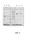


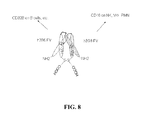


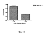


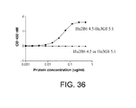

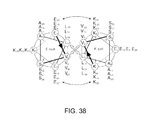


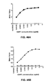
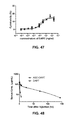
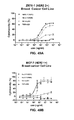


Classifications
-
- A—HUMAN NECESSITIES
- A61—MEDICAL OR VETERINARY SCIENCE; HYGIENE
- A61K—PREPARATIONS FOR MEDICAL, DENTAL OR TOILETRY PURPOSES
- A61K39/00—Medicinal preparations containing antigens or antibodies
- A61K39/395—Antibodies; Immunoglobulins; Immune serum, e.g. antilymphocytic serum
-
- A—HUMAN NECESSITIES
- A61—MEDICAL OR VETERINARY SCIENCE; HYGIENE
- A61K—PREPARATIONS FOR MEDICAL, DENTAL OR TOILETRY PURPOSES
- A61K39/00—Medicinal preparations containing antigens or antibodies
-
- A—HUMAN NECESSITIES
- A61—MEDICAL OR VETERINARY SCIENCE; HYGIENE
- A61P—SPECIFIC THERAPEUTIC ACTIVITY OF CHEMICAL COMPOUNDS OR MEDICINAL PREPARATIONS
- A61P1/00—Drugs for disorders of the alimentary tract or the digestive system
-
- A—HUMAN NECESSITIES
- A61—MEDICAL OR VETERINARY SCIENCE; HYGIENE
- A61P—SPECIFIC THERAPEUTIC ACTIVITY OF CHEMICAL COMPOUNDS OR MEDICINAL PREPARATIONS
- A61P1/00—Drugs for disorders of the alimentary tract or the digestive system
- A61P1/04—Drugs for disorders of the alimentary tract or the digestive system for ulcers, gastritis or reflux esophagitis, e.g. antacids, inhibitors of acid secretion, mucosal protectants
-
- A—HUMAN NECESSITIES
- A61—MEDICAL OR VETERINARY SCIENCE; HYGIENE
- A61P—SPECIFIC THERAPEUTIC ACTIVITY OF CHEMICAL COMPOUNDS OR MEDICINAL PREPARATIONS
- A61P1/00—Drugs for disorders of the alimentary tract or the digestive system
- A61P1/16—Drugs for disorders of the alimentary tract or the digestive system for liver or gallbladder disorders, e.g. hepatoprotective agents, cholagogues, litholytics
-
- A—HUMAN NECESSITIES
- A61—MEDICAL OR VETERINARY SCIENCE; HYGIENE
- A61P—SPECIFIC THERAPEUTIC ACTIVITY OF CHEMICAL COMPOUNDS OR MEDICINAL PREPARATIONS
- A61P11/00—Drugs for disorders of the respiratory system
-
- A—HUMAN NECESSITIES
- A61—MEDICAL OR VETERINARY SCIENCE; HYGIENE
- A61P—SPECIFIC THERAPEUTIC ACTIVITY OF CHEMICAL COMPOUNDS OR MEDICINAL PREPARATIONS
- A61P11/00—Drugs for disorders of the respiratory system
- A61P11/06—Antiasthmatics
-
- A—HUMAN NECESSITIES
- A61—MEDICAL OR VETERINARY SCIENCE; HYGIENE
- A61P—SPECIFIC THERAPEUTIC ACTIVITY OF CHEMICAL COMPOUNDS OR MEDICINAL PREPARATIONS
- A61P13/00—Drugs for disorders of the urinary system
- A61P13/12—Drugs for disorders of the urinary system of the kidneys
-
- A—HUMAN NECESSITIES
- A61—MEDICAL OR VETERINARY SCIENCE; HYGIENE
- A61P—SPECIFIC THERAPEUTIC ACTIVITY OF CHEMICAL COMPOUNDS OR MEDICINAL PREPARATIONS
- A61P15/00—Drugs for genital or sexual disorders; Contraceptives
-
- A—HUMAN NECESSITIES
- A61—MEDICAL OR VETERINARY SCIENCE; HYGIENE
- A61P—SPECIFIC THERAPEUTIC ACTIVITY OF CHEMICAL COMPOUNDS OR MEDICINAL PREPARATIONS
- A61P17/00—Drugs for dermatological disorders
-
- A—HUMAN NECESSITIES
- A61—MEDICAL OR VETERINARY SCIENCE; HYGIENE
- A61P—SPECIFIC THERAPEUTIC ACTIVITY OF CHEMICAL COMPOUNDS OR MEDICINAL PREPARATIONS
- A61P17/00—Drugs for dermatological disorders
- A61P17/02—Drugs for dermatological disorders for treating wounds, ulcers, burns, scars, keloids, or the like
-
- A—HUMAN NECESSITIES
- A61—MEDICAL OR VETERINARY SCIENCE; HYGIENE
- A61P—SPECIFIC THERAPEUTIC ACTIVITY OF CHEMICAL COMPOUNDS OR MEDICINAL PREPARATIONS
- A61P17/00—Drugs for dermatological disorders
- A61P17/06—Antipsoriatics
-
- A—HUMAN NECESSITIES
- A61—MEDICAL OR VETERINARY SCIENCE; HYGIENE
- A61P—SPECIFIC THERAPEUTIC ACTIVITY OF CHEMICAL COMPOUNDS OR MEDICINAL PREPARATIONS
- A61P17/00—Drugs for dermatological disorders
- A61P17/14—Drugs for dermatological disorders for baldness or alopecia
-
- A—HUMAN NECESSITIES
- A61—MEDICAL OR VETERINARY SCIENCE; HYGIENE
- A61P—SPECIFIC THERAPEUTIC ACTIVITY OF CHEMICAL COMPOUNDS OR MEDICINAL PREPARATIONS
- A61P19/00—Drugs for skeletal disorders
- A61P19/02—Drugs for skeletal disorders for joint disorders, e.g. arthritis, arthrosis
-
- A—HUMAN NECESSITIES
- A61—MEDICAL OR VETERINARY SCIENCE; HYGIENE
- A61P—SPECIFIC THERAPEUTIC ACTIVITY OF CHEMICAL COMPOUNDS OR MEDICINAL PREPARATIONS
- A61P19/00—Drugs for skeletal disorders
- A61P19/08—Drugs for skeletal disorders for bone diseases, e.g. rachitism, Paget's disease
-
- A—HUMAN NECESSITIES
- A61—MEDICAL OR VETERINARY SCIENCE; HYGIENE
- A61P—SPECIFIC THERAPEUTIC ACTIVITY OF CHEMICAL COMPOUNDS OR MEDICINAL PREPARATIONS
- A61P21/00—Drugs for disorders of the muscular or neuromuscular system
- A61P21/04—Drugs for disorders of the muscular or neuromuscular system for myasthenia gravis
-
- A—HUMAN NECESSITIES
- A61—MEDICAL OR VETERINARY SCIENCE; HYGIENE
- A61P—SPECIFIC THERAPEUTIC ACTIVITY OF CHEMICAL COMPOUNDS OR MEDICINAL PREPARATIONS
- A61P25/00—Drugs for disorders of the nervous system
-
- A—HUMAN NECESSITIES
- A61—MEDICAL OR VETERINARY SCIENCE; HYGIENE
- A61P—SPECIFIC THERAPEUTIC ACTIVITY OF CHEMICAL COMPOUNDS OR MEDICINAL PREPARATIONS
- A61P25/00—Drugs for disorders of the nervous system
- A61P25/28—Drugs for disorders of the nervous system for treating neurodegenerative disorders of the central nervous system, e.g. nootropic agents, cognition enhancers, drugs for treating Alzheimer's disease or other forms of dementia
-
- A—HUMAN NECESSITIES
- A61—MEDICAL OR VETERINARY SCIENCE; HYGIENE
- A61P—SPECIFIC THERAPEUTIC ACTIVITY OF CHEMICAL COMPOUNDS OR MEDICINAL PREPARATIONS
- A61P27/00—Drugs for disorders of the senses
- A61P27/02—Ophthalmic agents
-
- A—HUMAN NECESSITIES
- A61—MEDICAL OR VETERINARY SCIENCE; HYGIENE
- A61P—SPECIFIC THERAPEUTIC ACTIVITY OF CHEMICAL COMPOUNDS OR MEDICINAL PREPARATIONS
- A61P29/00—Non-central analgesic, antipyretic or antiinflammatory agents, e.g. antirheumatic agents; Non-steroidal antiinflammatory drugs [NSAID]
-
- A—HUMAN NECESSITIES
- A61—MEDICAL OR VETERINARY SCIENCE; HYGIENE
- A61P—SPECIFIC THERAPEUTIC ACTIVITY OF CHEMICAL COMPOUNDS OR MEDICINAL PREPARATIONS
- A61P3/00—Drugs for disorders of the metabolism
- A61P3/08—Drugs for disorders of the metabolism for glucose homeostasis
- A61P3/10—Drugs for disorders of the metabolism for glucose homeostasis for hyperglycaemia, e.g. antidiabetics
-
- A—HUMAN NECESSITIES
- A61—MEDICAL OR VETERINARY SCIENCE; HYGIENE
- A61P—SPECIFIC THERAPEUTIC ACTIVITY OF CHEMICAL COMPOUNDS OR MEDICINAL PREPARATIONS
- A61P31/00—Antiinfectives, i.e. antibiotics, antiseptics, chemotherapeutics
-
- A—HUMAN NECESSITIES
- A61—MEDICAL OR VETERINARY SCIENCE; HYGIENE
- A61P—SPECIFIC THERAPEUTIC ACTIVITY OF CHEMICAL COMPOUNDS OR MEDICINAL PREPARATIONS
- A61P31/00—Antiinfectives, i.e. antibiotics, antiseptics, chemotherapeutics
- A61P31/04—Antibacterial agents
-
- A—HUMAN NECESSITIES
- A61—MEDICAL OR VETERINARY SCIENCE; HYGIENE
- A61P—SPECIFIC THERAPEUTIC ACTIVITY OF CHEMICAL COMPOUNDS OR MEDICINAL PREPARATIONS
- A61P31/00—Antiinfectives, i.e. antibiotics, antiseptics, chemotherapeutics
- A61P31/10—Antimycotics
-
- A—HUMAN NECESSITIES
- A61—MEDICAL OR VETERINARY SCIENCE; HYGIENE
- A61P—SPECIFIC THERAPEUTIC ACTIVITY OF CHEMICAL COMPOUNDS OR MEDICINAL PREPARATIONS
- A61P31/00—Antiinfectives, i.e. antibiotics, antiseptics, chemotherapeutics
- A61P31/12—Antivirals
-
- A—HUMAN NECESSITIES
- A61—MEDICAL OR VETERINARY SCIENCE; HYGIENE
- A61P—SPECIFIC THERAPEUTIC ACTIVITY OF CHEMICAL COMPOUNDS OR MEDICINAL PREPARATIONS
- A61P31/00—Antiinfectives, i.e. antibiotics, antiseptics, chemotherapeutics
- A61P31/12—Antivirals
- A61P31/14—Antivirals for RNA viruses
-
- A—HUMAN NECESSITIES
- A61—MEDICAL OR VETERINARY SCIENCE; HYGIENE
- A61P—SPECIFIC THERAPEUTIC ACTIVITY OF CHEMICAL COMPOUNDS OR MEDICINAL PREPARATIONS
- A61P31/00—Antiinfectives, i.e. antibiotics, antiseptics, chemotherapeutics
- A61P31/12—Antivirals
- A61P31/14—Antivirals for RNA viruses
- A61P31/16—Antivirals for RNA viruses for influenza or rhinoviruses
-
- A—HUMAN NECESSITIES
- A61—MEDICAL OR VETERINARY SCIENCE; HYGIENE
- A61P—SPECIFIC THERAPEUTIC ACTIVITY OF CHEMICAL COMPOUNDS OR MEDICINAL PREPARATIONS
- A61P31/00—Antiinfectives, i.e. antibiotics, antiseptics, chemotherapeutics
- A61P31/12—Antivirals
- A61P31/14—Antivirals for RNA viruses
- A61P31/18—Antivirals for RNA viruses for HIV
-
- A—HUMAN NECESSITIES
- A61—MEDICAL OR VETERINARY SCIENCE; HYGIENE
- A61P—SPECIFIC THERAPEUTIC ACTIVITY OF CHEMICAL COMPOUNDS OR MEDICINAL PREPARATIONS
- A61P31/00—Antiinfectives, i.e. antibiotics, antiseptics, chemotherapeutics
- A61P31/12—Antivirals
- A61P31/20—Antivirals for DNA viruses
-
- A—HUMAN NECESSITIES
- A61—MEDICAL OR VETERINARY SCIENCE; HYGIENE
- A61P—SPECIFIC THERAPEUTIC ACTIVITY OF CHEMICAL COMPOUNDS OR MEDICINAL PREPARATIONS
- A61P31/00—Antiinfectives, i.e. antibiotics, antiseptics, chemotherapeutics
- A61P31/12—Antivirals
- A61P31/20—Antivirals for DNA viruses
- A61P31/22—Antivirals for DNA viruses for herpes viruses
-
- A—HUMAN NECESSITIES
- A61—MEDICAL OR VETERINARY SCIENCE; HYGIENE
- A61P—SPECIFIC THERAPEUTIC ACTIVITY OF CHEMICAL COMPOUNDS OR MEDICINAL PREPARATIONS
- A61P33/00—Antiparasitic agents
- A61P33/02—Antiprotozoals, e.g. for leishmaniasis, trichomoniasis, toxoplasmosis
-
- A—HUMAN NECESSITIES
- A61—MEDICAL OR VETERINARY SCIENCE; HYGIENE
- A61P—SPECIFIC THERAPEUTIC ACTIVITY OF CHEMICAL COMPOUNDS OR MEDICINAL PREPARATIONS
- A61P33/00—Antiparasitic agents
- A61P33/02—Antiprotozoals, e.g. for leishmaniasis, trichomoniasis, toxoplasmosis
- A61P33/06—Antimalarials
-
- A—HUMAN NECESSITIES
- A61—MEDICAL OR VETERINARY SCIENCE; HYGIENE
- A61P—SPECIFIC THERAPEUTIC ACTIVITY OF CHEMICAL COMPOUNDS OR MEDICINAL PREPARATIONS
- A61P35/00—Antineoplastic agents
-
- A—HUMAN NECESSITIES
- A61—MEDICAL OR VETERINARY SCIENCE; HYGIENE
- A61P—SPECIFIC THERAPEUTIC ACTIVITY OF CHEMICAL COMPOUNDS OR MEDICINAL PREPARATIONS
- A61P35/00—Antineoplastic agents
- A61P35/02—Antineoplastic agents specific for leukemia
-
- A—HUMAN NECESSITIES
- A61—MEDICAL OR VETERINARY SCIENCE; HYGIENE
- A61P—SPECIFIC THERAPEUTIC ACTIVITY OF CHEMICAL COMPOUNDS OR MEDICINAL PREPARATIONS
- A61P37/00—Drugs for immunological or allergic disorders
- A61P37/02—Immunomodulators
-
- A—HUMAN NECESSITIES
- A61—MEDICAL OR VETERINARY SCIENCE; HYGIENE
- A61P—SPECIFIC THERAPEUTIC ACTIVITY OF CHEMICAL COMPOUNDS OR MEDICINAL PREPARATIONS
- A61P37/00—Drugs for immunological or allergic disorders
- A61P37/02—Immunomodulators
- A61P37/06—Immunosuppressants, e.g. drugs for graft rejection
-
- A—HUMAN NECESSITIES
- A61—MEDICAL OR VETERINARY SCIENCE; HYGIENE
- A61P—SPECIFIC THERAPEUTIC ACTIVITY OF CHEMICAL COMPOUNDS OR MEDICINAL PREPARATIONS
- A61P37/00—Drugs for immunological or allergic disorders
- A61P37/08—Antiallergic agents
-
- A—HUMAN NECESSITIES
- A61—MEDICAL OR VETERINARY SCIENCE; HYGIENE
- A61P—SPECIFIC THERAPEUTIC ACTIVITY OF CHEMICAL COMPOUNDS OR MEDICINAL PREPARATIONS
- A61P39/00—General protective or antinoxious agents
- A61P39/02—Antidotes
-
- A—HUMAN NECESSITIES
- A61—MEDICAL OR VETERINARY SCIENCE; HYGIENE
- A61P—SPECIFIC THERAPEUTIC ACTIVITY OF CHEMICAL COMPOUNDS OR MEDICINAL PREPARATIONS
- A61P43/00—Drugs for specific purposes, not provided for in groups A61P1/00-A61P41/00
-
- A—HUMAN NECESSITIES
- A61—MEDICAL OR VETERINARY SCIENCE; HYGIENE
- A61P—SPECIFIC THERAPEUTIC ACTIVITY OF CHEMICAL COMPOUNDS OR MEDICINAL PREPARATIONS
- A61P5/00—Drugs for disorders of the endocrine system
- A61P5/14—Drugs for disorders of the endocrine system of the thyroid hormones, e.g. T3, T4
-
- A—HUMAN NECESSITIES
- A61—MEDICAL OR VETERINARY SCIENCE; HYGIENE
- A61P—SPECIFIC THERAPEUTIC ACTIVITY OF CHEMICAL COMPOUNDS OR MEDICINAL PREPARATIONS
- A61P5/00—Drugs for disorders of the endocrine system
- A61P5/38—Drugs for disorders of the endocrine system of the suprarenal hormones
-
- A—HUMAN NECESSITIES
- A61—MEDICAL OR VETERINARY SCIENCE; HYGIENE
- A61P—SPECIFIC THERAPEUTIC ACTIVITY OF CHEMICAL COMPOUNDS OR MEDICINAL PREPARATIONS
- A61P7/00—Drugs for disorders of the blood or the extracellular fluid
- A61P7/06—Antianaemics
-
- A—HUMAN NECESSITIES
- A61—MEDICAL OR VETERINARY SCIENCE; HYGIENE
- A61P—SPECIFIC THERAPEUTIC ACTIVITY OF CHEMICAL COMPOUNDS OR MEDICINAL PREPARATIONS
- A61P9/00—Drugs for disorders of the cardiovascular system
- A61P9/08—Vasodilators for multiple indications
-
- C—CHEMISTRY; METALLURGY
- C07—ORGANIC CHEMISTRY
- C07K—PEPTIDES
- C07K16/00—Immunoglobulins [IGs], e.g. monoclonal or polyclonal antibodies
- C07K16/18—Immunoglobulins [IGs], e.g. monoclonal or polyclonal antibodies against material from animals or humans
- C07K16/28—Immunoglobulins [IGs], e.g. monoclonal or polyclonal antibodies against material from animals or humans against receptors, cell surface antigens or cell surface determinants
- C07K16/2803—Immunoglobulins [IGs], e.g. monoclonal or polyclonal antibodies against material from animals or humans against receptors, cell surface antigens or cell surface determinants against the immunoglobulin superfamily
- C07K16/2809—Immunoglobulins [IGs], e.g. monoclonal or polyclonal antibodies against material from animals or humans against receptors, cell surface antigens or cell surface determinants against the immunoglobulin superfamily against the T-cell receptor (TcR)-CD3 complex
-
- C—CHEMISTRY; METALLURGY
- C07—ORGANIC CHEMISTRY
- C07K—PEPTIDES
- C07K16/00—Immunoglobulins [IGs], e.g. monoclonal or polyclonal antibodies
- C07K16/18—Immunoglobulins [IGs], e.g. monoclonal or polyclonal antibodies against material from animals or humans
- C07K16/28—Immunoglobulins [IGs], e.g. monoclonal or polyclonal antibodies against material from animals or humans against receptors, cell surface antigens or cell surface determinants
- C07K16/2803—Immunoglobulins [IGs], e.g. monoclonal or polyclonal antibodies against material from animals or humans against receptors, cell surface antigens or cell surface determinants against the immunoglobulin superfamily
- C07K16/283—Immunoglobulins [IGs], e.g. monoclonal or polyclonal antibodies against material from animals or humans against receptors, cell surface antigens or cell surface determinants against the immunoglobulin superfamily against Fc-receptors, e.g. CD16, CD32, CD64
-
- C—CHEMISTRY; METALLURGY
- C07—ORGANIC CHEMISTRY
- C07K—PEPTIDES
- C07K16/00—Immunoglobulins [IGs], e.g. monoclonal or polyclonal antibodies
- C07K16/18—Immunoglobulins [IGs], e.g. monoclonal or polyclonal antibodies against material from animals or humans
- C07K16/32—Immunoglobulins [IGs], e.g. monoclonal or polyclonal antibodies against material from animals or humans against translation products of oncogenes
-
- C—CHEMISTRY; METALLURGY
- C07—ORGANIC CHEMISTRY
- C07K—PEPTIDES
- C07K16/00—Immunoglobulins [IGs], e.g. monoclonal or polyclonal antibodies
- C07K16/44—Immunoglobulins [IGs], e.g. monoclonal or polyclonal antibodies against material not provided for elsewhere, e.g. haptens, metals, DNA, RNA, amino acids
-
- C—CHEMISTRY; METALLURGY
- C07—ORGANIC CHEMISTRY
- C07K—PEPTIDES
- C07K16/00—Immunoglobulins [IGs], e.g. monoclonal or polyclonal antibodies
- C07K16/46—Hybrid immunoglobulins
-
- A—HUMAN NECESSITIES
- A61—MEDICAL OR VETERINARY SCIENCE; HYGIENE
- A61K—PREPARATIONS FOR MEDICAL, DENTAL OR TOILETRY PURPOSES
- A61K39/00—Medicinal preparations containing antigens or antibodies
- A61K2039/505—Medicinal preparations containing antigens or antibodies comprising antibodies
-
- C—CHEMISTRY; METALLURGY
- C07—ORGANIC CHEMISTRY
- C07K—PEPTIDES
- C07K2317/00—Immunoglobulins specific features
- C07K2317/30—Immunoglobulins specific features characterized by aspects of specificity or valency
- C07K2317/31—Immunoglobulins specific features characterized by aspects of specificity or valency multispecific
-
- C—CHEMISTRY; METALLURGY
- C07—ORGANIC CHEMISTRY
- C07K—PEPTIDES
- C07K2317/00—Immunoglobulins specific features
- C07K2317/30—Immunoglobulins specific features characterized by aspects of specificity or valency
- C07K2317/34—Identification of a linear epitope shorter than 20 amino acid residues or of a conformational epitope defined by amino acid residues
-
- C—CHEMISTRY; METALLURGY
- C07—ORGANIC CHEMISTRY
- C07K—PEPTIDES
- C07K2317/00—Immunoglobulins specific features
- C07K2317/50—Immunoglobulins specific features characterized by immunoglobulin fragments
- C07K2317/52—Constant or Fc region; Isotype
-
- C—CHEMISTRY; METALLURGY
- C07—ORGANIC CHEMISTRY
- C07K—PEPTIDES
- C07K2317/00—Immunoglobulins specific features
- C07K2317/50—Immunoglobulins specific features characterized by immunoglobulin fragments
- C07K2317/56—Immunoglobulins specific features characterized by immunoglobulin fragments variable (Fv) region, i.e. VH and/or VL
-
- C—CHEMISTRY; METALLURGY
- C07—ORGANIC CHEMISTRY
- C07K—PEPTIDES
- C07K2317/00—Immunoglobulins specific features
- C07K2317/60—Immunoglobulins specific features characterized by non-natural combinations of immunoglobulin fragments
- C07K2317/62—Immunoglobulins specific features characterized by non-natural combinations of immunoglobulin fragments comprising only variable region components
- C07K2317/626—Diabody or triabody
-
- C—CHEMISTRY; METALLURGY
- C07—ORGANIC CHEMISTRY
- C07K—PEPTIDES
- C07K2317/00—Immunoglobulins specific features
- C07K2317/60—Immunoglobulins specific features characterized by non-natural combinations of immunoglobulin fragments
- C07K2317/64—Immunoglobulins specific features characterized by non-natural combinations of immunoglobulin fragments comprising a combination of variable region and constant region components
-
- C—CHEMISTRY; METALLURGY
- C07—ORGANIC CHEMISTRY
- C07K—PEPTIDES
- C07K2317/00—Immunoglobulins specific features
- C07K2317/70—Immunoglobulins specific features characterized by effect upon binding to a cell or to an antigen
- C07K2317/73—Inducing cell death, e.g. apoptosis, necrosis or inhibition of cell proliferation
-
- C—CHEMISTRY; METALLURGY
- C07—ORGANIC CHEMISTRY
- C07K—PEPTIDES
- C07K2317/00—Immunoglobulins specific features
- C07K2317/90—Immunoglobulins specific features characterized by (pharmaco)kinetic aspects or by stability of the immunoglobulin
- C07K2317/92—Affinity (KD), association rate (Ka), dissociation rate (Kd) or EC50 value
-
- C—CHEMISTRY; METALLURGY
- C07—ORGANIC CHEMISTRY
- C07K—PEPTIDES
- C07K2319/00—Fusion polypeptide
Definitions
- prophylactic agent and “prophylactic agents” refer to any agent(s) which can be used in the prevention of a disorder, or prevention of recurrence or spread of a disorder.
- a prophylactically effective amount may refer to the amount of prophylactic agent sufficient to prevent the recurrence or spread of hyperproliferative disease, particularly cancer, or the occurrence of such in a patient, including but not limited to those predisposed to hyperproliferative disease, for example those genetically predisposed to cancer or previously exposed to carcinogens.
- a prophylactically effective amount may also refer to the amount of the prophylactic agent that provides a prophylactic benefit in the prevention of disease.
- a prophylactically effective amount with respect to a prophylactic agent of the invention means that amount of prophylactic agent alone, or in combination with other agents, that provides a prophylactic benefit in the prevention of disease.
- the term "specifically binds an immune complex" and analogous terms refer to molecules that specifically bind to an immune complex and do not specifically bind to another molecule.
- a molecule that specifically binds to an immune complex may bind to other peptides or polypeptides with lower affinity as determined by, e.g. , immunoassays, BIAcore, or other assays known in the art.
- molecules that specifically bind an immune complex do not cross-react with other proteins.
- Molecules that specifically bind an immune complex can be identified, for example, by immunoassays, BIAcore, or other techniques known to those of skill in the art.
- a polypeptide chain of the invention comprises a hinge domain, which hinge domain is at the C-terminus of the polypeptide chain, wherein said polypeptide chain does not comprise an Fc domain.
- a polypeptide chain of the invention comprises a hinge-Fc domain, which hinge-Fc domain is at the C-terminus of the polypeptide chain.
- a polypeptide chain of the invention comprises a hinge-Fc domain, which hinge-Fc domain is at the N-terminus of the polypeptide chain.
- molecules of the invention are engineered to comprise an altered glycosylation pattern or an altered glycoform relative to the comparable portion of the template molecule.
- Engineered glycoforms may be useful for a variety of purposes, including, but not limited to, enhancing effector function.
- Engineered glycoforms may be generated by any method known to one skilled in the art, for example by using engineered or variant expression strains, by co-expression with one or more enzymes, for example, DI N-acetylglucosaminyltransferase III (GnTI11), by expressing a diabody of the invention in various organisms or cell lines from various organisms, or by modifying carbohydrate(s) after the diabody has been expressed and purified.
- DI N-acetylglucosaminyltransferase III GnTI11
- CD19 Troussard, X. et al. 1998 Hematol Cell Ther. 40(4):19-48
- CD20 Thomas, D.A. et al. 2006 Hematol Oncol Clin North Am. 20(5):1125-36
- CD22 Kreitman, R.J. 2006 AAPS J. 18;8(3):E532-51
- CD23 Rosati, S. et al. 2005 Curr Top Microbiol Immunol. 5;294:91-107
- CD25 Troussard, X. et al. 1998 Hematol Cell Ther. 40(4):139-48
- CD27 Bataille, R.
- coli Salmonella thyphimurium, Pseudomonas aeruginosa, Vibrio cholerae, Neisseria gonorrhoeae, Helicobacter pylori, Hemophilus influenzae, Shigella dysenteriae, Staphylococcus aureus, Mycobacterium tuberculosis and Streptococcus pneumoniae, fungal agents and parasites such as Giardi.
- variable domains derived from human, chimeric or humanized antibodies are particularly desirable for therapeutic treatment of human subjects.
- Human antibodies can be made by a variety of methods known in the art including phage display methods described above using antibody libraries derived from human immunoglobulin sequences. See also U.S. Patent Nos. 4,444,887 and 4,716,111 ; and International Publication Nos.
- Libraries can be constructed consisting of a pool of variant clones each of which differs by a single amino acid alteration in a single CDR and which contain variants representing each possible amino acid substitution for each CDR residue.
- Mutants with increased binding affinity for the antigen can be screened by contacting the immobilized mutants with labeled antigen. Any screening method known in the art can be used to identify mutant antibodies with increased avidity to the antigen (e.g ., ELISA) (See Wu et al. (1998) "Stepwise In Vitro Affinity Maturation Of Vitaxin, An Alphav Beta3-Specific Humanized mAb, " Proc Natl. Acad Sci. USA 95:6037-6042 ; Yelton et al.
- the binding domain agonizes Fc ⁇ RIIB-mediated inhibition of Fc ⁇ RI signaling.
- said binding domain inhibits Fc ⁇ RI-induced mast cell activation, calcium mobilization, degranulation, cytokine production, or serotonin release.
- the binding domains of the invention stimulate phosphorylation of Fc ⁇ RIIB, stimulate recruitment of SHIP, stimulate SHIP phosphorylation and its association with Shc, or inhibit activation of MAP kinase family members ( e.g ., Erk1, Erk2, JNK, p38, etc.).
- the binding domains are derived from humanized antibodies.
- a humanized Fc ⁇ RIIB specific antibody may comprise substantially all of at least one, and typically two, variable domains in which all or substantially all of the CDR regions correspond to those of a non-human immunoglobulin ( i.e., donor antibody) and all or substantially all of the framework regions are those of a human immunoglobulin consensus sequence.
- the framework regions of the humanized antibodies described herein does not necessarily consist of the precise amino acid sequence of the framework region of a natural occurring human antibody variable region, but contains various alterations, including but not limited to amino acid deletions, insertions, modifications that alter the property of the humanized antibody, for example, improve the binding properties of a humanized antibody region that is specific for the same target as the murine Fc ⁇ RIIB specific antibody.
- a minimal number of alterations are made to the framework region in order to avoid large-scale introductions of non-human framework residues and to ensure minimal immunogenicity of the humanized antibody in humans.
- the donor monoclonal antibody is preferably a monoclonal antibody produced by clones 2B6, 3H7, 1D5, 2E1, 2H9, 2D11, or 1F2.
- the antibodies can be characterized using assays for identifying the function of the antibodies of the invention, particularly the activity to modulate Fc ⁇ RIIB signaling.
- characterization assays of the invention can measure phosphorylation of tyrosine residues in the ITIM motif of Fc ⁇ RIIB, or measure the inhibition of B cell receptor-generated calcium mobilization.
- the characterization assays of the invention can be cell-based or cell-free assays.
- the anti-Fc ⁇ RIIB antibodies of the invention are characterized for their ability to modulate an IgE mediated response in a cell-based ⁇ -hexosaminidase (enzyme contained in the granules) release assay.
- ⁇ -hexosaminidase release from mast cells and basophils is a primary event in acute allergic and inflammatory condition ( Aketani et al. (2001) "Correlation Between Cytosolic Calcium Concentration And Degranulation In RBL-2H3 Cells In The Presence Of Various Concentrations Of Antigen-Specific IgEs, " Immunol. Lett. 75: 185-189 ; Aketani et al.
- Measuring Fc ⁇ RIIB-dependent activity can include, for example, measuring intracellular calcium mobilization by flow cytometry, measuring phosphorylation of Akt and/or Erk, measuring BCR-mediated accumulation of PI(3,4,5)P 3 , or measuring Fc ⁇ RIIB-mediated proliferation B cells.
- CD16A binding proteins which can be used as sources for light and heavy chain variable regions for covalent diabody production.
- CD16A binding proteins includes molecules comprising VL and VH domains of anti-CD16A antibodies, which VH and VL domains are used in the production of the diabodies of the present invention.
- the sequences of a number of CD16A binding proteins VL domains is shown in Table 6 .
- Light chains comprising these sequences fused to a human C ⁇ . constant domain were coexpressed with a Hu3G8VH heavy chain (described above) to form tetrameric antibodies, and the binding of the antibodies to CD16A was measured to assess the effect of amino acid substitutions compared to the Hu3G8VL-1 VL domain (SEQ ID NO: 71) .
- said amino acid modification and IgG hinge/Fc region display similar functionality, e.g ., increased affinity for Fc ⁇ RIIA, when separately assayed for Fc ⁇ R binding or effector function in the context of the diabody molecule or other Fc-containing molecule ( e.g . and immunoglobulin).
- the combination of said amino acid modification and selected IgG Fc region then act additively or, more preferably, synergistically to modify said functionality in the diabody molecule of the invention, relative to a diabody molecule of the invention comprising a wild-type Fc region.
- FACS fluorescence activated cell sorting
- Flow sorters are capable of rapidly examining a large number of individual cells that have been bound, e.g. , opsonized, by molecules of the invention (e.g. , 10-100 million cells per hour) (Shapiro et al. (1995) Practical Flow Cytometry ).
- specific parameters used for optimization of diabody behavior include but are not limited to, antigen concentration (i.e.
- the invention encompasses, effector cells, THP-1, expressing Fc ⁇ RI, Fc ⁇ RIIA and Fc ⁇ RIIB, and monocyte derived primary macrophages derived from whole human blood expressing both Fc ⁇ RIIIA and Fc ⁇ RIIB, to determine if heavy chain antibody mutants show increased ADCC activity and phagocytosis relative to wild type IgG2 antibodies.
- the resulting binding curves are globally fitted using computer algorithms supplied by the SPR instrument manufacturer, e.g. , BIAcore, Inc. (Piscataway, NJ). These algorithms calculate both the K on and K off , from which the apparent equilibrium binding constant, K d is deduced as the ratio of the two rate constants ( i.e. , K off /K on ). More detailed treatments of how the individual rate constants are derived can be found in the BIAevaluaion Software Handbook (BIAcore, Inc., Piscataway, NJ). The analysis of the generated data may be done using any method known in the art.
- the host cells used to express the molecules identified by the methods of the invention may be either bacterial cells such as Escherichia coli , or, preferably, eukaryotic cells, especially for the expression of whole recombinant immunoglobulin molecule.
- mammalian cells such as Chinese hamster ovary cells (CHO)
- CHO Chinese hamster ovary cells
- a vector such as the major intermediate early gene promoter element from human cytomegalovirus is an effective expression system for immunoglobulins ( Foecking et al. (1986) "Powerful And Versatile Enhancer-Promoter Unit For Mammalian Expression Vectors, " Gene 45:101-106 ; Cockett et al. (1990) "High Level Expression Of Tissue Inhibitor Of Metalloproteinases In Chinese Hamster Ovary Cells Using Glutamine Synthetase Gene Amplification," Biotechnology 8:662-667 ).
- yeast expression vectors containing sequences encoding the molecules identified by the methods of the invention
- insect cell systems infected with recombinant virus expression vectors e.g. , baclovirus
- plant cell systems infected with recombinant virus expression vectors e.g. , cauliflower mosaic virus (CaMV) and tobacco mosaic virus (TMV) or transformed with recombinant plasmid expression vectors ( e.g. , Ti plasmid) containing sequences encoding the molecules identified by the methods of the invention
- mammalian cell systems e.g.
- enterokinase which recognizes the amino acid sequence DDDDK ⁇ (SEQ ID NO: 91) ( Collins-Racie et al. (1995) "Production Of Recombinant Bovine Enterokinase Catalytic Subunit In Escherichia Coli Using The Novel Secretory Fusion Partner DsbA, " Biotechnology 13:982-987 hereby incorporated by reference herein in its entirety)
- furin which recognizes the amino acid sequence RXXR ⁇ , with a preference for RX(K/R)R ⁇ (SEQ ID NO: 92 and SEQ ID NO: 93, respectively) (additional R at P6 position appears to enhance cleavage)
- AcTEV which recognizes the amino acid sequence ENLYFQ ⁇ G (SEQ ID NO: 94) ( Parks et al.
- Different host cells have characteristic and specific mechanisms for the post-translational processing and modification of proteins and gene products. Appropriate cell lines or host systems can be chosen to ensure the correct modification and processing of the foreign protein expressed.
- eukaryotic host cells which possess the cellular machinery for proper processing of the primary transcript, glycosylation, and phosphorylation of the gene product may be used.
- mammalian host cells include but are not limited to CHO, VERY, BHK, HeLa, COS, MDCK, 293, 293T, 3T3, WI38, BT483, Hs578T, HTB2, BT20 and T47D, CRL7030 and Hs578Bst.
- antiviral agents include but are not limited to zidovudine, acyclovir, gangcyclovir, vidarabine, idoxuridine, trifluridine, and ribavirin, as well as foscarnet, amantadine, rimantadine, saquinavir, indinavir, amprenavir, lopinavir, ritonavir, the alpha-interferons; adefovir, clevadine, entecavir, pleconaril.
- a composition of the invention comprises nucleic acids encoding a fusion protein, said nucleic acids being a part of an expression vector that expresses the fusion protein in a suitable host.
- nucleic acids have promoters, preferably heterologous promoters, operably linked to the coding region of a fusion protein, said promoter being inducible or constitutive, and optionally, tissue-specific.
- nucleic acid molecules are used in which the coding sequence of the fusion protein and any other desired sequences are flanked by regions that promote homologous recombination at a desired site in the genome, thus providing for intrachromosomal expression of the fusion protein.
- the nucleic acid is introduced into a cell prior to administration in vivo of the resulting recombinant cell.
- introduction can be carried out by any method known in the art, including but not limited to, transfection, electroporation, microinjection, infection with a viral or bacteriophage vector, containing the nucleic acid sequences, cell fusion, chromosome-mediated gene transfer, microcellmediated gene transfer, spheroplast fusion, etc .
- Numerous techniques are known in the art for the introduction of foreign genes into cells (See, e.g., Loeffler et al. (1993) "Gene Transfer Into Primary And Established Mammalian Cell Lines With Lipopolyamine-Coated DNA, " Meth. Enzymol.
- a monospecific covalent diabody and a bispecific covalent diabody were constructed to assess the recombinant production, purification and binding characteristics of each.
- the affinity purified diabody molecules that were produced by the recombinant expression systems described herein were found by SDS-PAGE and SEC analysis to consist of a single dimerc species. ELISA and SPR analysis further revealed that the covalent bispecific diabody exhibited affinity for both target antigens and could bind both antigens simultaneously.
- pMGX0666 and pMGX0668 encoding constructs 3 and 4, respectively, were separately transfected into HEK-293 cells for expression of a covalent monospecific diabody (CMD), immunospecific for Fc ⁇ RIIIA (h3G8 diabody) and Fc ⁇ RIIB (h2B6 diabody), respectively. Following three days in culture, secreted products were purified from the conditioned media.
- CMD monospecific diabody
- Fc ⁇ RIIIA h3G8 diabody
- Fc ⁇ RIIB h2B6 diabody
- Co-transfection of these plasmids was designed to lead to the expression of a bispecific diabody (CBD) of tetravalency with IgG-like structure, immunospecific for both Fc ⁇ RIIB and Fc ⁇ RIIIA.
- CBD bispecific diabody
- An additional cotransfection was also performed: pMGX0674 and pMGX0676, encoding constructs 6 and 5, respectively.
- conditioned media was harvested.
- the amount of secreted product in the conditioned media was quantitiated by anti IgG Fc ELISA using purified Fc as a standard.
- the concentrations of product in the samples was then normalized based on the quantitation, and the normalized samples used for the remaining assays.
- Nucleic acid expression vectors were designed to produce modified versions of constructs 5, 6 and 8 presented in Example 6.2.
- Construct 9 (SEQ ID NO: 19) and construct 10 (SEQ ID NO: 20) (both shown schematically in FIG. 13 ) were analogous to constructs 8 and 6, with the exception that Fc domain or hinge-Fc domain, respectively, was shifted from the C-terminus of the polypeptide to the N-terminus. Additionally all Fc domains used were wild-type IgG1 Fc domains.
- Construct 11 SEQ ID NO: 21 , (shown schematically in FIG.
- the construct was re-amplified by using H9 and lgh646R as primers.
- the C-terminal LGGC linker was integrated in 1gh646R primer.
- the PCR product was then digested with Nhei/EcoRI restriction endonucleases, and cloned into pCIneo vector.
- CB3.1VL-8B5VH-LGGC The same strategy was used to create CB3.1VL-8B5VH-LGGC.
- the C-terminal LGGC linker was integrated in lgh648R primer and CB3.1 VL-8B5VH-FNRGEC was used as temperate.
- the PCR product was then digested with NheI/EcoRI restriction endonucleases, and cloned into pCIneo vector.
- HBCRCVL R45N-h8B5VH_LGGCGGGS-hG1 The hBCRCVL sequences are fused to the H8B5VH sequences by the linker GGGSGGGG (SEQ ID NO: 10) located at position 113-120.
- the H8B5VH sequences are fused to the Fc sequences by the linker LGGCGGGS (SEQ ID NO: 267) located at position 237-244 (both shown underlined above).
- separators of any amino acid sequence may be employed, it is preferable to employ separators that form an ⁇ helix coils, so as to maximally extend and project the Fc domain away from the variable domains ( Figure 37 ). Because the above-described coiled polypeptides of opposing charge additionally finction to promote heterodimer formation, such molecules are particularly preferred separators. Such coil-containing Fc-DART molecules provide benefits similar to those of Fc-DARTS, including improved serum half-life and effector function recruitment. The above-described E-coil and K-coil polypeptides are particularly preferred for this purpose.
- the DART has the ability to bind to HER2-expressing cells and to thereby attach to such cells a domain capable of binding to the T-cell receptor.
- T cells bind to this domain, they activate to initiate an immune response that leads to the killing of the HER2-expressing cells.
Abstract
Description
- This Application claims the benefit of
U.S. Patent Applications Serial Nos. 60/671,657 (filed April 15, 2005 11/409,339 (filed April 17, 2006 PCT/US06/014481 (filed April 17, 2006 ; expired);60/945,523 (filed June 21, 2007 61/019,051 (filed January 4, 2008 12/138,867 (filed June 13, 2008 PCT/US08/066957 (filed June 13, 2008 , pending),61/139,352 (filed December 19, 2008 61/156,035 (filed February 27, 2009 61/256,779 (filed October 30, 2009 - The present invention is directed to diabody molecules, otherwise referred to as "dual affinity retargeting reagents" ("DARTS"), and uses thereof in the treatment of a variety of diseases and disorders, including immunological disorders and cancers. The diabody molecules of the invention comprise at least two polypeptide chains that associate to form at least two epitope binding sites, which may recognize the same or different epitopes. Additionally, the epitopes may be from the same or different molecules or located on the same or different cells. The individual polypeptide chains of the diabody molecule may be covalently bound through non-peptide bond covalent bonds, such as, but not limited to, disulfide bonding of cysteine residues located within each polypeptide chain. In particular embodiments, the diabody molecules of the present invention further comprise an Fc region, which allows antibody-like functionality to be engineered into the molecule.
- The design of covalent diabodies is based on the single chain Fv construct (scFv) (Holliger et al. (1993) "'Diabodies': Small Bivalent And Bispecific Antibody Fragments," Proc. Natl. Acad. Sci. USA 90:6444-6448; herein incorporated by reference in its entirety). In an intact, unmodified IgG, the VL and VH domains are located on separate polypeptide chains, i.e., the light chain and the heavy chain, respectively. Interaction of an antibody light chain and an antibody heavy chain and, in particular, interaction of VL and VH domains forms one of the epitope binding sites of the antibody. In contrast, the scFv construct comprises a VL and VH domain of an antibody contained in a single polypeptide chain wherein the domains are separated by a flexible linker of sufficient length to allow self-assembly of the two domains into a functional epitope binding site. Where self assembly of the is impossible due to a linker of insufficient length (less than about 12 amino acid residues), two of the scFv constructs interact with each other to form a bivalent molecule, the VL of one chain associating with the VH of the other (reviewed in Marvin et al. (2005) "Recombinant Approaches To IgG-Like Bispecific Antibodies," Acta Pharmacol. Sin. 26:649-658). Moreover, addition of a cysteine residue to the c-terminus of the construct has been show to allow disulfide bonding of the polypeptide chains, stabilizing the resulting dimer without interfering with the binding characteristics of the bivalent molecule (see e.g., Olafsen et al. (2004) "Covalent Disulfide-Linked Anti-CEA Diabody Allows Site-Specific Conjugation And Radiolabeling For Tumor Targeting Applications, " Prot. Engr. Des. Sel. 17:21-27). Further, where VL and VH domains of differing specificity are selected, not only a bivalent, but also a bispecific molecule may be constructed.
- Bivalent diabodies have wide ranging applications including therapy and immunodiagnosis. Bivalency allows for great flexibility in the design and engineering of diabody in various applications, providing enhanced avidity to multimeric antigens, the cross-linking of differing antigens, and directed targeting to specific cell types relying on the presence of both target antigens. Due to their increased valency, low dissociation rates and rapid clearance from the circulation (for diabodies of small size, at or below ∼50 kDa), diabody molecules known in the art have also shown particular use in the filed of tumor imaging (Fitzgerald et al. (1997) "Improved Tumour Targeting By Disulphide Stabilized Diabodies Expressed In Pichia pastoris, " Protein Eng. 10:1221). Of particular importance is the cross linking of differing cells, for example the cross linking of cytotoxic T cells to tumor cells (Staerz et al. (1985) "Hybrid Antibodies Can Target Sites For Attack By T Cells, " Nature 314:628-631, and Holliger et al. (1996) "Specific Killing Of Lymphoma Cells By Cytotoxic T-Cells Mediated By A Bispecific Diabody, " Protein Eng. 9:299-305). Diabody epitope binding domains may also be directed to a surface determinant of any immune effector cell such as CD3, CD16, CD32, or CD64, which are expressed on T lymphocytes, natural killer (NK) cells or other mononuclear cells. In many studies, diabody binding to effector cell determinants, e.g., Fcγ receptors (FcγR), was also found to activate the effector cell (Halliger et al. (1996) "Specific Killing Of Lymphama Cells By Cytotoxic T-Cells Mediated By A Bispecific Diabody, " Protein Eng. 9:299-305; Holliger et al. (1999) "Carcinoembryonic Antigen (CEA)-Specific T-cell Activation In Colon Carcinoma Induced By Anti-CD3 x Anti-CEA Bispecific Diabodies And B7 x Anti-CEA Bispecific Fusion Proteins, " Cancer Res. 59:2909-2916). Normally, effector cell activation is triggered by the binding of an antigen bound antibody to an effector cell via Fc-FcγR interaction; thus, in this regard, diabody molecules of the invention may exhibit Ig-like functionality independent of whether they comprise an Fc domain (e.g., as assayed in any efferctor function assay known in the art or exemplified herein (e.g., ADCC assay)). By cross-linking tumor and effector cells, the diabody not only brings the effector cell within the proximity of the tumor cells but leads to effective tumor killing (see e.g., Cao et al. (2003) "Bispecific Antibody Conjugates In Therapeutics, " Adv. Drug. Deliv. Rev. 55:171-197, hereby incorporated by reference herein in its entirety).
- In traditional immune function the interaction of antibody-antigen complexes with cells of the immune system results in a wide array of responses, ranging from effector functions such as antibody-dependent cytotoxicity, mast cell degranulation, and phagocytosis to immunomodulatory signals such as regulating lymphocyte proliferation and antibody secretion. All these interactions are initiated through the binding of the Fc domain of antibodies or immune complexes to specialized cell surface receptors on hematopoietic cells. The diversity of cellular responses triggered by antibodies and immune complexes results from the structural heterogeneity of Fc receptors. Fc receptors share structurally related an antigen binding domains which presumably mediate intracellular signaling.
- The Fcγ receptors, members of the immunoglobulin gene superfamily of proteins, are surface glycoproteins that can bind the Fcγ portion of immunoglobulin molecules. Each member of the family recognizes immunoglobulins of one or more isotypes through a recognition domain on the alpha chain of the Fcγ receptor. Fcγ receptors are defined by their specificity for immunoglobulin subtypes. Fcγ receptors for IgG are referred to as FcγR, for IgE as FcεR, and for IgA as FcαR. Different accessory cells bear Fcγ receptors for antibodies of different isotype, and the isotype of the antibody determines which accessory cells will be engaged in a given response (reviewed by Ravetch J.V. et al. (1991) "Fc Receptors," Annu. Rev. Immunol. 9: 457-92; Gerber J.S. et al. (2001) "Stimulatory And Inhibitory Signals Originating From The Macrophage Fcgamma Receptors," Microbes and Infection, 3: 131-139; Billadeau D.D. et al. (2002), "ITAMs Versus ITIMs: Striking A Balance During Cell Regulation, " The Journal of Clinical Investigation, 2(109): 161-1681; Ravetch J.V. et al. (2000) "Immune Inhibitory Receptors, " Science, 290: 84-89; Ravetch J.V. et al., (2001) "IgG Fc Receptors," Annu. Rev. Immunol. 19:275-90; Ravetch J.V. (1994) "Fc Preceptors: Rubor Redux, " Cell, 78(4): 553-60). The different Fcγ receptors, the cells that express them, and their isotype specificity is summarized in Table 1 (adapted from Immunobiology: The Immune System in Health and Disease, 4th ed. 1999, Elsevier Science Ltd/Garland Publishing, New York).
- Each member of this family is an integral membrane glycoprotein, possessing extracellular domains related to a C2-set of immunoglobulin-related domains, a single membrane spanning domain and an intracytoplasmic domain of variable length. There are three known FcγRs, designated FcγRI(CD64), FcγRII(CD32), and FcγRIII(CD16). The three receptors are encoded by distinct genes; however, the extensive homology between the three family members suggest they arose from a common progenitor perhaps by gene duplication.
- FcγRII proteins are 40 kDa integral membrane glycoproteins which bind only the complexed IgG due to a low affinity for monomeric Ig (106 M-1). This receptor is the most widely expressed FcγR, present on all hematopoietic cells, including monocytes, macrophages, B cells, NK cells, neutrophils, mast cells, and platelets. FcγRII has only two immunoglobulin-like regions in its immunoglobulin binding chain and hence a much lower affinity for IgG than FcγRI. There are three human FcγRII genes (FcγRII-A, FcγRII-B, FcγRII-C), all of which bind IgG in aggregates or immune complexes.
- Distinct differences within the cytoplasmic domains of FcγRII-A and FcγRII-B create two functionally heterogenous responses to receptor ligation. The fundamental difference is that the A isoform initiates intracellular signaling leading to cell activation such as phagocytosis and respiratory burst, whereas the B isoform initiates inhibitory signals, e.g., inhibiting B-cell activation.
- Due to heterogeneity within this class, the size of FcγRIII ranges between 40 and 80 kDa in mouse and man. Two human genes encode two transcripts, FcγRIIIA, an integral membrane glycoprotein, and FcγRIIIB, a glycosylphosphatidyl-inositol (GPI)-linked version. One murine gene encodes an FcγRIII homologous to the membrane spanning human FcγRIIIA. The FcγRIII shares structural characteristics with each of the other two FcγRs. Like FcγRII, FcγRIII binds IgG with low affinity and contains the corresponding two extracellular Ig-like domains. FcγRIIIA is expressed in macrophages, mast cells and is the lone FcγR in NK cells. The GPI-linked FcγRIIIB is currently known to be expressed only in human neutrophils.
- Both activating and inhibitory signals are transduced through the FcγRs following ligation. These diametrically opposing functions result from structural differences among the different receptor isoforms. Two distinct domains within the cytoplasmic signaling domains of the receptor called immunoreceptor tyrosine based activation motifs (ITAMs) or immunoreceptor tyrosine based inhibitory motifs (ITIMS) account for the different responses. The recruitment of different cytoplasmic enzymes to these structures dictates the outcome of the FcγR-mediated cellular responses. ITAM-containing FcγR complexes include FcγRI, FcγRIIA, FcγRIIIA, whereas ITIM-containing complexes only include FcγRIIB.
- Human neutrophils express the FcγRIIA gene. FcγRIIA clustering via immune complexes or specific antibody cross-linking serves to aggregate ITAMs along with receptor-associated kinases which facilitate ITAM phosphorylation. ITAM phosphorylation serves as a docking site for Syk kinase, activation of which results in activation of downstream substrates (e.g., PI3K). Cellular activation leads to release of proinflammatory mediators.
- The FcγRIIB gene is expressed on B lymphocytes; its extracellular domain is 96% identical to FcγRIIA and binds IgG complexes in an indistinguishable manner. The presence of an ITIM in the cytoplasmic domain of FcγRIIB defines this inhibitory subclass of FcγR. Recently the molecular basis of this inhibition was established. When co-ligated along with an activating FcγR, the ITIM in FcγRIIB becomes phosphorylated and attracts the SH2 domain of the inosital polyphosphate 5'-phosphatase (SHIP), which hydrolyzes phosphoinositol messengers released as a consequence of ITAM-containing FcyR-mediated tyrosine kinase activation, consequently preventing the influx of intracellular Ca++. Thus crosslinking of FcγRIIB dampens the activating response to FcγR ligation and inhibits cellular responsiveness. B cell activation, B cell proliferation and antibody secretion is thus aborted.
TABLE 1. Receptors for the Fc Regions of Immunoglobulin Isotypes Receptor Binding Cell Type Effect of Ligation FcγRI (CD64) IgG1 108 M-1 Macrophages Uptake Stimulation Activation of respiratory burst Induction of killing Neutrophils Eosinophils Dendritic cells FcγRII-A (CD32) IgG1 2 x 10⁶M-1 Macrophages Uptake Granule Release Neutrophils Eosinophils Dendritic cells Platelets Langerhan cells FcγRII-B2 (CD32) IgG1 2 x 10⁶M-1 Macrophages Uptake Inhibition of Stimulation Neutrophils Eosinophils FcγRII-B1 (CD32) IgG1 2 x 10⁶ M-1 B cells No Uptake Inhibition of Stimulation Mast cells FcγRIII (CD16) IgG1 5 x 10⁵ M-1 NK cells Induction of Killing Eosinophil Macrophages Neutrophils Mast Cells FcεRI IgE 1010M-1 Mast cells Secretion of granules Eosinophil Basophils FcαRI (CD89) IgAl, IgA2 1017 M-1 Macrophages Uptake Induction of Killing Neutrophils Eosinophils - The present invention relates to covalent diabodies and/or covalent diabody molecules and to their use in the treatment of a variety of diseases and disorders including cancer, autoimmune disorders, allergy disorders and infectious diseases caused by bacteria, fungi or viruses. Preferably, the diabody of the present invention can bind to two different epitopes on two different cells wherein the first epitope is expressed on a different cell type than the second epitope, such that the diabody can bring the two cells together.
- In one embodiment, the present invention is directed to a covalent bispecific diabody, which diabody comprises a first and a second polypeptide chain, which first polypeptide chain comprises (i) a first domain comprising a binding region of a light chain variable domain of a first immunoglobulin (VL1) specific for a first epitope, (ii) a second domain comprising a binding region of a heavy chain variable domain of a second immunoglobulin (VH2) specific for a second epitope, and, optionally, (iii) a third domain comprising at least one cysteine residue, which first and second domains are covalently linked such that the first and second domains do not associate to form an epitope binding site; which second polypeptide chain comprises (i) a fourth domain comprising a binding region of a light chain variable domain of the second immunoglobulin (VL2), (ii) a fifth domain comprising a binding region of a heavy chain variable domain of the first immunoglobulin (VH1), and, optionally, (iii) a sixth domain comprising at least one cysteine residue, which fourth and fifth domains are covalently linked such that the fourth and fifth domains do not associate to form an epitope binding site; and wherein the first polypeptide chain and the second polypeptide chain are covalently linked, with the proviso that the covalent link is not a peptide bond; wherein the first domain and the fifth domain associate to form a first binding site (VL1)(VH1) that binds the first epitope; wherein the second domain and the fourth domain associate to form a second binding site (VL2)(VH2) that binds the second epitope.
- In another embodiment, the present invention is directed to a covalent bispecific diabody, which diabody comprises a first and a second polypeptide chain, which first polypeptide chain comprises (i) a first domain comprising a binding region of a light chain variable domain of a first immunoglobulin (VL1) specific for a first epitope, (ii) a second domain comprising a binding region of a heavy chain variable domain of a second immunoglobulin (VH2) specific for a second epitope and (iii) a third domain comprising an Fc domain or portion thereof, which first and second domains are covalently linked such that the first and second domains do not associate to form an epitope binding site; which second polypeptide chain comprises (i) a fourth domain comprising a binding region of a light chain variable domain of the second immunoglobulin (VL2), (ii) a fifth domain comprising a binding region of a heavy chain variable domain of the first immunoglobulin (VH1), which fourth and fifth domains are covalently linked such that the third and fourth domains do not associate to form an epitope binding site; and wherein the first polypeptide chain and the second polypeptide chain are covalently linked, with the proviso that the covalent link is not a peptide bond; wherein the first domain and the fifth domain associate to form a first binding site (VL1)(VH1) that binds the first epitope; wherein the second domain and the fourth domain associate to form a second binding site (VL2)(VH2) that binds the second epitope.
- In certain aspects, the present invention is directed to diabody molecule, which molecule comprises a first and a second polypeptide chain, which first polypeptide chain comprises (i) a first domain comprising a binding region of a light chain variable domain of a first immunoglobulin (VL1) specific for a first epitope, (ii) a second domain comprising a binding region of a heavy chain variable domain of a second immunoglobulin (VH2) specific for a second epitope and (iii) a third domain comprising an Fc domain or portion thereof, which first and second domains are covalently linked such that the first and second domains do not associate to form an epitope binding site; which second polypeptide chain comprises (i) a fourth domain comprising a binding region of a light chain variable domain of the second immunoglobulin (VL2), (ii) a fifth domain comprising a binding region of a heavy chain variable domain of the first immunoglobulin (VH1), and (iii) a sixth domain comprising at least one cysteine residue, which fourth and fifth domains are covalently linked such that the fourth and fifth domains do not associate to form an epitope binding site; and wherein the first polypeptide chain and the second polypeptide chain are covalently linked, with the proviso that the covalent link is not a peptide bond; wherein the first domain and the fifth domain associate to form a first binding site (VL1)(VH1) that binds the first epitope; wherein the second domain and the fourth domain associate to form a second binding site (VL2)(VH2) that binds the second epitope.
- In certain embodiments, the present invention is directed to a covalent bispecific diabody, which diabody is a dimer of diabody molecules, each diabody molecule comprising a first and a second polypeptide chain, which first polypeptide chain comprises (i) a first domain comprising a binding region of a light chain variable domain of a first immunoglobulin (VL1) specific for a first epitope, (ii) a second domain comprising a binding region of a heavy chain variable domain of a second immunoglobulin (VH2) specific for a second epitope and (iii) a third domain comprising an Fc domain or portion thereof, which first and second domains are covalently linked such that the first and second domains do not associate to form an epitope binding site; and which second polypeptide chain comprises (i) a fourth domain comprising a binding region of a light chain variable domain of the second immunoglobulin (VL2), (ii) a fifth domain comprising a binding region of a heavy chain variable domain of the first immunoglobulin (VH1), and (iii) a sixth domain comprising at least one cysteine residue, which fourth and fifth domains are covalently linked such that the fourth and fifth domains do not associate to form an epitope binding site; and wherein the first polypeptide chain and the second polypeptide chain of each diabody molecule are covalently linked, with the proviso that the covalent link is not a peptide bond; wherein the first domain and the fifth domain of each diabody molecule associate to form a first binding site (VL1)(VH1) that binds the first epitope; wherein the second domain and the fourth domain of each diabody molecule associate to form a second binding site (VL2)(VH2) that binds the second epitope.
- In yet other embodiments, the present invention is directed to a covalent tetrapecific diabody, which diabody is a dimer of diabody molecules, the first diabody molecule comprising a first and a second polypeptide chain, which first polypeptide chain comprises (i) a first domain comprising a binding region of a light chain variable domain of a first immunoglobulin (VL1) specific for a first epitope, (ii) a second domain comprising a binding region of a heavy chain variable domain of a second immunoglobulin (VH2) specific for a second epitope and (iii) a third domain comprising an Fc domain or portion thereof, which first and second domains are covalently linked such that the first and second domains do not associate to form an epitope binding site; and which second polypeptide chain comprises (i) a fourth domain comprising a binding region of a light chain variable domain of the second immunoglobulin (VL2), (ii) a fifth domain comprising a binding region of a heavy chain variable domain of the first immunoglobulin (VH1), and (iii) a sixth domain comprising at least one cysteine residue, which fourth and fifth domains are covalently linked such that the fourth and fifth domains do not associate to form an epitope binding site; and wherein the first polypeptide chain and the second polypeptide chain are covalently linked, with the proviso that the covalent link is not a peptide bond; wherein the first domain and the fifth domain associate to form a first binding site (VL1)(VH1) that binds the first epitope; wherein the second domain and the fourth domain associate to form a second binding site (VL2)(VH2) that binds the second epitope; and the second diabody molecule comprising a first and a second polypeptide chain, which first polypeptide chain comprises (i) a first domain comprising a binding region of a light chain variable domain of a third immunoglobulin (VL3) specific for a third epitope, (ii) a second domain comprising a binding region of a heavy chain variable domain of a fourth immunoglobulin (VH4) specific for a fourth epitope and (iii) a third domain comprising an Fc domain or portion thereof, which first and second domains are covalently linked such that the first and second domains do not associate to form an epitope binding site; and which second polypeptide chain comprises (i) a fourth domain comprising a binding region of a light chain variable domain of the fourth immunoglobulin (VL4), (ii) a fifth domain comprising a binding region of a heavy chain variable domain of the third immunoglobulin (VH3), and (iii) a sixth domain comprising at least one cysteine residue, which fourth and fifth domains are covalently linked such that the fourth and fifth domains do not associate to form an epitope binding site; and wherein the first polypeptide chain and the second polypeptide chain are covalently linked, with the proviso that the covalent link is not a peptide bond; wherein the first domain and the fifth domain associate to form a first binding site (VL3)(VH3) that binds the third epitope; wherein the second domain and the fourth domain associate to form a second binding site (VL4)(VH4) that binds the fourth epitope.
- In certain aspects of the invention the first epitope, second epitope, and where applicable, third epitope and fourth epitope can be the same. In other aspects, the first epitope, second epitope, and where applicable, third epitope and fourth epitope can each different from the other. In certain aspects of the invention comprising a third epitope binding domain, the first epitope and third epitope can be the same. In certain aspects of the invention comprising a fourth epitope binding domain, the first epitope and fourth epitope can be the same. In certain aspects of the invention comprising a third epitope binding domain, the second epitope and third epitope can be the same. In certain aspects of the invention comprising a fourth epitope binding domain, the second epitope and fourth epitope can be the same. In preferred aspects of the invention, the first eptitope and second epitope are different. In yet other aspects of the invention comprising a third epitope binding domain and a fourth epitope binding domain, the third epitope and fourth epitope can be different. It is to be understood that any combination of the foregoing is encompassed in the present invention.
- In particular aspects of the invention, the first domain and the fifth domain of the diabody or diabody molecule can be derived from the same immunoglobulin. In another aspect, the second domain and the fourth domain of the diabody or diabody molecule can be derived from the same immunoglobulin. In yet another aspect, the first domain and the fifth domain of the diabody or diabody molecule can be derived from a different immunoglobulin. In yet another aspect, the second domain and the fourth domain of the diabody or diabody molecule can be derived from a different immunoglobulin. It is to be understood that any combination of the foregoing is encompassed in the present invention.
- In certain aspects of the invention, the covalent linkage between the first polypeptide chain and second polypeptide chain of the diabody or diabody molecule can be via a disulfide bond between at least one cysteine residue on the first polypeptide chain and at least one cysteine residue on the second polypeptide chain. The cysteine residues on the first or second polypeptide chains that are responsible for disulfide bonding can be found anywhere on the polypeptide chain including within the first, second, third, fourth, fifth and sixth domains. In a specific embodiment the cysteine residue on the first polypeptide chain is found in the first domain and the cysteine residue on the second polypeptide chain is found in the fifth domain. The first, second, fourth and fifth domains correspond to the variable regions responsible for binding. In preferred embodiments, the cysteine residues responsible for the disulfide bonding between the first and second polypeptide chains are located within the third and sixth domains, respectively. In a particular aspect of this embodiment, the third domain of the first polypeptide chain comprises the C-
terminal 6 amino acids of the human kappa light chain, FNRGEC (SEQ ID NO: 23), which can be encoded by the amino acid sequence (SEQ ID NO: 17). In another aspect of this embodiment, the sixth domain of the second polypeptide chain comprises the C-terminal 6 amino acids of the human kappa light chain, FNRGEC (SEQ ID NO: 23), which can be encoded by the amino acid sequence (SEQ ID NO: 17). In still another aspect of this embodiment, the third domain of the first polypeptide chain comprises the amino acid sequence VEPKSC (SEQ ID NO: 77), derived from the hinge domain of a human IgG, and which can be encoded by the nucleotide sequence (SEQ ID NO: 78). In another aspect of this embodiment, the sixth domain of the second polypeptide chain comprises the amino acid sequence VEPKSC (SEQ ID NO: 77), derived from the hinge domain of a human IgG, and which can be encoded by the nucleotide sequence (SEQ ID NO: 78). In certain aspects of this embodiment, the third domain of the first polypeptide chain comprises the C-terminal 6 amino acids of the human kappa light chain, FNRGEC (SEQ ID NO: 23); and the sixth domain of the second polypeptide chain comprises the amino acid sequence VEPKSC (SEQ ID NO: 77). In other aspects of this embodiment, the sixth domain of the second polypeptide chain comprises the C-terminal 6 amino acids of the human kappa light chain, FNRGEC (SEQ ID NO: 23); and the third domain of the first polypeptide chain comprises the amino acid sequence VEPKSC (SEQ ID NO: 77). In yet other aspects of this embodiment, the third domain of the first polypeptide chain comprises the C-terminal 6 amino acids of the human kappa light chain, FNRGEC (SEQ ID NO: 23); and the sixth domain of the second polypeptide chain comprises a hinge domain. In other aspects of this embodiment, the sixth domain of the second polypeptide chain comprises the C-terminal 6 amino acids of the human kappa light chain, FNRGEC (SEQ ID NO: 23); and the third domain of the first polypeptide chain comprises the hinge domain. In yet other aspects of this embodiment, the third domain of the first polypeptide chain comprises the C-terminal 6 amino acids of the human kappa light chain, FNRGEC (SEQ ID NO: 23); and the sixth domain of the first polypeptide chain comprises an Fc domain, or portion thereof. In still other aspects of this embodiment, the sixth domain of the second polypeptide chain comprises the C-terminal 6 amino acids of the human kappa light chain, FNRGEC (SEQ ID NO: 23); and the third domain of the first polypeptide chain comprises an Fc domain, or portion thereof. - In other embodiments, the cysteine residues on the first or second polypeptide that are responsible for the disulfide bonding can be located outside of the first, second or third domains on the first polypeptide chain and outside of the fourth, fifth and sixth domain on the second polypeptide chain. In particular, the cysteine residue on the first polypeptide chain can be N-terminal to the first domain or can be C-terminal to the first domain. The cysteine residue on the first polypeptide chain can be N-terminal to the second domain or can be C-terminal to the second domain. The cysteine residue on the first polypeptide chain can be N-terminal to the third domain or can be C-terminal to the third domain. Further, the cysteine residue on the second polypeptide chain can be N-terminal to the fourth domain or can be C-terminal to the fourth domain. The cysteine residue on the second polypeptide chain can be N-terminal to the fifth domain or can be C-terminal to the fifth domain. Accordingly, the cysteine residue on the second polypeptide chain can be C-terminal to the sixth domain or can be N-terminal to the sixth domain. In a particular aspect, disulfide bond can between at least two cysteine residues on the first polypeptide chain and at least two cysteine residues on the second polypeptide chain. In a particular aspect, wherein the third domain and sixth domain do not comprise an Fc domain, or portion thereof, the cysteine residue can be at the C-terminus of the first polypeptide chain and at the C-terminus of the second polypeptide chain. It is to be understood that any combination of the foregoing is encompassed in the present invention.
- In specific embodiments of the invention described supra, the covalent diabody of the invention encompasses dimers of diabody molecules, wherein each diabody molecule comprises a first and second polypeptide chain. In certain aspects of this embodiment the diabody molecules can be covalently linked to form the dimer, with the proviso that the covalent linkage is not a peptide bond. In preferred aspects of this embodiment, the covalent linkage is a disulfide bond between at least one cysteine residue on the first polypeptide chain of each of the diabody molecules of the dimer. In yet more preferred aspects of this invention, the covalent linkage is a disulfide bond between at least one cysteine residue on the first polypeptide chain of each of the diabody molecules forming the dimer, wherein said at least one cysteine residue is located in the third domain of each first polypeptide chain.
- In certain aspects of the invention, the first domain on the first polypeptide chain can be N-terminal to the second domain or can be C-terminal to the second domain. The first domain on the first polypeptide chain can be N-terminal to the third domain or can be C-terminal to the third domain. The second domain on the first polypeptide chain can be N-terminal to the first domain or can be C-terminal to the first domain. Further, the second domain on the first polypeptide chain can be N-terminal to the third domain or can be C-terminal to the third domain. Accordingly, the third domain on the first polypeptide chain can be N-terminal to the first domain or can be C-terminal to the first domain. The third domain on the first polypeptide chain can be N-terminal to the second domain or can be C-terminal to the second domain. With respect to the second polypeptide chain, the fourth domain can be N-terminal to the fifth domain or can be C-terminal to the fifth domain. The fourth domain can be N-terminal to the sixth domain or can be C-terminal to the sixth domain. The fifth domain on the second polypeptide chain can be N-terminal to the fourth domain or can be C-terminal to the fourth domain. The fifth domain on the second polypeptide chain can be N-terminal to the sixth domain or can be C-terminal to the sixth domain. Accordingly the sixth domain on the second polypeptide chain can be N-terminal to the fourth domain or can be C-terminal to the fourth domain. The sixth domain on the second polypeptide chain can be N-terminal to the fifth domain or can be C-terminal to the fifth domain. It is to be understood that any combination of the foregoing is encompassed in the present invention.
- In certain embodiments, first domain and second domain can be located C-terminal to the third domain on the first polypeptide chain; or the first domain and second domain can be located N-terminal to the third domain on the first polypeptide chain. With respect to the second polypeptide chain, the fourth domain and fifth domain can be located C-terminal to the sixth domain, or the fourth domain and fifth domain can be located N-terminal to the sixth domain. In certain aspects of this embodiment, the present invention is directed to a covalent bispecific diabody, which diabody is a dimer of diabody molecules, each diabody molecule comprising a first and a second polypeptide chain, which first polypeptide chain comprises (i) a first domain comprising a binding region of a light chain variable domain of a first immunoglobulin (VL1) specific for a first epitope, (ii) a second domain comprising a binding region of a heavy chain variable domain of a second immunoglobulin (VH2) specific for a second epitope and (iii) a third domain comprising an Fc domain or portion thereof, which first and second domains are covalently linked such that the first and second domains do not associate to form an epitope binding site and wherein the third domain is located N-terminal to both the first domain and second domain; and which second polypeptide chain comprises (i) a fourth domain comprising a binding region of a light chain variable domain of the second immunoglobulin (VL2), (ii) a fifth domain comprising a binding region of a heavy chain variable domain of the first immunoglobulin (VH1), and (iii) a sixth domain comprising at least one cysteine residue, which fourth and fifth domains are covalently linked such that the fourth and fifth domains do not associate to form an epitope binding site; and wherein the first polypeptide chain and the second polypeptide chain of each diabody molecule are covalently linked, with the proviso that the covalent link is not a peptide bond; wherein the first domain and the fifth domain of each diabody molecule associate to form a first binding site (VL1)(VH1) that binds the first epitope; wherein the second domain and the fourth domain of each diabody molecule associate to form a second binding site (VL2)(VH2) that binds the second epitope.
- In yet another embodiment, the present invention is directed to a covalent tetrapecific diabody, which diabody is a dimer of diabody molecules, the first diabody molecule comprising a first and a second polypeptide chain, which first polypeptide chain comprises (i) a first domain comprising a binding region of a light chain variable domain of a first immunoglobulin (VL1) specific for a first epitope, (ii) a second domain comprising a binding region of a heavy chain variable domain of a second immunoglobulin (VH2) specific for a second epitope and (iii) a third domain comprising an Fc domain or portion thereof, which first and second domains are covalently linked such that the first and second domains do not associate to form an epitope binding site and wherein the third domain is located N-terminal to both the first domain and second domain; and which second polypeptide chain comprises (i) a fourth domain comprising a binding region of a light chain variable domain of the second immunoglobulin (VL2), (ii) a fifth domain comprising a binding region of a heavy chain variable domain of the first immunoglobulin (VH1), and (iii) a sixth domain comprising at least one cysteine residue, which fourth and fifth domains are covalently linked such that the fourth and fifth domains do not associate to form an epitope binding site; and wherein the first polypeptide chain and the second polypeptide chain are covalently linked, with the proviso that the covalent link is not a peptide bond; wherein the first domain and the fifth domain associate to form a first binding site (VL1)(VH1) that binds the first epitope; wherein the second domain and the fourth domain associate to form a second binding site (VL2)(VH2) that binds the second epitope; and the second diabody molecule comprises a first and a second polypeptide chain, which first polypeptide chain comprises (i) a first domain comprising a binding region of a light chain variable domain of a third immunoglobulin (VL3) specific for a third epitope, (ii) a second domain comprising a binding region of a heavy chain variable domain of a fourth immunoglobulin (VH4) specific for a fourth epitope and (iii) a third domain comprising an Fc domain or portion thereof, which first and second domains are covalently linked such that the first and second domains do not associate to form an epitope binding site and wherein the third domain is located N-terminal to both the first domain and second domain; and which second polypeptide chain comprises (i) a fourth domain comprising a binding region of a light chain variable domain of the fourth immunoglobulin (VL4), (ii) a fifth domain comprising a binding region of a heavy chain variable domain of the third immunoglobulin (VH3), and (iii) a sixth domain comprising at least one cysteine residue, which fourth and fifth domains are covalently linked such that the fourth and fifth domains do not associate to form an epitope binding site; and wherein the first polypeptide chain and the second polypeptide chain are covalently linked, with the proviso that the covalent link is not a peptide bond; wherein the first domain and the fifth domain associate to form a first binding site (VL3)(VH3) that binds the third epitope; wherein the second domain and the fourth domain associate to form a second binding site (VL4)(VH4) that binds the fourth epitope.
- As discussed above, the domains on the individual polypeptide chains are covalently linked. In specific aspects, the covalent link between the first and second domain, first and third domain, second and third domain, fourth and fifth domain, fourth and sixth domain, and/or fifth and sixth domain can be a peptide bond. In particular, the first and second domains, and the fourth and fifth domains can be separated by the third domain and sixth domain, respectively, or by additional amino acid residues, so long as the first and second, and fourth and fifth domains do not associate to form a binding site. The number of amino acid residues can be 0, 1, 2, 3, 4, 5, 6, 7, 8 or 9 amino acid residues. In one preferred aspect, the number of amino acid residues between the domains is 8.
- In certain aspects of the invention, the domains of the first and second polypeptid chain comprising an Fc domain, i.e., optionally, the third and sixth domains, respectively, can further comprise a hinge domain such that the domain comprises a hinge-Fc region. In alternative embodiments, the first polypeptide chain or the second polypeptide chain can comprise a hinge domain without also comprising an Fc domain. The heavy chains, light chains, hinge regions, Fc domains, and/or hinge-Fc domains for use in the invention can be derived from any immunoglobulin type including IgA, IgD, IgE, IgG or IgM. In a preferred aspect, the immunoglobulin type is IgG, or any subtype thereof, i.e., IgG1, IgG2, IgG3 or IgG4. In other aspects, the immunoglobulin from which the light and heavy chains are derived is humanized or chimerized.
- Further, the first epitope and second epitopes, and, where applicable, third epitope and fourth epitope, to which the diabody or diabody molecule binds can be different epitopes from the same antigen or can be different epitopes from different antigens. The antigens can be any molecule to which an antibody can be generated. For example, proteins, nucleic acids, bacterial toxins, cell surface markers, autoimmune markers, viral proteins, drugs, etc. In particular aspects, at least one epitope binding site of the diabody is specific for an antigen on a particular cell, such as a B-cell, a T-cell, a phagocytic cell, a natural killer (NK) cell or a dendritic cell.
- In certain aspects of the present embodiment, at least one epitope binding site of the diabody or diabody molecule is specific for a Fc receptor, which Fc receptor can be an activating Fc receptor or an inhibitory Fc receptor. In particular aspects, the Fc receptor is a Fcγ receptor, and the Fcγ receptor is a FcγRI, FcγRII or FcγRIII receptor. In more preferred aspects, the FcγRIII receptor is the FcγRIIIA (CD16A) receptor or the FcγRIIIB (CD16B) receptor, and, more preferably, the FcγRIII receptor is the FcγRIIIA (CD16A) receptor. In another preferred aspect, the FcγRII receptor is the FcγRIIA (CD32A) receptor or the FcγRIIB (CD32B) receptor, and more preferably the FcγRIIB (CD32B) receptor. In a particularly preferred aspect, one binding site of the diabody is specific for CD32B and the other binding site is specific for CD16A. In a specific embodiment of the invention, at least one epitope binding site of the diabody or diabody molecule is specific for an activating Fc receptor and at least one other site is specific for an inhibitory Fc receptor. In certain aspects of this embodiment the activating Fc receptor is CD32A and the inhibitory Fc receptor is CD32B. In other aspects of this embodiment the activating Fc receptor is BCR and the inhibitory Fc receptor is CD32B. In still other aspects of this embodiment, the activating Fc receptor is IgERI and the inhibitory Fc receptor is CD32B.
- In cases where one epitope binding site is specific for CD16A, the VL and VH domains can be the same as or similar to the VL and VH domains of the mouse antibody 3G8, the sequence of which has been cloned and is set forth herein. In other cases where one epitope binding site is specific for CD32A, the VL and VH domains can be the same as or similar to the VL and VH domains of the mouse antibody IV.3. In yet other cases where one epitope binding site is specific for CD32B, the VL and VH domains can be the same as or similar to the VL and VH domains of the mouse antibody 2B6, the sequence of which has been cloned and is set forth herein. It is to be understood that any of the VL or VH domains of the 3G8, 2B6 and IV.3 antibodies can be used in any combination. The present invention is also directed to a bispecific diabody or diabody molecule wherein the first epitope is specific for CD32B, and the second epitope is specific for CD16A.
- In other aspects, an epitope binding site can be specific for a pathogenic antigen. As used herein, a pathogenic antigen is an antigen involved in a specific pathogenic disease, including cancer, infection and autoimmune disease. Thus, the pathogenic antigen can be a tumor antigen, a bacterial antigen, a viral antigen, or an autoimmune antigen. Exemplary pathogenic antigens include, but are not limited to lipopolysaccharide, viral antigens selected from the group consisting of viral antigens from human immunodeficiency virus, Adenovirus, Respiratory Syncitial Virus, West Nile Virus (e.g., E16 and/or E53 antigens) and hepatitis virus, nucleic acids (DNA and RNA) and collagen. Preferably, the pathogenic antigen is a neutralizing antigen. In a preferred aspect, where one epitope binding site is specific for CD16A or CD32A, the other epitope binding site is specific for a pathogenic antigen excluding autoimmune antigens. In yet another preferred aspect, where one epitope binding site is specific for CD32B, the other epitope binding site is specific for any pathogenic antigen. In specific embodiments, the diabody molecule of the invention binds two different antigens on the same cell, for example, one antigen binding site is specific for an activating Fc receptor while the other is specific for an inhibitory Fc receptor. In other embodiments, the diabody molecule binds two distinct viral neutralizing epitopes, for example, but not limited to, E16 and E53 of West Nile Virus.
- In yet another embodiment of the present invention, the diabodies of the invention can be used to treat a variety of diseases and disorders. Accordingly, the present invention is directed to a method for treating a disease or disorder comprising administering to a patient in need thereof an effective amount of a covalent diabody or diabody molecule of the invention in which at least one binding site is specific for a pathogenic antigen, such as an antigen expressed on the surface of a cancer cell or on the surface of a bacterium or virion and at least one other binding site is specific for a Fc receptor, e.g., CD16A.
- In yet another embodiment, the invention is directed to a method for treating a disease or disorder comprising administering to a patient in need thereof an effective amount of a diabody or diabody molecule of the invention, in which at least one binding site is specific for CD32B and at least one other binding site is specific for CD16A.
- In yet another embodiment, the invention is directed to a method for inducing immune tolerance to a pathogenic antigen comprising administering to a patient in need there an effective amount of a covalent diabody or dovalent diabody molecule of the invention, in which at least one binding site is specific for CD32B and at least one other binding site is specific for said pathogenic antigen. In aspects of this embodiment, the pathogenic antigen can be an allergen or another molecule to which immune tolerance is desired, such as a protein expressed on transplanted tissue.
- In yet another embodiment, the present invention is directed to a method for detoxification comprising administering to a patient in need thereof an effective amount of a covalent diabody or diabody molecule of the invention, in which at least one binding site is specific for a cell surface marker and at least one other binding site is specific for a toxin. In particular aspects, the diabody of the invention administered is one where one binding site is specific for a cell surface marker such as an Fc and the other binding site is specific for a bacterial toxin or for a drug. In one aspect, the cell surface marker is not found on red blood cells.
- Unless otherwise defined, all terms of art, notations and other scientific terms or terminology used herein are intended to have the meanings commonly understood by those of skill in the art to which this invention pertains. In some cases, terms with commonly understood meanings are defined herein for clarity and/or for ready reference, and the inclusion of such definitions herein should not necessarily be construed to represent a substantial difference over what is generally understood in the art. The practice of the present invention will employ, unless otherwise indicated, conventional techniques of molecular biology (including recombinant techniques), microbiology, cell biology, biochemistry, nucleic acid chemistry, and immunology, which are within the skill of the art. Such techniques are explained fully in the literature, such as, Current Protocols in Immunology (J. E. Coligan et al., eds., 1999, including supplements through 2001); Current Protocols in Molecular Biology (F. M. Ausubel et al., eds., 1987, including supplements through 2001); Molecular Cloning: A Laboratory Manual, third edition (Sambrook and Russel, 2001); PCR: The Polymerase Chain Reaction, (Mullis et al., eds., 1994); The Immunoassay Handbook (D. Wild, ed., Stockton Press NY, 1994); Bioconjugate Techniques (Greg T. Hermanson, ed., Academic Press, 1996); Methods of Immunological Analysis (R. Masseyeff, W. H. Albert, and N. A. Staines, eds., Weinheim: VCH Verlags gesellschaft mbH, 1993), Harlow and Lane Using Antibodies: A Laboratory Manual Cold Spring Harbor Laboratory Press, Cold Spring Harbor, N.Y., 1999; and Beaucage et al. eds., Current Protocols in Nucleic Acid Chemistry John Wiley & Sons, Inc., New York, 2000).
- As used herein, the terms "antibody" and "antibodies" refer to monoclonal antibodies, multispecific antibodies, human antibodies, humanized antibodies, synthetic antibodies, chimeric antibodies, polyclonal antibodies, camelized antibodies, single-chain Fvs (scFv), single chain antibodies, Fab fragments, F(ab') fragments, disulfide-linked bispecific Fvs (sdFv), intrabodies, and anti-idiotypic (anti-Id) antibodies (including, e.g., anti-Id and anti-anti-Id antibodies to antibodies of the invention), and epitope-binding fragments of any of the above. In particular, antibodies include immunoglobulin molecules and immunologically active fragments of immunoglobulin molecules, i.e., molecules that contain an antigen binding site. Immunoglobulin molecules can be of any type (e.g., IgG, IgE, IgM, IgD, IgA and IgY), class (e.g., IgG1, IgG2, IgG3, IgG4, IgA1 and IgA2) or subclass.
- As used herein, the terms "immunospecifically binds," "immunospecifically recognizes," "specifically binds," "specifically recognizes" and analogous terms refer to molecules that specifically bind to an antigen (e.g., eptiope or immune complex) and do not specifically bind to another molecule. A molecule that specifically binds to an antigen may bind to other peptides or polypeptides with lower affinity as determined by, e.g., immunoassays, BIAcore, or other assays known in the art. Preferably, molecules that specifically bind an antigen do not cross-react with other proteins. Molecules that specifically bind an antigen can be identified, for example, by immunoassays, BIAcore, or other techniques known to those of skill in the art.
- As used herein, "immune complex" refers to a structure which forms when at least one target molecule and at least one heterologous Fcγ region-containing polypeptide bind to one another forming a larger molecular weight complex. Examples of immune complexes are antigen-antibody complexes which can be either soluble or particulate (e.g., an antigen/antibody complex on a cell surface.).
- As used herein, the terms "heavy chain," "light chain," "variable region," "framewark region," "constant domain," and the like, have their ordinary meaning in the immunology art and refer to domains in naturally occurring immunoglobulins and the corresponding domains of synthetic (e.g., recombinant) binding proteins (e.g., humanized antibodies, single chain antibodies, chimeric antibodies, etc.). The basic structural unit of naturally occurring immunoglobulins (e.g., IgG) is a tetramer having two light chains and two heavy chains, usually expressed as a glycoprotein of about 150,000 Da. The amino-terminal ("N") portion of each chain includes a variable region of about 100 to 110 or more amino acids primarily responsible for antigen recognition. The carboxy-terminal ("C") portion of each chain defines a constant region, with light chains having a single constant domain and heavy chains usually having three constant domains and a hinge region. Thus, the structure of the light chains of an IgG molecule is n-VL-CL-c and the structure of IgG heavy chains is n-VH-CH1-H-CH2-CH3-c (where H is the hinge region). The variable regions of an IgG molecule consist of the complementarity determining regions (CDRs), which contain the residues in contact with antigen and non-CDR segments, referred to as framework segments, which in general maintain the structure and determine the positioning of the CDR loops (although certain framework residues may also contact antigen). Thus, the VL and VH domains have the structure n-FR1, CDR1, FR2, CDR2, FR3, CDR3, FR4-c..
- When referring to binding proteins or antibodies (as broadly defined herein), the assignment of amino acids to each domain is in accordance with the definitions of Kabat, Sequences of Proteins of Immunological Interest (National Institutes of Health, Bethesda, Md., 1987 and 1991). Amino acids from the variable regions of the mature heavy and light chains of immunoglobulins are designated by the position of an amino acid in the chain. Kabat described numerous amino acid sequences for antibodies, identified an amino acid consensus sequence for each subgroup, and assigned a residue number to each amino acid. Kabat's numbering scheme is extendible to antibodies not included in his compendium by aligning the antibody in question with one of the consensus sequences in Kabat by reference to conserved amino acids. This method for assigning residue numbers has become standard in the field and readily identifies amino acids at equivalent positions in different antibodies, including chimeric or humanized variants. For example, an amino acid at
position 50 of a human antibody light chain occupies the equivalent position to an amino acid atposition 50 of a mouse antibody light chain. - As used herein, the term "heavy chain" is used to define the heavy chain of an IgG antibody. In an intact, native IgG, the heavy chain comprises the immunoglobulin domains VH, CH1, hinge, CH2 and CH3. Throughout the present specification, the numbering of the residues in an IgG heavy chain is that of the EU index as in Kabat et al., Sequences of Proteins of Immunological Interest, 5th Ed. Public Health Service, NH1, MD (1991), expressly incorporated herein by references. The "EU index as in Kabat" refers to the numbering of the human IgG1 EU antibody. Examples of the amino acid sequences containing human IgG1 hinge, CH2 and CH3 domains are shown in
FIGS. 1A and1B as described, intra.FIGS. 1A and1B also set forth amino acid sequences of the hinge, CH2 and CH3 domains of the heavy chains of IgG2, IgG3 and IgG4. The amino acid sequences of IgG2, IgG3 and IgG4 isotypes are aligned with the IgG1 sequence by placing the first and last cysteine residues of the respective hinge regions, which form the inter-heavy chain S-S bonds, in the same positions. For the IgG2 and IgG3 hinge region, not all residues are numbered by the EU index. - The "hinge region" or "hinge domain" is generally defined as stretching from Glu216 to Pro230 of human IgG1. An example of the amino acid sequence of the human IgG1 hinge region is shown in
FIG. 1A (amino acid residues inFIG. 1A are numbered according to the Kabat system). Hinge regions of other IgG isotypes may be aligned with the IgG1 sequence by placing the first and last cysteine residues forming inter-heavy chain S-S binds in the same positions as shown inFIG. 1A . - As used herein, the term "Fc region," "Fc domain" or analogous terms are used to define a C-terminal region of an IgG heavy chain. An example of the amino acid sequence containing the human IgG1 is shown in
FIG. 1B . Although boundaries may vary slightly, as numbered according to the Kabat system, the Fc domain extends fromamino acid 231 to amino acid 447 (amino acid residues inFIG. 1B are numbered according to the Kabat system).FIG. 1B also provides examples of the amino acid sequences of the Fc regions of IgG isotypes IgG2, IgG3, and IgG4. - The Fc region of an IgG comprises two constant domains, CH2 and CH3. The CH2 domain of a human IgG Fc region usually extends from
amino acids 231 to amino acid 341 according to the numbering system of Kabat (FIG. 1B ). The CH3 domain of a human IgG Fc region usually extends from amino acids 342 to 447 according to the numbering system of Kabat (FIG. 1B ). The CH2 domain of a human IgG Fc region (also referred to as "Cγ2" domain) is unique in that it is not closely paired with another domain. Rather, two N-linked branched carbohydrate chains are interposed between the two CH2 domains of an intact native IgG. - As used herein the terms "FcγR binding protein," "FcγR antibody," and "anti-FcγR antibody", are used interchangeably and refer to a variety of immunoglobulin-like or immunoglobulin-derived proteins. "FcγR binding proteins" bind FcγR via an interaction with VL and/or VH domains (as distinct from Fcγ-mediated binding). Examples of FcγR binding proteins include fully human, polyclonal, chimeric and humanized antibodies (e.g., comprising 2 heavy and 2 light chains), fragments thereof (e.g., Fab, Fab', F(ab')2, and Fv fragments), bifunctional or multifunctional antibodies (see, e.g., Lanzavecchia et al. (1987) "The Use Of Hybrid Hybridomas To Target Human Cytotoxic T Lymphocytes, " Eur. J. Immunol. 17:105-111), single chain antibodies (see, e.g., Bird et al. (1988) "Single-Chain Antigen-Binding Proteins, " Science 242:423-26), fusion proteins (e.g., phage display fusion proteins), "minibodies" (see, e.g.,
U.S. Patent No. 5,837,821 ) and other antigen binding proteins comprising a VL and/or VH domain or fragment thereof. In one aspect, the FcγRIIIA binding protein is a "tetrameric antibody" i.e., having generally the structure of a naturally occurring IgG and comprising variable and constant domains, i.e., two light chains comprising a VL domain and a light chain constant domain and two heavy chains comprising a VH domain and a heavy chain hinge and constant domains. - As used herein the term "FcγR antagonists" and analogous terms refer to protein and non-proteinacious substances, including small molecules which antagonize at least one biological activity of an FcγR, e.g., block signaling. For example, the molecules of the invention block signaling by blocking the binding of IgGs to an FcγR.
- As used herein, the term "derivative" in the context of polypeptides or proteins refers to a polypeptide or protein that comprises an amino acid sequence which has been altered by the introduction of amino acid residue substitutions, deletions or additions. The term "derivative" as used herein also refers to a polypeptide or protein which has been modified, i. e, by the covalent attachment of any type of molecule to the polypeptide or protein. For example, but not by way of limitation, an antibody may be modified, e.g., by glycosylation, acetylation, pegylation, phosphorylation, amidation, derivatization by known protecting/blocking groups, proteolytic cleavage, linkage to a cellular an antigen or other protein, etc. A derivative polypeptide or protein may be produced by chemical modifications using techniques known to those of skill in the art, including, but not limited to specific chemical cleavage, acetylation, formylation, metabolic synthesis of tunicamycin, etc. Further, a derivative polypeptide or protein derivative possesses a similar or identical function as the polypeptide or protein from which it was derived.
- As used herein, the term "derivative" in the context of a non-proteinaceous derivative refers to a second organic or inorganic molecule that is formed based upon the structure of a first organic or inorganic molecule. A derivative of an organic molecule includes, but is not limited to, a molecule modified, e.g., by the addition or deletion of a hydroxyl, methyl, ethyl, carboxyl or amine group. An organic molecule may also be esterified, alkylated and/or phosphorylated.
- As used herein, the term "diabody molecule" refers to a complex of two or more polypeptide chains or proteins, each comprising at least one VL and one VH domain or fragment thereof, wherein both domains are comprised within a single polypeptide chain. In certain embodiments "diabody molecule" includes molecules comprising an Fc or a hinge-Fc domain. Said polypeptide chains in the complex may be the same or different, i.e., the diabody molecule may be a homo-multimer or a hetero-multimer. In specific aspects, "diabody molecule" includes dimers or tetramers or said polypeptide chains containing both a VL and VH domain. The individual polypeptide chains comprising the multimeric proteins may be covalently joined to at least one other peptide of the multimer by interchain disulfide bonds.
- As used herein, the terms "disorder" and "disease" are used interchangeably to refer to a condition in a subject. In particular, the term "autoimmune disease" is used interchangeably with the term "autoimmune disorder" to refer to a condition in a subject characterized by cellular, tissue and/or organ injury caused by an immunologic reaction of the subject to its own cells, tissues and/or organs. The term "inflammatory disease" is used interchangeably with the term "inflammatory disorder" to refer to a condition in a subject characterized by inflammation, preferably chronic inflammation. Autoimmune disorders may or may not be associated with inflammation. Moreover, inflammation may or may not be caused by an autoimmune disorder. Thus, certain disorders may be characterized as both autoimmune and inflammatory disorders.
- "Identical polypeptide chains" as used herein also refers to polypeptide chains having almost identical amino acid sequence, for example, including chains having one or more amino acid differences, preferably conservative amino acid substitutions, such that the activity of the two polypeptide chains is not significantly different
- As used herein, the term "cancer" refers to a neoplasm or tumor resulting from abnormal uncontrolled growth of cells. As used herein, cancer explicitly includes, leukemias and lymphomas. In some embodiments, cancer refers to a benign tumor, which has remained localized. In other embodiments, cancer refers to a malignant tumor, which has invaded and destroyed neighboring body structures and spread to distant sites. In some embodiments, the cancer is associated with a specific cancer antigen.
- As used herein, the term "immunomodulatory agent" and variations thereof refer to an agent that modulates a host's immune system. In certain embodiments, an immunomodulatory agent is an immunosuppressant agent. In certain other embodiments, an immunomodulatory agent is an immunostimulatory agent. Immunomodatory agents include, but are not limited to, small molecules, peptides, polypeptides, fusion proteins, antibodies, inorganic molecules, mimetic agents, and organic molecules.
- As used herein, the term "epitope" refers to a fragment of a polypeptide or protein or a non-protein molecule having antigenic or immunogenic activity in an animal, preferably in a mammal, and most preferably in a human. An epitope having immunogenic activity is a fragment of a polypeptide or protein that elicits an antibody response in an animal. An epitope having antigenic activity is a fragment of a polypeptide or protein to which an antibody immunospecifically binds as determined by any method well-known to one of skill in the art, for example by immunoassays. Antigenic epitopes need not necessarily be immunogenic.
- As used herein, the term "fragment" refers to a peptide or polypeptide comprising an amino acid sequence of at least 5 contiguous amino acid residues, at least 10 contiguous amino acid residues, at least 15 contiguous amino acid residues, at least 20 contiguous amino acid residues, at least 25 contiguous amino acid residues, at least 40 contiguous amino acid residues, at least 50 contiguous amino acid residues, at least 60 contiguous amino residues, at least 70 contiguous amino acid residues, at least contiguous 80 amino acid residues, at least contiguous 90 amino acid residues, at least contiguous 100 amino acid residues, at least contiguous 125 amino acid residues, at least 150 contiguous amino acid residues, at least contiguous 175 amino acid residues, at least contiguous 200 amino acid residues, or at least contiguous 250 amino acid residues of the amino acid sequence of another polypeptide. In a specific embodiment, a fragment of a polypeptide retains at least one function of the polypeptide.
- As used herein, the terms "nucleic acids" and "nucleotide sequences" include DNA molecules (e.g., cDNA or genomic DNA), RNA molecules (e.g., mRNA), combinations of DNA and RNA molecules or hybrid DNA/RNA molecules, and analogs of DNA or RNA molecules. Such analogs can be generated using, for example, nucleotide analogs, which include, but are not limited to, inosine or tritylated bases. Such analogs can also comprise DNA or RNA molecules comprising modified backbones that lend beneficial attributes to the molecules such as, for example, nuclease resistance or an increased ability to cross cellular membranes. The nucleic acids or nucleotide sequences can be single-stranded, double-stranded, may contain both single-stranded and double-stranded portions, and may contain triple-stranded portions, but preferably is double-stranded DNA.
- As used herein, a "therapeutically effective amount" refers to that amount of the therapeutic agent sufficient to treat or manage a disease or disorder. A therapeutically effective amount may refer to the amount of therapeutic agent sufficient to delay or minimize the onset of disease, e.g., delay or minimize the spread of cancer. A therapeutically effective amount may also refer to the amount of the therapeutic agent that provides a therapeutic benefit in the treatment or management of a disease. Further, a therapeutically effective amount with respect to a therapeutic agent of the invention means the amount of therapeutic agent alone, or in combination with other therapies, that provides a therapeutic benefit in the treatment or management of a disease.
- As used herein, the terms "prophylactic agent" and "prophylactic agents" refer to any agent(s) which can be used in the prevention of a disorder, or prevention of recurrence or spread of a disorder. A prophylactically effective amount may refer to the amount of prophylactic agent sufficient to prevent the recurrence or spread of hyperproliferative disease, particularly cancer, or the occurrence of such in a patient, including but not limited to those predisposed to hyperproliferative disease, for example those genetically predisposed to cancer or previously exposed to carcinogens. A prophylactically effective amount may also refer to the amount of the prophylactic agent that provides a prophylactic benefit in the prevention of disease. Further, a prophylactically effective amount with respect to a prophylactic agent of the invention means that amount of prophylactic agent alone, or in combination with other agents, that provides a prophylactic benefit in the prevention of disease.
- As used herein, the terms "prevent", "preventing" and "prevention" refer to the prevention of the recurrence or onset of one or more symptoms of a disorder in a subject as result of the administration of a prophylactic or therapeutic agent.
- As used herein, the term "in combination" refers to the use of more than one prophylactic and/or therapeutic agents. The use of the term "in combination" does not restrict the order in which prophylactic and/or therapeutic agents are administered to a subject with a disorder. A first prophylactic or therapeutic agent can be administered prior to (e.g., 5 minutes, 15 minutes, 30 minutes, 45 minutes, 1 hour, 2 hours, 4 hours, 6 hours, 12 hours, 24 hours, 48 hours, 72 hours, 96 hours, 1 week, 2 weeks, 3 weeks, 4 weeks, 5 weeks, 6 weeks, 8 weeks, or 12 weeks before), concomitantly with, or subsequent to (e.g., 5 minutes, 15 minutes, 30 minutes, 45 minutes, 1 hour, 2 hours, 4 hours, 6 hours, 12 hours, 24 hours, 48 hours, 72 hours, 96 hours, 1 week, 2 weeks, 3 weeks, 4 weeks, 5 weeks, 6 weeks, 8 weeks, or 12 weeks after) the administration of a second prophylactic or therapeutic agent to a subject with a disorder.
- "Effector function" as used herein is meant a biochemical event that results from the interaction of an antibody Fc region with an Fc receptor or an antigen. Effector functions include but are not limited to antibody dependent cell mediated cytotoxicity (ADCC), antibody dependent cell mediated phagocytosis (ADCP), and complement dependent cytotoxicity (CDC). Effector functions include both those that operate after the binding of an antigen and those that operate independent of antigen binding.
- "Effector cell" as used herein is meant a cell of the immune system that expresses one or more Fc receptors and mediates one or more effector functions. Effector cells include but are not limited to monocytes, macrophages, neutrophils, dendritic cells, eosinophils, mast cells, platelets, B cells, large granular lymphocytes, Langerhans' cells, natural killer (NK) cells, and may be from any organism including but not limited to humans, mice, rats, rabbits, and monkeys.
- As used herein, the term "specifically binds an immune complex" and analogous terms refer to molecules that specifically bind to an immune complex and do not specifically bind to another molecule. A molecule that specifically binds to an immune complex may bind to other peptides or polypeptides with lower affinity as determined by, e.g., immunoassays, BIAcore, or other assays known in the art. Preferably, molecules that specifically bind an immune complex do not cross-react with other proteins. Molecules that specifically bind an immune complex can be identified, for example, by immunoassays, BIAcore, or other techniques known to those of skill in the art.
- A "stable fusion protein" as used herein refers to a fusion protein that undergoes minimal to no detectable level of degradation during production and/or storage as assessed using common biochemical and functional assays known to one skilled in the art, and can be stored for an extended period of time with no loss in biological activity, e.g., binding to FcγR.
-
-
FIGS. 1A-B AMINO ACID SEQUENCE OF HUMAN IgG CH1, HINGE and Fc REGIONS
Figure 1 provides the amino acid sequences of human IgG1, IgG2, IgG3 and IgG4 hinge (A) and Fc (B) domains. (IgG1 hinge domain (SEQ ID NO: 1); IgG2 hinge domain (SEQ ID NO: 2); IgG3 hinge domain (SEQ ID NO: 3); IgG4 hinge domain (SEQ ID NO: 4); IgG1 Fc domain (SEQ ID NO: 5); IgG2 Fc domain (SEQ ID NO: 6); IgG3 Fc domain (SEQ ID NO: 7); IgG1 Fc domain (SEQ ID NO: 8)). The amino acid residues shown inFIGS. 1A and1B are numbered according to the numbering system of Kabat EU. Isotype sequences are aligned with the IgG1 sequence by placing the first and last cysteine residues of the respective hinge regions, which form the inter-heavy chain S-S bonds, in the same positions. ForFigure 1B , residues in the CH2 domain are indicated by +, while residues in the CH3 domain are indicated by -. -
FIG. 2 SCHEMATIC REPRESENTATION OF POLYPEPTIDE CHAINS OF COVALENT BIFUNCTIONAL DIABODIES
Polypeptides of a covalent, bifunctional diabody consist of an antibody VL and an antibody VH domain separated by a short peptide linker. The 8 amino acid residue linker prevents self assembly of a single polypeptide chain into scFv constructs, and, instead, interactions between the VL and VH domains of differing polypeptide chains predominate. 4 constructs were created (each construct is described from the amino terminus ("n"), left side of the construct, to the carboxy terminus ("c"), right side of figure): construct (1) (SEQ ID NO: 9) comprised, n-the VL domain Hu2B6 - linker (GGGSGGGG (SEQ ID NO: 10)) - the VH domain of Hu3G8 - and a C-terminal sequence (LGGC)-c; construct (2) (SEQ ID NO: 11) comprised n-the VL domain Hu3G8 - linker (GGGSGGGG (SEQ ID NO: 10)) - the VH domain of Hu2B6 - and a C-terminal sequence (LGGC)-c; construct (3) (SEQ ID NO: 12) comprised n-the VL domain Hu3G8 - linker (GGGSGGGG (SEQ ID NO: 10)) - the VH domain of Hu3G8 - and a C-terminal sequence (LGGC)-c; construct (4) (SEQ ID NO: 13) comprised n-the VL domain Hu2B6 - linker (GGGSGGGG (SEQ ID NO: 10)) - the VH domain of Hu2B6 - and a C-terminal sequence (LGGC)-c. -
FIG. 3 SDS-PAGE ANALYSIS OF AFFINITY PURIFIED DIABODIES
Affinity purified diabodies were subjected to SDS-PAGE analysis under reducing (lanes 1-3) or non-reducing (lanes 4-6) conditions. Approximate molecular weights of the standard (in betweenlanes 3 and 4) are indicated.Lanes Lanes Lanes -
FIGS. 4A-B SEC ANALYSIS OF AFFINITY PURIFIED DIABODIES
Affinity purified diabodies were subjected to SEC analysis. (A) Elution profile of known standards: full-length IgG (∼150 kDa), Fab fragment of IgG (-50 kDa), and scFv (∼30 kDa); (B) Elution profile of h2b6 CMD, h3G8 CMD, and h2B6-h3G8 CBD. -
FIG. 5 BINDING OF h2B6-h3G8 CBD TO sCD32B AND sCD16A
The binding of h2B6-h3G8 CBD to sCD32B and sCD16A was assayed in a sandwich ELISA. sCD32B was used as the target protein. The secondary probe was HRP conjugated sCD16A. h3G8 CMD, which binds CD16A, was used as control. -
FIGS. 6A-C BIACORE ANALYSIS OF DIABODY BINDING TO sCD16A, sCD32B AND sCD32B
The binding of h2B6-h3G8 CBD, h2B6 CMD and h3G8 CMD to sCD16A, sCD32B, and sCD32A (negative control) was assayed by SPR analysis. h3G8 scFv was also tested as a control. (A) Binding to sCD16; (B) Binding to sCD32B and (C) Binding to sCD32A. Diabodies were injected at a concentration of 100 NM, and scFv at a concentration of 200 nM, over receptor surfaces at a flow rate of 50 ml/min for 60 sec. -
FIGS. 7A-C BIACORE ANALYSIS OF DIABODY BINDING TO sCD16A and sCD32B
The binding of h2B6-h3G8 CBD, h2B6 CMD and h3G8 CMD to sCD16A, and sCD32B was assayed by SPR analysis. h3G8 scFv was also tested as a control. (A) Binding of to h3G8 CMD sCD16A; (B) Binding of h2B6-h3G8 CBD to sCD16A; (C) Binding of h3G8 scFv to sCD16A; (D) Binding of h2B6 CMD to sCD32B; and (E) Binding of h2B6-h3G8 CBD to sCD32B. Diabodies were injected at concentrations of 6.25-200 nM over receptor surfaces at a flow rate of 70 ml/min for 180 sec. -
FIG. 8 SCHEMATIC DEPICTING THE INTERACTION OF POLYPEPTIDE CHAINS COMPRISING VL AND VH DOMAINS TO FORM A COVALENT BISPECIFIC DIABODY MOLECULE
NH2 and COOH represent the amino-terminus and carboxy terminus, respectively of each polypeptide chain. S represents the C-terminal cysteine residue on each polypeptide chain. VL and VH indicate the variable light domain and variable heavy domain, respectively. Dotted and dashed lines are to distinguish between the two polypeptide chains and, in particular, represent the linker portions of said chains. h2B6 Fv and h3G8 Fv indicate an epitope binding site specific for CD32B and CD16, respectively. -
FIG. 9 SCHEMATIC REPRESENTATION OF POLYPEPTIDE CHAINS CONTAINING Fc DOMAINS OF COVALENT BISPECIFIC DIABODIES
Representation of polypeptide constructs of the diabody molecules of the invention (each construct is described from the amino terminus ("n"), left side of the construct, to the carboxy terminus ("c"), right side of figure). Construct (5) (SEQ ID NO: 14) comprised, n-VL domain Hu2B6 - a first linker (GGGSGGGG (SEQ ID NO: 10)) - the VH domain of Hu3G8 - a second linker (LGGC)- and a C-terminal Fc domain of human IgG1-c; construct (6) (SEQ ID NO: 15) comprised n-the VL domain Hu3G8 - linker (GGGSGGGG (SEQ ID NO: 10)) - the VH domain of Hu2B6 - and second linker (LGGC)-and a C-terminal Fc domain of human IgG1-c; construct (7) (SEQ ID NO: 16) comprised n-the VL domain Hu2B6 - a first linker (GGGSGGGG (SEQ ID NO: 10)) - the VH domain of Hu3G8 - and a C-terminal sequence (LGGCFNRGEC) (SEQ ID NO: 17)-c; construct (8) (SEQ ID NO: 18) comprised n-the VL domain Hu3G8 - linker (GGGSGGGG (SEQ ID NO: 10)) - the VH domain of Hu2B6 - and second linker (LGGC)-and a C-terminal hinge/Fc domain of human IgG1 (with amino acid substitution A215V)-c. -
FIG.10 BINDING OF DIABODY MOLECULES COMPRISING Fc DOMAINS TO sCD32B AND sCD16A
The binding of diabody molecules comprising Fc domains to sCD32B and sCD16A was assayed in a sandwich ELISA. Diabodies assayed were produced by 3 recombinant expression systems: cotransfection of pMGX669 and pMGX674, expressingconstructs constructs constructs -
FIG. 11 SCHEMATIC DEPICTING THE INTERACTION OF TWO POLYPEPTIDE CHAINS EACH COMPRISING AN Fc DOMAIN TO FORM A BIVALENT, COVALENT DIABODY
NH2 and COOH represent the amino-terminus and carboxy terminus, respectively of each polypeptide chain. S represents the at least one disulfide bond between a cysteine residue in the second linker sequence of each polypeptide chain. VL and VH indicate the variable light domain and variable heavy domain, respectively. Dotted and dashed lines are to distinguish between the two polypeptide chains and, in particular, represent the first linker portions of said chains. CH2 and CH3 represent the CH2 and CH3 constant domains of an Fc domain. h2B6 Fv and h3G8 Fv indicate an epitope binding site specific for CD32B and CD16, respectively. -
FIG.12 BINDING OF DIABODY MOLECULES COMPRISING HINGE/Fc DOMAINS TO sCD32B AND sCD16A
The binding of diabody molecules comprising Fc domains to sCD32B and sCD16A was assayed in a sandwich ELISA. Diabodies assayed were produced by 4 recombinant expression systems: cotransfection of pMGX669 + pMGX674, expressingconstructs constructs constructs constructs -
FIG. 13 SCHEMATIC DEPICTING THE INTERACTION OF POLYPEPTIDE CHAINS TO FORM A TETRAMERIC DIABODY MOLECULE
NH2 and COOH represent the amino-terminus and carboxy terminus, respectively of each polypeptide chain. S represents the at least one disulfide bond between a cysteine residue in the second linker sequence the Fc bearing, 'heavier,' polypeptide chain and a cysteine residue in the C-terminal sequence of the non-Fc bearing, 'lighter,' polypeptide chain. VL and VH indicate the variable light domain and variable heavy domain, respectively. Dotted and dashed lines are to distinguish between polypeptide chains and, in particular, represent the first linker portions of said heavier chains or the linker of said lighter chains. CH2 and CH3 represent the CH2 and CH3 constant domains of an Fc domain. h2B6 Fv and h3G8 Fv indicate an epitope binding site specific for CD32B and CD16, respectively. -
FIG. 14 SCHEMATIC REPRESENTATION OF POLYPEPTIDES CHAINS CONTAINING Fc DOMAINS WHICH Form COVALENT BISPECIFIC DIABODIES
Representation of polypeptide constructs which form the diabody molecules of the invention (each construct is described from the amino terminus ("n"), left side of the construct, to the carboxy terminus ("c"), right side of figure). Construct (9) (SEQ ID NO: 19) comprised n-a Hinge/Fc domain of human IgG1 - the VL domain Hu3G8 - linker (GGGSGGGG (SEQ ID NO: 10)) - the VH domain of Hu2B6 - linker (GGGSGGGG (SEQ ID NO: 10))- and a C-terminal LGGC sequence-c; construct (10) (SEQ ID NO: 20) comprised n-an Fc domain of human IgG1 - the VL domain Hu3G8 - linker (GGGSGGGG (SEQ ID NO: 10)) - the VH domain of Hu2B6 - linker (GGGSGGGG (SEQ ID NO: 10))- and a C-terminal LGGC sequence-c; construct (11) (SEQ ID NO: 21) comprised n-the VL domain Hu2B6 (G105C) - linker (GGGSGGGG (SEQ ID NO: 10)) - the VH domain of Hu3G8 - and a C-terminal hinge/Fc domain of human IgG1 with amino acid substitution A215V-c; construct (12) (SEQ ID NO: 22) comprised n-the VL domain Hu3G8 - linker (GGGSGGGG (SEQ ID NO: 10)) - the VH domain of Hu2B6 (G44C) - and a C-terminal FNRGEC (SEQ ID NO: 23) sequence-c. -
FIG. 15A-B SDS-PAGE AND WESTERN BLOT ANALYSIS OF AFFINITY TETRAMERIC DIABODIES
Diabodies produced by recombinant expression systems cotransfected withvectors expressing constructs molecules comprising constructs lanes -
FIG.16 BINDING OF DIABODY MOLECULES COMPRISING Fc DOMAINS AND ENGINEERED INTERCHAIN DISULFIDE BONDS TO sCD32B AND sCD16A
The binding of diabody molecules comprising Fc domains and engineered disulfide bonds between the 'lighter' and 'heavier' polypeptide chains to sCD32B and sCD16A was assayed in a sandwich ELISA. Diabodies assayed were produced by 3 recombinant expression systems: expressingconstructs constructs constructs -
FIG. 17 SCHEMATIC REPRESENTATION OF POLYPROTEIN PRECURSOR OF DIABODY MOLECULE AND SHCEMATIC REPRESENTATION OF POLYPEPTIDE CHAINS CONTAINING LAMBDA LIGHT CHAIN AND/OR HINGE DOMAINS
Representation of polypeptide constructs which comprise the diabody molecules of the invention (each construct is described from the amino terminus ("n"), left side of the construct, to the carboxy terminus ("c"), right side of figure). Construct (13) (SEQ ID NO: 95) comprised, n-VL domain 3G8 - a first linker (GGGSGGGG (SEQ ID NO: 10)) - the VH domain of 2.4G2VH - a second linker (LGGC)- furin recognition site (RAKR (SEQ ID NO: 93))-VL domain of 2.4G2- a third linker (GGGSGGG (SEQ ID NO: 10)-VH domain of 3G8- and a C-terminal LGGC domain; (nucleotide sequence encoding SEQ ID NO: 95 is provided in SEQ ID NO: 96). Construct (14) (SEQ ID NO: 97) comprised, n-VL domain 3G8 - a first linker (GGGSGGGG (SEQ ID NO: 10)) - the VH domain of 2.4G2VH - a second linker (LGGC)- furin recognition site (RAKR (SEQ ID NO: 93))-FMD (Foot and Mouth Disease Virus Protease C3) site-VL domain of 2.4G2- a third linker (GGGSGGG (SEQ ID NO: 10)-VH domain of 3G8- and a C-terminal LGGC domain; (nucleotide sequence encoding SEQ ID NO: 97 is provided in SEQ ID NO: 98). Construct (15) (SEQ ID NO: 99) comprised, n-VL domain Hu2B6 - a linker (GGGSGGGG (SEQ ID NO: 10)) - the VH domain of Hu3G8-and a C-terminal FNRGEC (SEQ ID NO: 23) domain; (nucleotide sequence encoding SEQ ID NO: 99 is provided in SEQ ID NO: 100). Construct (16) (SEQ ID NO: 101) comprised, n-VL domain Hu3G8 - a linker (GGGSGGGG (SEQ ID NO: 10)) - the VH domain of Hu2B6-and a C-terminal VEPKSC (SEQ ID NO: 77) domain; (nucleotide sequence encoding SEQ ID NO: 101 is provided in SEQ ID NO: 102). -
FIG. 18 BINDING OF DIABODY MOLECULES DERIVED FROM A POLYPROTEIN PRECURSOR MOLECULE TO mCD32B AND sCD16A
The binding of diabody molecules derived from the polyprotein precursor molecule construct 13 (SEQ ID NO: 95) to murine CD32B (mCD32B) and soluble CD16A (sCD16A) was assayed in a sandwich ELISA. mCD32B was used as the target protein. The secondary probe was biotin conjugated sCD16A. -
FIG.19 BINDING OF DIABODY MOLECULES COMPRISING LAMBDA CHAIN AND/OR HINGE DOMAINS TO sCD32B AND sCD16A
The binding of diabody molecules comprising domains derived from the C-terminus of the human lambda light chain and/or the hinge domain of IgG to sCD32B and sCD16A was assayed and compared to thediabody comprising constructs 1 and 2 (FIG. 5 ) in a sandwich ELISA. Diabodies assayed were produced by the recombinant expressionsystem expressing constructs 15 and 16 (SEQ ID NO: 99 and SEQ ID NO: 101, respectively). sCD32B was used as the target protein. The secondary probe was HRP conjugated sCD16A. Bars with small boxes represent theconstruct 15/16 combination while bars with large boxes representconstruct 1/2 combination. -
FIG. 20 SCHEMATIC REPRESENTATION OF 2B6/4420 DART BOUND TO CD32B LOCATED AT THE SURFACE OF A CELL AND A FLUORESCEIN-CONJUGATED MOLECULE
The diagram shows the flexibility of the "universal adaptor" anti-fluorescein arm of the DART as well as the possibility of substituting other specificities for the 2B6 arm. V-regions are shown as boxes, GGGSGGGG (SEQ ID NO: 10) linkers are shown as lines, the disulfide bond is shown connecting the two chains. The constituents of one chain are shown in blue while the other is colored pink. N, amino terminus; C, carboxy terminus; FL, fluorescein, VL, light chain variabile region; VH, heavy chain variable region. -
FIG.21 (PANELS A AND B) THE 2B6/4420 DART BINDS SPECIFICALLY TO FLUORESCEIN-CONJUGATED MOLECULES AND CAN SIMULTANEOUSLY BIND CD32B.
(A) 2B6/4420 or 2B6/3G8 were bound to ELISA plates coated with FITC-S Protein. Binding and function of the 2B6 arm were detected by engagement of soluble CD32B, followed by an antibody specific for CD32B and a secondary detecting antibody conjugated to HRP. (B) 2B6/4420 or 2B6/3G8 were bound to ELISA plates coated with HuIgG or FITC-HuIgG (fluorescein-conjugated). Binding was detected by engagement with a polyclonal serum specific for 2B6 Fv followed by an HRP-conjugated secondary antibody. -
FIG. 22 (PANELS A AND B) ACTIVATION OF PURIFIED B CELLS USING ANTI-HUMAN CD79B ANTIBODIES.
Purified B cells were activated using increasing concentrations of anti-human CD79b antibodies FITC-conjugated, CB3.1-FITC (A) or CB3.2-FITC (B) and 50µg/ml of F(ab')2 fragment of GAM IgG Fc specific(x-axis). B cells were activate in the presence of PBS (white bars) or 5µg/ml of either αFITCαCD32BDART (black bars) or αCD16αCD32BDART (grey bars). The reactions were performed in triplicate and standard deviations were calculated. -
FIG. 23 (PANELS A AND B) ACTIVATION OF PURIFIED B CELLS
Purified B cells from a second healthy donor were activated as described inFIG. 22 , Panel B. The proliferation index was measured in cells activated in the presence of the anti CD79b antibody FITC-conjugated CB3.2-FITC (A) and compared to the proliferation index of cells activated in the presence of the unlabeled CB3.2 antibody (B). -
FIG. 24 (Upper and Lower Panels) IN VIVO MOUSE B CELL DEPLETION IN HCD16A/B TRANSGENIC MICE USING MGD261
mCD32-/- hCD16A+ C57B1/6, mCD32-/- hCD32B+ C57B1/6 and mCD32-/- hCD16A+ hCD32B+ C57B1/6 mice from MacroGenics breeding colony were injected IV atdays -
FIG. 25 IN VIVO MOUSE B CELL DEPLETION IN HCD16A/B TRANSGENIC MICE USING 2.4G2-3G8 DB
mCD16-/-, mCD16-/- hCD16A+ C57B1/6, mCD16-/- hCD16B+ and mCD16-/- hCD16A+ hCD16B+ mice from MacroGenics breeding colony were injected IP atdays -
FIG. 26 DEMONSTRATION OF ANTI-TUMOR ACTIVITY OF MGD261 USING AN INTRAVENOUS (IV) MODEL OF THE HUMAN TUMOR CELL LINE RAJI.
Twelve-twenty week old mCD16-/-, hCD16A+, RAG1-/- C57B1/6 mice from MacroGenics breeding colony were injected IV atday 0 with 5x10⁶ Raji cells. AtDays -
FIG. 27 DART EXPRESSION IN A NON-MAMMALIAN HOST
BL21DE3 cells (Novagen) were transformed with the pET25b(+) T7-lac+ 3G8/3G8 plasmid and an amp-resistant colony was used to seed broth culture. When the culture reached 0.5 OD600 units, 0.5mM IPTG was added to induce expression. The culture was grown at 30°C for 2 hours and the cell-free medium was collected. -
FIG. 28 DART ELISA
h3G8-h3G8 DART binding ELISA were conducted using 96-well Maxisorp plates. After reaction, the plate was washed with PBS-T three times and developed with 80 µl/well of TMB substrate. After 5 minutes incubation, the reaction was stopped by 40 µl/well of 1% H2SO4. The OD450 nm was read using a 96-well plate reader and SOFTmax software. The read out was plotted using GraphPadPrism 3.03 software. -
FIG. 29 DART-INDUCED HUMAN B-CELL DEATH
Human PBMC were incubated overnight with the indicated molecules. Apoptosis was assayed by FACS analysis as the percentage of PI+Annexin-V+ population of B cells (CD20+ cells) on the total FSC/SSC ungated population. -
FIG. 30 8B5-CB3.1 DART CONSTRUCTS
Multiple 8B5-CB3.1 DART constructs were produced to illustrate the present invention. Theconstruct 9and 10, encoded expression plasmids were co-transfected into HEK-293 cells to express 8B5-CB3.1 DART with or without anti flag tag using Lipofectamine 2000 (Invitrogen). The conditioned medium was harvested in every three days for three times. The conditioned medium was then purified using CD32B affinity column. -
FIG. 31 8B5-CB3.1 DART ELISA
8B5-CB3.1 DART/ch8B5 competition ELISA were conducted using 96-well Maxisorp plates. After reaction, the plate was washed with PBS-T three times and developed with 80 µl/well of TMB substrate. After 5 minutes incubation, the reaction was stopped by 40 µl/well of 1% H2SO4. The OD450 nm was read using a 96-well plate reader and SOFTmax software. The read out was plotted using GraphPadPrism 3.03 software. -
FIG. 32 SCHEMATIC ILLUSTRATION OF TETRAVALENT DART STRUCTURE
Illustrates the general structure of a DART species produced through the assembly of four polypeptide chains. The four antigen-binding domains of the Ig-like DART are shown as striped and dark grey ellipses. -
FIG. 33 Ig-LIKE TETRAVALENT DART
Provides a schematic of the epitope binding sites of an Ig-like tetravalent DART. -
FIG. 34 mCD32-hCD16A BINDING ELISA
Provides the results of ELISAs that demonstrate that the Ig-like tetravalent DART species of Example 6.10 binds antigen with greater affinity than control (ch-mCD32 mAb) antibody or other DART species. -
FIG. 35 SCHEMATIC ILLUSTRATION OF Ig DART MOLECULES
Provides a schematic of Ig DART molecules. Specificity is indicated by shading, pattern or white colored regions, constant regions are shown in black, and disulfide bonds are indicated by dotted black lines. The N-termini of all protein chains are oriented toward the top of the figure, while the C-termini of all protein chains are oriented toward the bottom of the Figure. Illustations A-E are bispecific and Illustations F-J are trispecific. Illustations A and E are tetravalent. Illustations B, C, F, I, and J are hexavalent. Illustations D, G, and H are octavalent. Refer toFigures 1 ,2 ,9 ,14 and17 and to Section 3.1 for detailed descriptions of the individual domains. -
FIG. 36 BINDING ABILITY OF HU2B6 4.5-HU3G8 5.1 BIOSPECIFIC DIABODY
Figure 36 shows the ability of the Hu2B6 4.5-Hu3G8 5.1 bispecific diabody (squares) to bind CD32b and CD16a relative to Hu2B6 4.5 or Hu3G8 5.1 diabodies (triangles). -
FIG. 37 SCHEMATIC OF E-COIL AND K-COIL DART DERIVATIVES
Figure 37 illustrates the general conformation of E-coil and K-coil DART derivatives. -
FIG. 38 HELIX ARRANGEMENT OF PREFERRED E-COIL AND K-COIL SEPARATORS
Figure 38 shows the helix arrangement of preferred "E-coil" sequence (EVAALEK) 4 (SEQ ID NO: 299) and preferred "K-coil" sequence (KVAALKE) 4 (SEQ ID NO: 300). -
FIG. 39 E-COIL AND K-COIL Fc-CONTAINING DART DERIVATIVES
Figure 39 illustrates the different species of E-coil and K-coil Fc-containing DART derivatives that can be formed via chain swapping. -
FIG. 40 SIZE EXCLUSION CHROMATOGRAPHY ON E-COIL AND/OR K-COIL DERIVATIVES AND E-COIL AND/OR K-COIL FC-CONTAINING DERIVATIVES OF h2B6YAhCB3 DARTS
Figure 40 shows the results of size exclusion chromatography on E-coil and/or K-coil derivatives and E-coil and/or K-coil Fc-containing derivatives of h2B6YAhCB3 DARTS. Four species of such molecules were analyzed; all had an E-coli and a K-coil: EK (no Fc region), 2.1 mg; EFc/K (Fc linked to E-coil), 2.7 mgs; E/KFc (Fc linked to K-coil), 1.8 mgs; EFc/KFc (Fc linked to the K-coil and the E-coil of the same DART), 2.0 mg -
FIG. 41 STRUCTURE OF PRODUCED DIMER MOLECULES
Figure 41 shows the possible structure of the produced dimer molecule identified in the size exclusion chromatograph ofFigure 40 . -
FIG. 42 SDS-POLYACRYLAMIDE GEL ELECTROPHORETIC ANALYSIS OF THE E-COIL AND/OR K-COIL DERIVATIVES AND E-COIL AND/OR K-COIL FC-CONTAINING DERIVATIVES OF h2B6YAhCB3 DARTS
Figure 42 shows the results of an SDS polyacrylamide gel electrophoretic analysis of the fractions obtained from size exclusion chromatography (Figure 40 ) of E-coil and/or K-coil derivatives and E-coil and/or K-coil Fc-containing derivatives of h2B6YAhCB3 DARTs. Flanking lanes: molecular marker controls; Lane 1: EK (no Fc region); Lane 2: EFc/K, aggregate fraction; Lane 3: EFc/K, monomer fraction; Lane 4: E/KFc, aggregate fraction; Lane 5: E/KFc, monomer fraction; Lane 6: EFc/KFc, aggregate fraction; Lane 7: EFc/KFc, dimer fraction; Lane 8: EFc/KFc, monomer fraction. -
FIG. 43 BISPECIFIC BINDING ELISA ANALYSIS OF
Figure 43 shows the result of a bispecific binding ELISA comparing E-coil / K-coil Fc-containing h2B6YAhCB3 DART derivatives (EFc/K or E/KFc), h2B6YAhCB3 DART, control and an EFc/KFc h2B6YAhCB3 DART derivative. -
FIG. 44 ABILITY OF THE E-COIL AND/OR K-COIL DERIVATIVES AND E-COIL AND/OR K-COIL FC-CONTAINING DERIVATIVES OF h2B6YAhCB3 DARTS TO INHIBIT T-CELL PROLIFERATION
Figure 44 shows the ability of the E-coil and/or K-coil derivatives and E-coil and/or K-coil Fc-containing derivatives of h2B6YAhCB3 DARTs to inhibit T-cell proliferation. -
FIG. 45 hCD16-hCD32B ABD-DART
Figure 45 shows a schematic of a recombinant antibody molecule, hCD16-hCD32B ABD-DART composed of the the ABD3 domain of streptococcal protein G fused to a recombinant bispecific DART that is immunoreactive with hCD16 and hCD32B antigens. -
FIG. 46A/46B BINDING AFFINITY OF hCD16-hCD32B ABD-DART USING DUAL SPECIFIC ELISA
ELISA plates were coated with either CD16 antigen (Figure 46A ) or human serum albumin (Figure 46B ) at a concentration of 2µg/mL. Varying concentrations of hCD16-hCD32B ABD-DART (■) and control hCD16-hCD32B DART (ο) starting with 2µg/mL were bound. Biotinylated sCD32B antigen was added to the plate followed by HRP conjugated Streptavidin for detection. -
FIG. 47 PBMC MEDIATED CYTOTOXICITY OF DART PROTEINS
PBMC mediated cytotoxicity of DART proteins. ADCC assays were performed using human B-cell lines, Daudi as target cells incubated with PBMC as effector cells. Individual assays were done in triplicate at an effector-to-target ratio of 20:1 and a titration of antibodies: hCD16A-hCD32B DART (●) and hCD16A -hCD32B ABD DART (■). Cell mediated cytotoxicity was measured by LDH release assay. The lower curve at 10° is hCD16A-hCD32B ABD DART (■). -
FIG. 48 IMPROVED PHARMACOKINETIC PROPERTIES OF hCD16-hCD32B ABD-DART IN C57B1/6 MICE
Mice (n=3) were injected with a single intravenous injection of (A) hCD16-hCD32B ABD-DART (●) and (B) hCD16-hCD32B DART (A) at 5mg/kg. Mouse serum was collected at various time points and concentrations of protein in serum were quantified by ELISA. Pharmacokinetic calculations were performed using WinNonlin Professional 5.1. -
FIG. 49A-E HER2 x TCRb DART ACTIVITY ON PANEL OF HER2 LOW EXPRESSING CELL LINES
DART molecules having Her2 and T-cell receptor (TCR) binding domains were tested for their ability to mediate cytotoxicity in multiple breast cancer, colon cancer and bladder cancer cell lines that had been previously characterized as exhibiting low levels of HER2 expression (and thus being refractory to treatment with the anti-Her2/neu antibody, Herceptin®. The tested breast cancer cell lines are ZR75-1 (HER2 2+) (FIG. 49A ), MCF-7 (HER2 1+) (FIG. 49B ) and MDA-MB468 (HER2-ve) (FIG. 49C ). The non-breast cancer cell lines tested are HT-29 (colon cancer cell line) (FIG. 49D ) and SW780 (bladder cancer cell line) (FIG. 49E ). - Each polypeptide chain of the diabody molecule comprises a VL domain and a VH domain, which are covalently linked such that the domains are constrained from self assembly. Interaction of two of the polypeptide chains will produce two VL-VH pairings, forming two eptipoe binding sites, i.e., a bivalent molecule. Neither the VH or VL domain is constrained to any position within the polypeptide chain, i.e., restricted to the amino (N) or carboxy (C) teminus, nor are the domains restricted in their relative positions to one another, i.e., the VL domain may be N-terminal to the VH domain and vice-versa. The only restriction is that a complimentary polypeptide chain be available in order to form functional diabody. Where the VL and VH domains are derived from the same antibody, the two complimentary polypeptide chains may be identical. For example, where the binding domains are derived from an antibody specific for epitope A (i.e., the binding domain is formed from a VLA-VHA interaction), each polypeptide will comprise a VHA and a VLA. Homodimerization of two polypeptide chains of the antibody will result in the formation two VLA-VHA binding sites, resulting in a bivalent monospecific antibody. Where the VL and VH domains are derived from antibodies specific for different antigens, formation of a functional bispecific diabody requires the interaction of two different polypeptide chains, i.e., formation of a heterodimer. For example, for a bispecific diabody, one polypeptide chain will comprise a VLA and a VLB; homodimerization of said chain will result in the formation of two VLA-VHB binding sites, either of no binding or of unpredictable binding. In contrast, where two differing polypeptide chains are free to interact, e.g., in a recombinant expression system, one comprising a VLA and a VHB and the other comprising a VLB and a VHA, two differing binding sites will form: VLA-VHA and VLB-VHB. For all diabody polypeptide chain pairs, the possibly of misalignment or mis-binding of the two chains is a possibility, i.e., interaction of VL-VL or VH-VH domains; however, purification of functional diabodies is easily managed based on the immunospecificity of the properly dimerized binding site using any affinity based method known in the are or exemplified herein, e.g., affinity chromatography.
- In other embodiments, one or more of the polypeptide chains of the diabody comprises an Fc domain. Fc domains in the polypeptide chains of the diabody molecules preferentially dimerize, resulting in the formation of a diabody molecule that exhibits immunoglobulin-like properties, e.g., Fc-FcγR, interactions. Fc comprising diabodies may be dimers, e.g., comprised of two polypeptide chains, each comprising a VH domain, a VL domain and an Fc domain. Dimerization of said polypeptide chains results in a bivalent diabody comprising an Fc domain, albeit with a structure distinct from that of an unmodified bivalent antibody (
FIG.11 ). Such diabody molecules will exhibit altered phenotypes relative to a wild-type immunoglobulin, e.g., altered serum half-life, binding properties, etc. In other embodiments, diabody molecules comprising Fc domains may be tetramers. Such tertramers comprise two 'heavier' polypeptide chains, i.e. a polypeptide chain comprising a VL, aVH and an Fc domain, and two 'lighter' polypeptide chains, i.e., polypeptide chain comprising a VL and a VH. Said lighter and heavier chains interact to form a monomer, and said monomers interact via their unpaired Fc domains to form an Ig-like molecule. Such an Ig-like diabody is tetravalent and may be monospecific, bispecific or tetraspecific. - The at least two binding sites of the diabody molecule can recognize the same or different epitopes. Different epitopes can be from the same antigen or epitopes from different antigens. In one embodiment, the epitopes are from different cells. In another embodiment, the epitopes are cell surface antigens on the same cell or virus. The epitopes binding sites can recognize any antigen to which an antibody can be generated. For example, proteins, nucleic acids, bacterial toxins, cell surface markers, autoimmune markers, viral proteins, drugs, etc. In particular aspects, at least one epitope binding site of the diabody is specific for an antigen on a particular cell, such as a B-cell or T-cell, a phagocytotic cell, a natural killer (NK) cell or a dendritic cell.
- Each domain of the polypeptide chain of the diabody, i.e., the VL, VH and FC domain may be separated by a peptide linker. The peptide linker may be 0, 1, 2, 3, 4, 5, 6, 7, 8, or 9. amino acids. In certain embodiments the amino acid linker sequence is GGGSGGGG (SEQ ID NO: 10) encoded by the nucleic acid sequence (SEQ ID NO: 74).
- In certain embodiments, each polypeptide chain of the diabody molecule is engineered to comprise at least one cysteine residue that will interact with a counterpart at least one cysteine residue on a second polypeptide chain of the invention to form an interchain disulfide bond. Said interchain disulfide bonds serve to stabilize the diabody molecule, improving expression and recovery in recombinant systems, resulting in a stable and consistent formulation as well as improving the stability of the isolated and/or purified product in vivo. Said at least one cysteine residue may be introduced as a single amino acid or as part of larger amino-acid sequence, e.g. hinge domain, in any portion of the polypeptide chain. In a specific embodiment, said at least one cysteine residue is engineered to occur at the C-terminus of the polypeptide chain. In some embodiments, said at least one cysteine residue in introduced into the polypeptide chain within the amino acid sequence LGGC. In a specific embodiment, the C-terminus of the polypeptide chain comprising the diabody molecule of the invention comprises the amino acid sequence LGGC. In another embodiment, said at least one cysteine residue is introduced into the polypeptide within an amino acid sequence comprising a hinge domain, e.g. SEQ ID NO: 1 or SEQ ID NO: 4. In a specific embodiment, the C-terminus of a polypeptide chain of the diabody molecule of the invention comprises the amino acid sequence of an IgG hinge domain, e.g. SEQ ID NO: 1. In another embodiment, the C-terminus of a polypeptide chain of a diabody molecule of the invention comprises the amino acid sequence VEPKSC (SEQ ID NO: 77), which can be encoded by nucleotide sequence (SEQ ID NO: 78). In other embodiments, said at least one cysteine residue in introduced into the polypeptide chain within the amino acid sequence LGGCFNRGEC (SEQ ID NO: 17), which can be encoded by the nucleotide sequence (SEQ ID NO: 76). In a specific embodiment, the C-terminus of a polypeptide chain comprising the diabody of the invention comprises the amino acid sequence LGGCFNRGEC (SEQ ID NO: 17), which can be encoded by the nucleotide sequence (SEQ ID NO: 76). In yet other embodiments, said at least one cysteine residue in introduced into the polypeptide chain within the amino acid sequence FNRGEC (SEQ ID NO: 23), which can be encoded by the nucleotide sequence (SEQ ID NO: 75). In a specific embodiment, the C-terminus of a polypeptide chain comprising the diabody of the invention comprises the amino acid sequence FNRGEC (SEQ ID NO: 23), which can be encoded by the nucleotide sequence (SEQ ID NO: 75).
- In certain embodiments, the diabody molecule comprises at least two polypeptide chains, each of which comprise the amino acid sequence LGGC and are covalently linked by a disulfide bond between the cysteine residues in said LGGC sequences. In another specific embodiment, the diabody molecule comprises at least two polypeptide chains, one of which comprises the sequence FNRGEC (SEQ ID NO: 23) while the other comprises a hinge domain (containing at least one cysteine residue), wherein said at least two polypeptide chains are covalently linked by a disulfide bond between the cysteine residue in FNRGEC (SEQ ID NO: 23) and a cysteine residue in the hinge domain. In particular aspects, the cysteine residue responsible for the disulfide bond located in the hinge domain is Cys-128 (as numbered according to Kabat EU; located in the hinge domain of an unmodified, intact IgG heavy chain) and the counterpart cysteine residue in SEQ ID NO: 23 is Cys-214 (as numbered according to Kabat EU; located at the C-terminus of an unmodified, intact IgG light chain) (Elkabetz et al. (2005) "Cysteines In CH1 Underlie Retention Of Unassembled Ig Heavy Chains, " J. Biol. Chem. 280:14402-14412; hereby incorporated by reference herein in its entirety). In yet other embodiments, the at least one cysteine residue is engineered to occur at the N-terminus of the amino acid chain. In still other embodiments, the at least one cysteine residue is engineered to occur in the linker portion of the polypeptide chain of the diabody molecule. In further embodiments, the VH or VL domain is engineered to comprise at least one amino acid modification relative to the parental VH or VL domain such that said amino acid modification comprises a substitution of a parental amino acid with cysteine.
- The invention encompasses diabody molecules comprising an Fc domain or portion thereof (e.g. a CH2 domain, or CH3 domain). The Fc domain or portion thereof may be derived from any immunoglobulin isotype or allotype including, but not limited to, IgA, IgD, IgG, IgE and IgM. In preferred embodiments, the Fc domain (or portion thereof) is derived from IgG. In specific embodiments, the IgG isotype is IgG1, IgG2, IgG3 or IgG4 or an allotype thereof. In one embodiment, the diabody molecule comprises an Fc domain, which Fc domain comprises a CH2 domain and CH3 domain independently selected from any immunoglobulin isotype (i.e. an Fc domain comprising the CH2 domain derived from IgG and the CH3 domain derived form IgE, or the CH2 domain derived from IgG1 and the CH3 domain derived from IgG2, etc.). Said Fc domain may be engineered into a polypeptide chain comprising the diabody molecule of the invention in any position relative to other domains or portions of said polypeptide chain (e.g., the Fc domain, or portion thereof, may be c-terminal to both the VL and VH domains of the polypeptide of the chain; may be n-terminal to both the VL and VH domains; or may be N-terminal to one domain and c-terminal to another (i.e., between two domains of the polypeptide chain)).
- The present invention also encompasses molecules comprising a hinge domain. The hinge domain be derived from any immunoglobulin isotype or allotype including IgA, IgD, IgG, IgE and IgM. In preferred embodiments, the hinge domain is derived from IgG, wherein the IgG isotype is IgG1, IgG2, IgG3 or IgG4, or an allotpye thereof. Said hinge domain may be engineered into a polypeptide chain comprising the diabody molecule together with an Fc domain such that the diabody molecule comprises a hinge-Fc domain. In certain embodiments, the hinge and Fc domain are independently selected from any immunoglobulin isotype known in the art or exemplified herein. In other embodiments the hinge and Fc domain are separated by at least one other domain of the polypeptide chain, e.g., the VL domain. The hinge domain, or optionally the hinge-Fc domain, may be engineered in to a polypeptide of the invention in any position relative to other domains or portions of said polypeptide chain. In certain embodiments, a polypeptide chain of the invention comprises a hinge domain, which hinge domain is at the C-terminus of the polypeptide chain, wherein said polypeptide chain does not comprise an Fc domain. In yet other embodiments, a polypeptide chain of the invention comprises a hinge-Fc domain, which hinge-Fc domain is at the C-terminus of the polypeptide chain. In further embodiments, a polypeptide chain of the invention comprises a hinge-Fc domain, which hinge-Fc domain is at the N-terminus of the polypeptide chain.
- As discussed above, the invention encompasses multimers of polypeptide chains, each of which polypeptide chains comprise a VH and VL domain. In certain aspects, the polypeptide chains in said multimers further comprise an Fc domain. Dimerization of the Fc domains leads to formation of a diabody molecule that exhibits immunoglobulin-like functionality, i.e., Fc mediated function (e.g., Fc-FcγR interaction, complement binding, etc.). In certain embodiments, the VL and VH domains comprising each polypeptide chain have the same specificity, and said diabody molecule is bivalent and monospecific. In other embodiments, the VL and VH domains comprising each polypeptide chain have differing specificity and the diabody is bivalent and bispecific.
- In yet other embodiments, diabody molecules of the invention encompass tetramers of polypeptide chains, each of which polypeptide chain comprises a VH and VL domain. In certain embodiments, two polypeptide chains of the tetramer further comprise an Fc domain. The tetramer is therefore comprised of two 'heavier' polypeptide chains, each comprising a VL, VH and Fc domain, and two 'lighter' polypeptide chains, comprising a VL and VH domain. Interaction of a heavier and lighter chain into a bivalent monomer coupled with dimerization of said monomers via the Fc domains of the heavier chains will lead to formation of a tetravalent immunoglobulin-like molecule (exemplified in Example 6.2 and Example 6.3). In certain aspects the monomers are the same, and the tetravalent diabody molecule is monospecific or bispecific. In other aspects the monomers are different, and the tetra valent molecule is bispecific or tetraspecific.
- Formation of a tetraspecific diabody molecule as described supra requires the interaction of four differing polypeptide chains. Such interactions are difficult to achieve with efficiency within a single cell recombinant production system, due to the many variants of potential chain mispairings. One solution to increase the probability of mispairings, is to engineer "knobs-into-holes" type mutations into the desired polypeptide chain pairs. Such mutations favor heterodimerization over homodimerization. For example, with respect to Fc-Fc-interactions, an amino acid substitution (preferably a substitution with an amino acid comprising a bulky side group forming a 'knob', e.g., tryptophan) can be introduced into the CH2 or CH3 domain such that steric interference will prevent interaction with a similarly mutated domain and will obligate the mutated domain to pair with a domain into which a complementary, or accommodating mutation has been engineered, i.e., 'the hole' (e.g., a substitution with glycine). Such sets of mutations can be engineered into any pair of polypeptides comprising the diabody molecule, and further, engineered into any portion of the polypeptides chains of said pair. Methods of protein engineering to favor heterodimerization over homodimerization are well known in the art, in particular with respect to the engineering of immunoglobulin-like molecules, and are encompassed herein (see e.g., Ridgway et al. (1996) "'Knobs-Into-Holes' Engineering Of Antibody CH3 Domains For Heavy Chain Heterodimerization, " Protein Engr. 9:617-621, Atwell et al. (1997) "Stable Heterodimers From Remodeling The Domain Interface Of A Homodimer Using A Phage Display Library, " J. Mol. Biol. 270: 26-35, and Xie et al. (2005) "A New Format Of Bispecific Antibody: Highly Efficient Heterodimerization, Expression And Tumor Cell Lysis, " J. Immunol. Methods 296:95-101; each of which is hereby incorporated herein by reference in its entirety).
- The invention also encompasses diabody molecules comprising variant Fc or variant hinge-Fc domains (or portion thereof), which variant Fc domain comprises at least one amino acid modification (e.g. substitution, insertion deletion) relative to a comparable wild-type Fc domain or hinge-Fc domain (or portion thereof). Molecules comprising variant Fc domains or hinge-Fc domains (or portion thereof) (e.g., antibodies) normally have altered phenotypes relative to molecules comprising wild-type Fc domains or hinge-Fc domains or portions thereof. The variant phenotype may be expressed as altered serum half-life, altered stability, altered susceptibility to cellular enzymes or altered effector function as assayed in an NK dependent or macrophage dependent assay. Fc domain variants identified as altering effector function are disclosed in International Application
WO04/063351 U.S. Patent Application Publications 2005/0037000 and2005/0064514 ,U.S. Provisional Applications 60/626,510, filed November 10, 200460/636,663, filed December 15, 2004 60/781,564, filed March 10, 2006 U.S. Patent Applications 11/271, 140, filed November 10, 200511/305,787, filed December 15, 2005 - The bispecific diabodies of the invention can simultaneously bind two separate and distinct epitopes. In certain embodiments the epitopes are from the same antigen. In other embodiments, the epitopes are from different antigens. In preferred embodiments, at least one epitope binding site is specific for a determinant expressed on an immune effector cell (e.g. CD3, CD16, CD32, CD64, etc.) which are expressed on T lymphocytes, natural killer (NK) cells or other mononuclear cells. In one embodiment, the diabody molecule binds to the effector cell determinant and also activates said effector cell. In this regard, diabody molecules of the invention may exhibit Ig-like functionality independent of whether they further comprise an Fc domain (e.g., as assayed in any effector function assay known in the art or exemplified herein (e.g., ADCC assay). In certain embodiments the bispecific diabody of the invention binds both a cancer antigen on a tumor cell and an effector cell determinant while activating said cell. In alternative embodiments, the bispecific diabody or diabody molecule of the invention may inhibit activation of a target, e.g., effector, cell by simultaneously binding, and thus linking, an activating and inhibitory receptor on the same cell (e.g., bind both CD32A and CD32B, BCR and CD32B, or IgERI and CD32B) as described supra (see, Background Section). In a further aspect of this embodiment, the bispecific diabody may exhibit anti-viral properties by simultaneously binding two neutralizing epitopes on a virus (e.g., RSV epitopes; WNV epitopes such as E16 and E53).
- In certain embodiments, bispecific diabody molecules of the invention offer unique opportunities to target specific cell types. For example, the bispecific diabody or diabody molecule can be engineered to comprise a combination of epitope binding sites that recognize a set of antigens unique to a target cell or tissue type. Additionally, where either or both of the individual antigens is/are fairly common separately in other tissue and/or cell types, low affinity biding domains can be used to construct the diabody or diabody molecule. Such low affinity binding domains will be unable to bind to the individual epitope or antigen with sufficient avidity for therapeutic purposes. However, where both epitopes or antigens are present on a single target cell or tissue, the avidity of the diabody or diabody molecule for the cell or tissue, relative to a cell or tissue expressing only one of the antigens, will be increased such that said cell or tissue can be effectively targeted by the invention. Such a bispecific molecule can exhibit enhanced binding to one or both of its target antigens on cells expressing both of said antigens relative to a monospecific diabody or an antibody with a specificity to only one of the antigens.
- Preferably, the binding properties of the diabodies of the invention are characterized by in vitro functional assays for determining binding activity and/or one or more FcγR mediator effector cell functions (mediated via Fc-FcγR interactions or by the immunospecific binding of a diabody molecule to an FcγR) (See Section 5.4.2 and 5.4.3). The affinities and binding properties of the molecules, e.g., diabodies, of the invention for an FcγR can be determined using in vitro assays (biochemical or immunological based assays) known in the art for determining binding domain-antigen or Fc-FcγR interactions, i.e., specific binding of an antigen to a binding domain or specific binding of an Fc region to an FcγR, respectively, including but not limited to ELISA assay, surface plasmon resonance assay, immunoprecipitation assays (See Section 5.4.2). In most preferred embodiments, the molecules of the invention have similar binding properties in in vivo models (such as those described and disclosed herein) as those in in vitro based assays. However, the present invention does not exclude molecules of the invention that do not exhibit the desired phenotype in in vitro based assays but do exhibit the desired phenotype in vivo.
- In some embodiments, molecules of the invention are engineered to comprise an altered glycosylation pattern or an altered glycoform relative to the comparable portion of the template molecule. Engineered glycoforms may be useful for a variety of purposes, including, but not limited to, enhancing effector function. Engineered glycoforms may be generated by any method known to one skilled in the art, for example by using engineered or variant expression strains, by co-expression with one or more enzymes, for example, DI N-acetylglucosaminyltransferase III (GnTI11), by expressing a diabody of the invention in various organisms or cell lines from various organisms, or by modifying carbohydrate(s) after the diabody has been expressed and purified. Methods for generating engineered glycoforms are known in the art, and include but are not limited to those described in Umana et al. (1999) "Engineered Glycoforms Of An Antineuroblastoma IgG1 With Optimized Antibody-Dependent Cellular Cytotoxic Activity, " Nat. Biotechnol 17:176-180; Davies et al. (2001) "Expression Of GnTIII In A Recombinant Anti-CD20 CHO Production Cell Line: Expression Of Antibodies With Altered Glycoforms Leads To An Increase In Adcc Through Higher Affinity For Fc Gamma RIII, " Biotechnol Bioeng 74:288-294; Shields et al. (2002) "Lack Of Fucose On Human IgG1 N-Linked Oligosaccharide Improves Binding To Human Fcgamma RIII And Antibody-Dependent Cellular Toxicity," J Biol Chem 277:26733-26740; Shinkawa et al. (2003) "The Absence Of Fucose But Not The Presence Of Galactose Or Bisecting N-Acetylglucosamine Of Human IgG1 Complex-Type Oligosaccharides Shows The Critical Role Of Enhancing Antibody-Dependent Cellular Cytotoxicity, " J Biol Chem 278:3466-3473)
US 6,602,684 ;USSN 10/277,370USSN 10/113,929PCT WO 00/61739A1 PCT WO 01/292246A1 PCT WO 02/311140A1 PCT WO 02/30954A1 WO 00061739 EA01229125 US 20030115614 ; Okazaki et al. (2004) "Fucose Depletion From Human IgG1 Oligosaccharide Enhances Binding Enthalpy And Association Rate Between IgG1 And FcGammaRIIIA, " JMB, 336: 1239-49 each of which is incorporated herein by reference in its entirety. - The invention further encompasses incorporation of unnatural amino acids to generate the diabodies of the invention. Such methods are known to those skilled in the art such as those using the natural biosynthetic machinery to allow incorporation of unnatural amino acids into proteins, see, e.g., Wang et al. (2002) "Expanding The Genetic Code, " Chem. Comm. 1: 1-11; Wang et al. (2001) "Expanding The Genetic Code Of Escherichia coli, " Science, 292: 498-500; van Hest et al. (2001) "Protein-Based Materials, Toward A New Level Of Structural Control, " Chem. Comm. 19: 1897-1904, each of which is incorporated herein by reference in its entirety. Alternative strategies focus on the enzymes responsible for the biosynthesis of amino acyl-tRNA, see, e.g., Tang et al. (2001) "Biosynthesis Of A Highly Stable Coiled-Coil Protein Containing Hexafluoroleucine In An Engineered Bacterial Host, " J. Am. Chem. Soc. 123(44): 11089-11090; Kiick et al. (2001) "Identification Of An Expanded Set Of Translationally Active Methionine Analogues In Escherichia coli, " FEBS Lett. 502(1-2):25-30; each of which is incorporated herein by reference in its entirety.
- In some embodiments, the invention encompasses methods of modifying a VL, VH or Fc domain of a molecule of the invention by adding or deleting a glycosylation site. Methods for modifying the carbohydrate of proteins are well known in the art and encompassed within the invention, see, e.g.,
U.S. Patent No. 6,218,149 ;EP 0 359 096 B1US 2002/0028486 ;WO 03/035835 U.S. Publication No. 2003/0115614 ;U.S. Patent No. 6,218,149 ;U.S. Patent No. 6,472,511 ; all of which are incorporated herein by reference in their entirety. - The diabody molecules of the present invention may be constructed to comprise a domain that is a binding ligand for the Natural Killer Group 2D (NKG2D) receptor. Such binding ligands, and particularly those which are not expressed on normal cells, include the histocompatibility 60 (H60) molecule, the product of the retinoic acid early inducible gene-1 (RAE-1), and the murine UL16-binding proteinlike transcript 1 (MULT1) (Raulet D.H. (2003) "Roles Of The NKG2D Immunoreceptor And Its Ligands," Nature Rev. Immunol. 3:781-790; Coudert, J.D. et al. (2005) "Altered NKG2D Function In NK Cells Induced By Chronic Exposure To Altered NKG2D Ligand-Expressing Tumor Cells," Blood 106:1711-1717). Additional ligands reactive with human NKG2D include the polymorphic MHC class I chain-related molecules MICA and MICB (Diefenbach, A. et al. (1999) "Natural Killer Cells: Stress Out, Turn On, Tune In," Curr. Biol. 9(22):R851-R8533; Bauer, S. et al. (1999) "Activation Of NK Cells And T Cells By NKG2D, A Receptor For Stress-Inducible MICA," Science 285(5428):727-729; Stephens, H.A. (2001) "MICA and MICB genes: can the enigma of their polymorphism be resolved?," Trends Immunol. 22:378-385.
- The sequence of MICA is SEQ ID NO: 311:

- The sequence of MICB is SEQ ID NO: 312:

- Antibodies that specifically bind to the T-cell Receptor include the anti-TCR antibody BMA 031 (Kurrle, R. et al. (1989) "BMA 031 - A TCR-Specific Monoclonal Antibody For Clinical Application," Transplant Proc. 21(1 Pt 1):1017-1019; Nashan, B. et al. (1987) "Fine Specificity Of A Panel Of Antibodies Against The TCR/CD3 Complex," Transplant Proc. 19(5):4270-4272; Shearman, C.W. et al. (1991) "Construction, Expression, And Biologic Activity Of Murine/Human Chimeric Antibodies With Specificity For The Human α/β T Cell," J. Immunol. 146(3):928-935; Shearman, C.W. et al. (1991) "Construction, Expression And Characterization of Humanized Antibodies Directed Against The Human α/β T Cell Receptor," J. Immunol. 147(12):4366-4373). Antibodies that specifically bind to the NKG2D Receptor include KYK-2.0 (Kwong, KY et al. (2008) "Generation, Affinity Maturation, And Characterization Of A Human Anti-Human NKG2D Monoclonal Antibody With Dual Antagonistic And Agonistic Activity," J. Mol. Biol. 384:1143-1156; and
PCT/US09/54911 ). - Through the use of such a diabody, the target cell is now redirected to be a cell that can be bound by cells that array the (NKG2D) receptor. The NKG2D receptor is expressed on all human (and other mammalian) Natural Killer cells (Bauer, S. et al. (1999) "Activation Of NK Cells And T Cells By NKG2D, A Receptor For Stress-Inducible MICA," Science 285(5428):727-729; Jamieson, A.M. et al. (2002) "The Role Of The NKG2D Immunoreceptor In Immune Cell Activation And Natural Killing," Immunity 17(1):19-29) as well as on all CD8+ T cells (Groh, V. et al. (2001) "Costimulation Of CD8αβ T Cells By NKG2D Via Engagement By MIC Induced On Virus-Infected Cells," Nat. Immunol. 2(3):255-260; Jamieson, A.M. et al. (2002) "The Role Of The NKG2D Immunoreceptor In Immune Cell Activation And Natural Killing," Immunity 17(1):19-29).
- Alternatively, the diabody molecules of the present invention may be constructed to comprise a domain that is a binding ligand for the T-cell receptor ("TCR"). The TCR is natively expressed by CD4+ or CD8+ T-cells, and permits such cells to recognize antigenic peptides that are bound and presented by class I or class II MHC proteins of antigen-presenting cells. Recognition of a pMHC (peptide-MHC) complex by a TCR initiates the propagation of a cellular immune response that leads to the production of cytokines and the lysis of the antigen-presenting cell (see, e.g., Armstrong, K.M. et al. (2008) "Conformational Changes And Flexibility In T-Cell Receptor Recognition Of Peptide-MHC Complexes," Biochem. J. 415(Pt 2):183-196; Willemsen, R. (2008) "Selection Of Human Antibody Fragments Directed Against Tumor T-Cell Epitopes For Adoptive T-Cell Therapy," Cytometry A. 73(11):1093-1099; Beier, K.C. et al. (2007) "Master Switches Of T-Cell Activation And Differentiation," Eur. Respir. J. 29:804-812; Mallone, R. et al. (2005) "Targeting T Lymphocytes For Immune Monitoring And Intervention In Autoimmune Diabetes," Am. J. Ther. 12(6):534-550).
- By constructing such diabody molecules to additionally comprise at least one epitope-binding domain capable of binding to, for example, a receptor present on the surface of a target cell, such diabody molecules will be DART molecules and thus be capable of binding to the target cells and thereby cause the target cells to display the binding ligand for the Natural Killer Group 2D (NKG2D) receptor or to the TCR (whichever is present on the target cell-bound diabody) (see, e.g., Germain, C. et al. (2008) "Redirecting NK Cells Mediated Tumor Cell Lysis By A New Recombinant Bifunctional Protein," Prot. Engineer. Design Selection 21(11):665-672).
- Such diabodies can be used to redirect any desired target cell into a cell that is a target ofNK cell-mediated cell lysis or T-cell mediated cytotoxicity. In one embodiment, the epitope-binding domain of the diabody capable of binding to a receptor present on the surface of a target cell is an epitope that binds to a tumor-associated antigen so as to redirect such cancer cells into substrates for NK cell-mediated cell lysis or T-cell mediated cytotoxicity. Of particular interest is a tumor-associated antigens that is a breast cancer antigen, an ovarian cancer antigen, a prostate cancer antigen, a cervical cancer antigen, a pancreatic carcinoma antigen, a lung cancer antigen, a bladder cancer antigen, a colon cancer antigen, a testicular cancer antigen, a glioblastoma cancer antigen, an antigen associated with a B cell malignancy, an antigen associated with multiple myeloma, an antigen associated with non-Hodgkins lymphoma, or an antigen associated with chronic lymphocytic leukemia.
- Suitable tumor-associated antigens for such use include A33 (a colorectal carcinoma antigen; Almqvist, Y. 2006, Nucl Med Biol. Nov;33(8):991-998); B1 (Egloff, A.M. et al. 2006, Cancer Res. 66(1):6-9); BAGE (Bodey, B. 2002 Expert Opin Biol Ther. 2(6):577-84); beta-catenin (Prange W. et al. 2003 JPathol. 201(2):250-9); CA125 (Bast, R.C. Jr. et al. 2005 Int J Gynecol Cancer 15 Suppl 3:274-81); CD5 (Calin, G.A. et al. 2006 Semin Oncol. 33(2):167-73; CD19 (Troussard, X. et al. 1998 Hematol Cell Ther. 40(4):19-48); CD20 (Thomas, D.A. et al. 2006 Hematol Oncol Clin North Am. 20(5):1125-36); CD22 (Kreitman, R.J. 2006 AAPS J. 18;8(3):E532-51); CD23 (Rosati, S. et al. 2005 Curr Top Microbiol Immunol. 5;294:91-107); CD25 (Troussard, X. et al. 1998 Hematol Cell Ther. 40(4):139-48); CD27 (Bataille, R. 2006 Haematologica 91(9):1234-40); CD28 (Bataille, R. 2006 Haematologica 91(9):1234-40); CD36 (Ge, Y. 2005 Lab Hematol. 11(1):31-7); CD40/CD154 (Messmer, D. et al. 2005 Ann N YAcad Sci. 1062:51-60); CD45 (Jurcic, J.G. 2005 Curr Oncol Rep. 7(5):339-46); CD56 (Bataille, R. 2006 Haematologica 91(9):1234-40); CD79a/CD79b (Troussard, X. et al. 1998 Hematol Cell Ther. 40(4):139-48; Chu, P.G. et al. 2001 Appl Immunohistochem Mol Morphol. 9(2):97-106); CD103 (Troussard, X. et al. 1998 Hematol Cell Ther. 40(4):139-48); CDK4 (Lee, Y.M. et al. 2006 Cell Cycle 5(18):2110-4); CEA (carcinoembryonic antigen; Mathelin, C. 2006 Gynecol Obstet Fertil. 34(7-8):638-46; Tellez-Avila, F.I. et al. 2005 Rev Invest Clin. 57(6):814-9); CTLA4 (Peggs, K.S. et al. 2006 Curr Opin Immunol. 18(2):206-13); EGF-R (epidermal growth factor receptor; Adenis, A. et al. 2003 Bull Cancer. 90 Spec No:S228-32); Erb (ErbB1; ErbB3; ErbB4; Zhou, H. et al. 2002 Oncogene 21(57):8732-40; Rimon, E. et al. 2004 Int J Oncol. 24(5):1325-38); GAGE (GAGE-1; GAGE-2; Akcakanat, A. et al. 2006 Int J Cancer. 118(1):123-8); GD2/GD3/GM2 (Livingston, P.O. et al. 2005 Cancer Immunol Immunother. 54(10):1018-25); gp100 (Lotem, M. et al. 2006 J Immunother. 29(6):616-27); HER-2/neu (Kumar, Pal S et al. 2006 Semin Oncol. 33(4):386-91); human papillomavirus-E6/human papillomavirus-E7 (DiMaio, D. et al. 2006 Adv Virus Res. 66:125-59; KSA (17-1A) (Ragupathi, G. 2005 Cancer Treat Res. 123:157-80); MAGE (MAGE-1; MAGE-3; (Bodey, B. 2002 Expert Opin Biol Ther. 2(6):577-84); MART (Kounalakis, N. et al. 2005 Curr Oncol Rep. 7(5):377-82; MUC-1 (Mathelin, C. 2006 Gynecol Obstet Fertil. 34(7-8):63 8-46); MUM-1 (Castelli, C. et al. 2000 J Cell Physiol. 182(3):323-3 1); N-acetylglucosaminyltransferase (Dennis, J.W. 1999 Biochim Biophys Acta. 6;1473(1):21-34); p15 (Gil, J. et al. 2006 Nat Rev Mol Cell Biol. 7(9):667-77); PSA (prostate specific antigen; Cracco, C.M. et al. 2005 Minerva Urol Nefrol. 57(4):301-11); PSMA (Ragupathi, G. 2005 Cancer Treat Res. 123:157-80); sTn (Holmberg, L.A. 2001 Export Opin Biol Ther. 1(5):881-91); TNF-receptor (TNF-α receptor, TNF-β receptor; or TNF-γ receptor; van Horssen, R. et al. 2006 Oncologist. 11(4):397-408; Gardnerova, M. et al. 2000 Curr Drug Targets. 1(4):327-64); or VEGF receptor (O'Dwyer. P.J. 2006 Oncologist. 11(9):992-8).
- Additional tumor-associated antigens for such use (and publications disclosing specifically reactive antibodies for such antigens) include ADAM-9 (
United States Patent Publication No. 2006/0172350 ;PCT Publication No.WO 06/084075 PCT Publication No.WO 03/093443 United States Patent Publication No. 2006/0166291 ); CD46 (United States Patent No. 7,148,038 ;PCT Publication No.WO 03/032814 PCT Publication No.WO 03/024191 United States Patent No. 7,569,672 ;PCT Publication No.WO 06/084226 PCT Publication No.WO 03/087340 PCT Publication No.WO 06/084078 PCT Publication No.WO 05/028498 PCT Publication No.WO 06/076584 United States Patent Publication No. 2006/0172349 ;PCT Publication No.WO 06/083852 United States Patent No. 7,572,896 ;PCT Publication No.WO 06/084092 United States Patent No. 7,405,061 ;PCT Publication No.WO 04/043239 United States Patent No. 7,527,969 ;PCT Publication No.WO 041001381 United States Patent No. 5,843,749 ); TES7 (PCT Publication No.WO 08/066691 United States Patent No. 7,572,895 ;PCT Publication No.WO 05/121179 - Also of interest are antigens specific to particular infectious agents, e.g., viral agents including, but not limited to human immunodeficiency virus (HIV), hepatitis B virus (HBV), influenza, human papilloma virus (HPV), foot and mouth (coxsackieviruses), the rabies virus, herpes simplex virus (HSV), and the causative agents of gastroenteritis, including rotaviruses, adenoviruses, caliciviruses, astroviruses and Norwalk virus; bacterial agents including, but not limited to E. coli, Salmonella thyphimurium, Pseudomonas aeruginosa, Vibrio cholerae, Neisseria gonorrhoeae, Helicobacter pylori, Hemophilus influenzae, Shigella dysenteriae, Staphylococcus aureus, Mycobacterium tuberculosis and Streptococcus pneumoniae, fungal agents and parasites such as Giardi.
- Alternatively, such epitope may bind to an Fc receptor (e.g., FcγRI or FcγRII), so as to, for example redirect acute monocytic leukemic cells into substrates for NK cell-mediated cell lysis.
- The diabodies of the present invention comprise antigen binding domains generally derived from immunoglobulins or antibodies. The antibodies from which the binding domains used in the methods of the invention are derived may be from any animal origin including birds and mammals (e.g., human, non-human primate, murine, donkey, sheep, rabbit, goat, guinea pig, camel, horse, or chicken). Preferably, the antibodies are human or humanized monoclonal antibodies. As used herein, "human" antibodies include antibodies having the amino acid sequence of a human immunoglobulin and include antibodies isolated from human immunoglobulin libraries or libraries of synthetic human immunoglobulin coding sequences or from mice that express antibodies from human genes.
- The invention contemplates the use of any antibodies known in the art for the treatment and/or prevention of cancer, autoimmune disease, inflammatory disease or infectious disease as source of binding domains for the diabodies of the invention. Non-limiting examples of known cancer antibodies are provided in section 5.7.1 as well as other antibodies specific for the listed target antigens and antibodies against the cancer antigens listed in section 5.6.1; nonlimiting examples of known antibodies for the treatment and/or prevention of autoimmune disease and inflammatory disease are provided in section 5.7.2. as well as antibodies against the listed target antigens and antibodies against the antigens listed in section 5.6.2; in other embodiments antibodies against epitopes associated with infectious diseases as listed in Section 5.6.3 can be used. In certain embodiments, the antibodies comprise a variant Fc region comprising one or more amino acid modifications, which have been identified by the methods of the invention to have a conferred effector function and/or enhanced affinity for FcγRIIB and a decreased affinity for FcγRIIIA relative to a comparable molecule comprising a wild type Fc region. A non-limiting example of the antibodies that are used for the treatment or prevention of inflammatory disorders which can be engineered according to the invention is presented in Table 9, and a non-limiting example of the antibodies that are used for the treatment or prevention of autoimmune disorder is presented in Table 10.
- For some uses, including in vivo use of antibodies in humans and in vitro detection assays, it may be preferable to use diabodies with variable domains derived from human, chimeric or humanized antibodies. Variable domains from completely human antibodies are particularly desirable for therapeutic treatment of human subjects. Human antibodies can be made by a variety of methods known in the art including phage display methods described above using antibody libraries derived from human immunoglobulin sequences. See also
U.S. Patent Nos. 4,444,887 and4,716,111 ; and International Publication Nos.WO 98/46645 WO 98/50433 WO 98/24893 WO 98/16654 WO 96/34096 WO 96/33735 WO 91/10741 - A humanized antibody is an antibody, a variant or a fragment thereof which is capable of binding to a predetermined antigen and which comprises a framework region having substantially the amino acid sequence of a human immunoglobulin and a CDR having substantially the amino acid sequence of a non-human immunoglobulin. A humanized antibody may comprise substantially all of at least one, and typically two, variable domains in which all or substantially all of the CDR regions correspond to those of a non-human immunoglobulin (i.e., donor antibody) and all or substantially all of the framework regions are those of a human immunoglobulin consensus sequence.
- The framework and CDR regions of a humanized antibody need not correspond precisely to the parental sequences, e.g., the donor CDR or the consensus framework may be mutagenized by substitution, insertion or deletion of at least one residue so that the CDR or framework residue at that site does not correspond to either the consensus or the donor antibody. Such mutations, however, are preferably not extensive. Usually, at least 75% of the humanized antibody residues will correspond to those of the parental framework region (FR) and CDR sequences, more often 90%, and most preferably greater than 95%. Humanized antibodies can be produced using variety of techniques known in the art, including but not limited to, CDR-grafting (European Patent No.
EP 239,400 WO 91/09967 U.S. Patent Nos. 5,225,539 ,5,530,101 , and5,585,089 ), veneering or resurfacing (European Patent Nos.EP 592,106 EP 519,596 U.S. Patent No. 5,565,332 ), and techniques disclosed in, e.g.,U.S. Patent Nos. 6,407,213 ,5,766,886 ,5,585,089 , International Publication No.WO 9317105 Queen et al., U.S. Patent No. 5,585,089 ;U.S. Publication Nos. 2004/0049014 and2003/0229208 ;U.S. Patent Nos. 6,350,861 ;6,180,370 ;5,693,762 ;5,693,761 ;5,585,089 ; and5,530,101 and Riechmann et al. (1988) "Reshaping Human Antibodies For Therapy, " Nature 332:323-327, all of which are incorporated herein by reference in their entireties.). - In a most preferred embodiment, the humanized binding domain specifically binds to the same epitope as the donor murine antibody. It will be appreciated by one skilled in the art that the invention encompasses CDR grafting of antibodies in general. Thus, the donor and acceptor antibodies may be derived from animals of the same species and even same antibody class or sub-class. More usually, however, the donor and acceptor antibodies are derived from animals of different species. Typically the donor antibody is a non-human antibody, such as a rodent mAb, and the acceptor antibody is a human antibody.
- In some embodiments, at least one CDR from the donor antibody is grafted onto the human antibody. In other embodiments, at least two and preferably all three CDRs of each of the heavy and/or light chain variable regions are grafted onto the human antibody. The CDRs may comprise the Kabat CDRs, the structural loop CDRs or a combination thereof. In some embodiments, the invention encompasses a humanized FcγRIIB antibody comprising at least one CDR grafted heavy chain and at least one CDR-grafted light chain.
- The diabodies used in the methods of the invention include derivatives that are modified, i.e., by the covalent attachment of any type of molecule to the diabody. For example, but not by way of limitation, the diabody derivatives include diabodies that have been modified, e.g., by glycosylation, acetylation, pegylation, phosphorylation, amidation, derivatizatian by known protecting/blocking groups, proteolytic cleavage, linkage to a cellular ligand or other protein, etc. Any of numerous chemical modifications may be carried out by known techniques, including, but not limited to, specific chemical cleavage, acetylation, formylation, metabolic synthesis of tunicamycin, etc. Additionally, the derivative may contain one or more non-classical amino acids.
- A chimeric antibody is a molecule in which different portions of the antibody are derived from different immunoglobulin molecules such as antibodies having a variable region derived from a non-human antibody and a human immunoglobulin constant region. Methods for producing chimeric antibodies are known in the art. See e.g., Morrison (1985) "Transfectomas Provide Novel Chimeric Antibodies, " Science 229:1202-1207; Oi et al. (1986) "Chimeric Antibodies, " BioTechniques 4:214-221; Gillies et al. (1989) "High-Level Expression Of Chimeric Antibodies Using Adapted cDNA Variable Region Cassettes, " J. Immunol. Methods 125:191-202; and
U.S. Patent Nos. 6,311,415 ,5,807,715 ,4,816,567 , and4,816,397 , which are incorporated herein by reference in their entirety. - Often, framework residues in the framework regions will be substituted with the corresponding residue from the CDR donor antibody to alter, preferably improve, antigen binding. These framework substitutions are identified by methods well known in the art, e.g., by modeling of the interactions of the CDR and framework residues to identify framework residues important for antigen binding and sequence comparison to identify unusual framework residues at particular positions. (See, e.g.,
U.S. Patent No. 5,585,089 ; and Riechmann et al. (1988) "Reshaping Human Antibodies For Therapy, " Nature 332:323-327, which are incorporated herein by reference in their entireties.) - Monoclonal antibodies from which binding domains of the diabodies of the invention can be prepared using a wide variety of techniques known in the art including the use of hybridoma, recombinant, and phage display technologies, or a combination thereof. For example, monoclonal antibodies can be produced using hybridoma techniques including those known in the art and taught, for example, in Harlow et al., Antibodies: A Laboratory Manual, (Cold Spring Harbor Laboratory Press, 2nd ed. 1988); Hammerling, et al., in: Monoclonal Antibodies and T-Cell Hybridomas, pp. 563-681 (Elsevier, N.Y., 1981) (both of which are incorporated by reference in their entireties). The term "monoclonal antibody" as used herein is not limited to antibodies produced through hybridoma technology. The term "monoclonal antibody" refers to an antibody that is derived from a single clone, including any eukaryotic, prokaryotic, or phage clone, and not the method by which it is produced.
- Methods for producing and screening for specific antibodies using hybridoma technology are routine and well known in the art. In a non-limiting example, mice can be immunized with an antigen of interest or a cell expressing such an antigen. Once an immune response is detected, e.g., antibodies specific for the antigen are detected in the mouse serum, the mouse spleen is harvested and splenocytes isolated. The splenocytes are then fused by well known techniques to any suitable myeloma cells. Hybridomas are selected and cloned by limiting dilution. The hybridoma clones are then assayed by methods known in the art for cells that secrete antibodies capable of binding the antigen. Ascites fluid, which generally contains high levels of antibodies, can be generated by inoculating mice intraperitoneally with positive hybridoma clones. Antigens of interest include, but are not limited to, antigens associated with the cancers provided in section 5.8.1, antigens associated with the autoimmune diseases and inflammatory diseases provided in section 5.8.2, antigens associated with the infectious diseases provided in section 5.8.3, and the toxins provided in section 5.8.4.
- Antibodies can also be generated using various phage display methods known in the art. In phage display methods, functional antibody domains are displayed on the surface of phage particles which carry the polynucleotide sequences encoding them. In a particular embodiment, such phage can be utilized to display antigen binding domains, such as Fab and Fv or disulfide-bond stabilized Fv, expressed from a repertoire or combinatorial antibody library (e.g., human or murine). Phage expressing an antigen binding domain that binds the antigen of interest can be selected or identified with antigen, e.g., using labeled antigen or antigen bound or captured to a solid surface or bead. Phage used in these methods are typically filamentous phage, including fd and M13. The antigen binding domains are expressed as a recombinantly fused protein to either the phage gene III or gene VIII protein. Examples of phage display methods that can be used to make the immunoglobulins, or fragments thereof, of the present invention include those disclosed in Brinkmann et al. (1995) "Phage Display Of Disulfide-Stabilized Fv Fragments, " J. Immunol. Methods, 182:41-50; Ames et al. (1995) "Conversion Of Murine Fabs Isolated From A Combinatorial Phage Display Library To Full Length Immunoglobulins, " J. Immunol. Methods, 184:177-186; Kettleborough et al. (1994) "Isolation Of Tumor Cell-Specific Single-Chain Fv From Immunized Mice Using Phage-Antibody Libraries And The Re-Construction Of Whole Antibodies From These Antibody Fragments, " Eur. J. Immunol., 24:952-958; Persic et al. (1997) "An Integrated Vector System For The Eukaryotic Expression Of Antibodies Or Their Fragments After Selection From Phage Display Libraries, " Gene, 187:9-18; Burton et al. (1994) "Human Antibodies From Combinatorial Libraries, " Advances in Immunology, 57:191-280; PCT Application No.
PCT/GB91/01134 PCT Publications WO 90/02809 WO 91/10737 WO 92/01047 WO 92/18619 WO 93/11236 WO 95/15982 WO 95/20401 U.S. Patent Nos. 5,698,426 ;5,223,409 ;5,403,484 ;5,580,717 ;5,427,908 ;5,750,753 ;5,821,047 ;5,571,698 ;5,427,908 ;5,516,637 ;5,780,225 ;5,658,727 ;5,733,743 and5,969,108 ; each of which is incorporated herein by reference in its entirety. - Phage display technology can be used to increase the affinity of an antibody for its antigen. This technique would be useful in obtaining high affinity antibodies. The technology, referred to as affinity maturation, employs mutagenesis or CDR walking and re-selection using the cognate antigen to identify antibodies that bind with higher affinity to the antigen when compared with the initial or parental antibody (See, e.g.,Glaser et al. (1992) "Dissection Of The Combining Site In A Humanized Anti-Tac Antibody, " J. Immunology 149:2607-2614). Mutagenizing entire codons rather than single nucleotides results in a semi-randomized repertoire of amino acid mutations. Libraries can be constructed consisting of a pool of variant clones each of which differs by a single amino acid alteration in a single CDR and which contain variants representing each possible amino acid substitution for each CDR residue. Mutants with increased binding affinity for the antigen can be screened by contacting the immobilized mutants with labeled antigen. Any screening method known in the art can be used to identify mutant antibodies with increased avidity to the antigen (e.g., ELISA) (See Wu et al. (1998) "Stepwise In Vitro Affinity Maturation Of Vitaxin, An Alphav Beta3-Specific Humanized mAb, " Proc Natl. Acad Sci. USA 95:6037-6042; Yelton et al. (1995) "Affinity Maturation Of The Br96 Anti-Carcinoma Antibody By Codon-Based Mutagenesis, " J. Immunology 155:1994-2004). CDR walking which randomizes the light chain is also possible (See Schier et al. (1996) "Isolation Of Picomolar Affinity Anti-C-ErbB-2 Single-Chain Fv By Molecular Evolution Of The Complementarity Determining Regions In The Center Of The Antibody Binding Site, " J. Mol. Bio. 263:551-567).
- The present invention also encompasses the use of binding domains comprising the amino acid sequence of any of the binding domains described herein or known in the art with mutations (e.g., one or more amino acid substitutions) in the framework or CDR regions. Preferably, mutations in these binding domains maintain or enhance the avidity and/or affinity of the binding domains for FcγRIIB to which they immunospecifically bind. Standard techniques known to those skilled in the art (e.g., immunoassays) can be used to assay the affinity of an antibody for a particular antigen.
- Standard techniques known to those skilled in the art can be used to introduce mutations in the nucleotide sequence encoding an antibody, or fragment thereof, including, e.g., site-directed mutagenesis and PCR-mediated mutagenesis, which results in amino acid substitutions. Preferably, the derivatives include less than 15 amino acid substitutions, less than 10 amino acid substitutions, less than 5 amino acid substitutions, less than 4 amino acid substitutions, less than 3 amino acid substitutions, or less than 2 amino acid substitutions relative to the original antibody or fragment thereof. In a preferred embodiment, the derivatives have conservative amino acid substitutions made at one or more predicted non-essential amino acid residues.
- In a particular embodiment, at least one of the binding domains of the diabodies of the invention agonizes at least one activity of FcγRIIB. In one embodiment of the invention, said activity is inhibition of B cell receptor-mediated signaling. In another embodiment, the binding domain inhibits activation of B cells, B cell proliferation, antibody production, intracellular calcium influx of B cells, cell cycle progression, or activity of one or more downstream signaling molecules in the FcγRIIB signal transduction pathway. In yet another embodiment, the binding domain enhances phosphorylation of FcγRIIB or SHIP recruitment. In a further embodiment of the invention, the binding domain inhibits MAP kinase activity or Akt recruitment in the B cell receptor-mediated signaling pathway. In another embodiment, the binding domain agonizes FcγRIIB-mediated inhibition of FcεRI signaling. In a particular embodiment, said binding domain inhibits FcεRI-induced mast cell activation, calcium mobilization, degranulation, cytokine production, or serotonin release. In another embodiment, the binding domains of the invention stimulate phosphorylation of FcγRIIB, stimulate recruitment of SHIP, stimulate SHIP phosphorylation and its association with Shc, or inhibit activation of MAP kinase family members (e.g., Erk1, Erk2, JNK, p38, etc.). In yet another embodiment, the binding domains of the invention enhance tyrosine phosphorylation of p62dok and its association with SHIP and rasGAP. In another embodiment, the binding domains of the invention inhibit FcγR-mediated phagocytosis in monocytes or macrophages.
- In another embodiment, the binding domains antagonize at least one activity of FcγRIIB. In one embodiment, said activity is activation of B cell receptor-mediated signaling. In a particular embodiment, the binding domains enhance B cell activity, B cell proliferation, antibody production, intracellular calcium influx, or activity of one or more downstream signaling molecules in the FcγRIIB signal transduction pathway. In yet another particular embodiment, the binding domains decrease phosphorylation of FcγRIIB or SHIP recruitment. In a further embodiment of the invention, the binding domains enhance MAP kinase activity or Akt recruitment in the B cell receptor mediated signaling pathway. In another embodiment, the binding domains antagonize FcγRIIB-mediated inhibition of FcεRI signaling. In a particular embodiment, the binding domains enhance FcεRI-induced mast cell activation, calcium mobilization, degranulation, cytokine production, or serotonin release. In another embodiment, the binding domains inhibit phosphorylation of FcγRIIB, inhibit recruitment of SHIP, inhibit SHIP phosphorylation and its association with Shc, enhance activation of MAP kinase family members (e.g., Erk1, Erk2, JNK, p38, etc.). In yet another embodiment, the binding domains inhibit tyrosine phosphorylation of p62dok and its association with SHIP and rasGAP. In another embodiment, the binding domains enhance FcγR-mediated phagocytosis in monocytes or macrophages. In another embodiment, the binding domains prevent phagocytosis, clearance of opsonized particles by splenic macrophages.
- In other embodiments, at least one of the binding domains can be used to target the diabodies of the invention to cells that express FcγRIIB.
- In one particular embodiment, one of the binding domains is derived from a mouse monoclonal antibody produced by clone 2B6 or 3H7, having ATCC accession numbers PTA-4591 and PTA-4592, respectively. Hybridomas producing antibodies 2B6 and 3H7 have been deposited with the American Type Culture Collection (10801 University Blvd., Manassas, VA. 20110-2209) on August 13, 2002 under the provisions of the Budapest Treaty on the International Recognition of the Deposit of Microorganisms for the Purposes of Patent Procedures, and assigned accession numbers PTA-4591 (for hybridoma producing 2B6) and PTA-4592 (for hybridoma producing 3H7), respectively, and are incorporated herein by reference. In a preferred embodiment, the binding domains are human or have been humanized, preferably are derived from a humanized version of the antibody produced by clone 3H7 or 2B6.
- The invention also encompasses diabodies with binding domains from other antibodies, that specifically bind FcγRIIB, preferably human FcγRIIB, more preferably native human FcγRIIB, that are derived from clones including but not limited to 1D5, 2E1, 2H9, 2D11, and 1F2 having ATCC Accession numbers, PTA-5958, PTA-5961, PTA-5962, PTA-5960, and PTA-5959, respectively. Hybridomas producing the above-identified clones were deposited under the provisions of the Budapest Treaty with the American Type Culture Collection (10801 University Blvd., Manassas, VA. 20110-2209) on May 7, 2004, and are incorporated herein by reference. In preferred embodiments, the binding domains from the antibodies described above are humanized.
- In a specific embodiment, the binding domains used in the diabodies of the present invention are from an antibody or an antigen-binding fragment thereof (e.g., comprising one or more complementarily determining regions (CDRs), preferably all 6 CDRs) of the antibody produced by clone 2B6, 3H7, 1D5, 2E1, 2H9, 2D11, or 1F2. In another embodiment, the binding domain binds to the same epitope as the mouse monoclonal antibody produced from clone 2B6, 3H7, 1D5, 2E1, 2H9, 2D11, or 1F2, respectively and/or competes with the mouse monoclonal antibody produced from clone 2B6, 3H7, 1D5, 2E1, 2H9, 2D11, or 1F2 as determined, e.g., in an ELISA assay or other appropriate competitive immunoassay, and also binds FcγRIIB with a greater affinity than the binding domain binds FcγRIIA.
- The present invention also encompasses diabodies with binding domains comprising an amino acid sequence of a variable heavy chain and/or variable light chain that is at least 45%, at least 50%, at least 55%, at least 60%, at least 65%, at least 70%, at least 75%, at least 80%, at least 85%, at least 90%, at least 95%, or at least 99% identical to the amino acid sequence of the variable heavy chain and/or light chain of the mouse monoclonal antibody produced by clone 2B6, 3H7, 1D5, 2E1, 2H9, 2D11, or 1F2. The present invention further encompasses diabodies with binding domains that specifically bind FcγRIIB with greater affinity than said antibody or fragment thereof binds FcγRIIA, and that comprise an amino acid sequence of one or more CDRs that is at least 45%, at least 50%, at least 55%, at least 60%, at least 65%, at least 70%, at least 75%, at least 80%, at least 85%, at least 90%, at least 95%, or at least 99% identical to the amino acid sequence of one or more CDRs of the mouse monoclonal antibody produced by clone 2B6, 3H7, 1D5, 2E1, 2H9, 2D11, or 1F2. The determination of percent identity of two amino acid sequences can be determined by any method known to one skilled in the art, including BLAST protein searches.
- The present invention also encompasses the use of diabodies containing binding domains that specifically bind FcγRIIB with greater affinity than binding domain binds FcγRIIA, which are encoded by a nucleotide sequence that hybridizes to the nucleotide sequence of the mouse monoclonal antibody produced by clone 2B6, 3H7, 1D5, 2E1, 2H9, 2D11, or 1F2 under stringent conditions. In a preferred embodiment, the binding domain specifically binds FcγRIIB with greater affinity than FcγRIIA, and comprises a variable light chain and/or variable heavy chain encoded by a nucleotide sequence that hybridizes under stringent conditions to the nucleotide sequence of the variable light chain and/or variable heavy chain of the mouse monoclonal antibody produced by clone 2B6, 3H7, 1D5, 2E1, 2H9, 2D11, or 1F2 under stringent conditions. In another preferred embodiment, the binding domains specifically bind FcγRIIB with greater affinity than FcγRIIA, and comprise one or more CDRs encoded by a nucleotide sequence that hybridizes under stringent conditions to the nucleotide sequence of one or more CDRs of the mouse monoclonal antibody produced by clone 2B6, 3H7, 1D5, 2E1, 2H9, 2D11, or 1F2. Stringent hybridization conditions include, but are not limited to, hybridization to filter-bound DNA in 6X sodium chloride/sodium citrate (SSC) at about 45°C followed by one or more washes in 0.2X SSC/0.1% SDS at about 50-65°C, highly stringent conditions such as hybridization to filter-bound DNA in 6X SSC at about 45°C followed by one or more washes in 0.1X SSC/0.2% SDS at about 60°C, or any other stringent hybridization conditions known to those skilled in the art (see, for example, Ausubel, F.M. et al., eds. 1989 Current Protocols in Molecular Biology, vol. 1, Green Publishing Associates, Inc. and John Wiley and Sons, Inc., NY at pages 6.3.1 to 6.3.6 and 2.10.3, incorporated herein by reference).
- The present invention also encompasses the use of binding domains comprising the amino acid sequence of any of the binding domains described above with mutations (e.g., one or more amino acid substitutions) in the framework or CDR regions. Preferably, mutations in these binding domains maintain or enhance the avidity and/or affinity of the binding domains for FcγRIIB to which they immunospecifically bind. Standard techniques known to those skilled in the art (e.g., immunoassays) can be used to assay the affinity of an antibody for a particular antigen.
- Standard techniques known to those skilled in the art can be used to introduce mutations in the nucleotide sequence encoding an antibody, or fragment thereof, including, e.g., site-directed mutagenesis and PCR-mediated mutagenesis, which results in amino acid substitutions. Preferably, the derivatives include less than 15 amino acid substitutions, less than 10 amino acid substitutions, less than 5 amino acid substitutions, less than 4 amino acid substitutions, less than 3 amino acid substitutions, or less than 2 amino acid substitutions relative to the original antibody or fragment thereof. In a preferred embodiment, the derivatives have conservative amino acid substitutions made at one or more predicted non-essential amino acid residues.
- In preferred embodiments, the binding domains are derived from humanized antibodies. A humanized FcγRIIB specific antibody may comprise substantially all of at least one, and typically two, variable domains in which all or substantially all of the CDR regions correspond to those of a non-human immunoglobulin (i.e., donor antibody) and all or substantially all of the framework regions are those of a human immunoglobulin consensus sequence.
- The diabodies of present invention comprise humanized variable domains specific for FcγRIIB in which one or more regions of one or more CDRs of the heavy and/or light chain variable regions of a human antibody (the recipient antibody) have been substituted by analogous parts of one or more CDRs of a donor monoclonal antibody which specifically binds FcγRIIB, with a greater affinity than FcγRIIA, e.g., a monoclonal antibody produced by clone 2B6, 3H7, 1D5, 2E1, 2H9, 2D11, or 1F2. In other embodiments, the humanized antibodies bind to the same epitope as 2B6, 3H7, 1D5, 2E1, 2H9, 2D11, or 1F2, respectively.
- In a preferred embodiment, the CDR regions of the humanized FcγRIIB binding domain are derived from a murine antibody specific for FcγRIIB. In some embodiments, the humanized antibodies described herein comprise alterations, including but not limited to amino acid deletions, insertions, modifications, of the acceptor antibody, i.e., human, heavy and/or light chain variable domain framework regions that are necessary for retaining binding specificity of the donor monoclonal antibody. In some embodiments, the framework regions of the humanized antibodies described herein does not necessarily consist of the precise amino acid sequence of the framework region of a natural occurring human antibody variable region, but contains various alterations, including but not limited to amino acid deletions, insertions, modifications that alter the property of the humanized antibody, for example, improve the binding properties of a humanized antibody region that is specific for the same target as the murine FcγRIIB specific antibody. In most preferred embodiments, a minimal number of alterations are made to the framework region in order to avoid large-scale introductions of non-human framework residues and to ensure minimal immunogenicity of the humanized antibody in humans. The donor monoclonal antibody is preferably a monoclonal antibody produced by clones 2B6, 3H7, 1D5, 2E1, 2H9, 2D11, or 1F2.
- In a specific embodiment, the binding domain encompasses variable domains of a CDR-grafted antibody which specifically binds FcγRIIB with a greater affinity than said antibody binds FcγRIIA, wherein the CDR-grafted antibody comprises a heavy chain variable region domain comprising framework residues of the recipient antibody and residues from the donor monoclonal antibody, which specifically binds FcγRIIB with a greater affinity than said antibody binds FcγRIIA, e.g., monoclonal antibody produced from clones 2B6, 3H7, 1D5, 2E1, 2H9, 2D11, or 1F2. In another specific embodiment, the diabodies of the invention comprise variable domains from a CDR-grafted antibody which specifically binds FcγRIIB with a greater affinity than said antibody binds FcγRIIA, wherein the CDR-grafted antibody comprises a light chain variable region domain comprising framework residues of the recipient antibody and residues from the donor monoclonal antibody, which specifically binds FcγRIIB with a greater affinity than said antibody binds FcγRIIA, e.g., monoclonal antibody produced from clones 2B6, 3H7, 1D5, 2E1, 2H9, 2D11, or 1F2.
- The humanized anti- FcγRIIB variable domains used in the invention may have a heavy chain variable region comprising the amino acid sequence of CDR1 (SEQ ID NO: 24 or SEQ ID NO: 25) and/or CDR2 (SEQ ID NO: 26 or SEQ ID NO: 27) and/or CDR3 (SEQ ID NO: 28 or SEQ ID NO: 29) and/or a light chain variable region comprising the amino acid sequence of CDR1 (SEQ ID NO: 30 or SEQ ID NO: 31) and/or a CDR2 (SEQ ID NO: 32, SEQ ID NO: 33, SEQ ID NO: 34, or SEQ ID NO: 35) and/or CDR3 (SEQ ID NO: 36 or SEQ ID NO: 37).
- In one specific embodiment, the diabody comprises variable domains from a humanized 2B6 antibody, wherein the VH region consists of the FR segments from the human germline VH segment VH1-18 (Matsuda et al. (1998) "The Complete Nucleotide Sequence Of The Human Immunoglobulin Heavy Chain Variable Region Locus, " J. Exp. Med. 188:2151-2162) and JH6 (Ravetch et al. (1981) "Structure Of The Human Immunoglobulin Mu Locus: Characterization Of Embryonic And Rearranged J And D Genes, " Cell 27(3 Pt. 2): 583-91), and one or more CDR regions of the 2B6 VH, having the amino acid sequence of SED ID NO:24, SEQ ID NO: 26, or SEQ ID NO: 28. In one embodiment, the 2B6 VH has the amino acid sequence of SEQ ID NO: 38. In another embodiment the 2B6 VH domain has the amino acid sequence of Hu2B6VH, SEQ ID NO: 85, and can be encoded by the nucleotide sequence of SEQ ID NO: 86. In another specific embodiment, the diabody further comprises a VL region, which consists of the FR segments of the human germline VL segment VK-A26 (Lautner-Rieske et al. (1992) "The Human Immunoglobulin Kappa Locus. Characterization Of The Duplicated A Regions, " Eur. J. Immunol. 22:1023-1029) and JK4 (Hieter et al. (1982) "Evolution Of Human Immunoglobulin Kappa J Region Genes, " J. Biol. Chem. 257:1516-22), and one or more CDR regions of 2B6VL, having the amino acid sequence of SEQ ID NO: 30, SEQ ID NO: 32, SEQ ID NO: 33, SEQ ID NO: 34, and SEQ ID NO: 36. In one embodiment, the 2B6 VL has the amino acid sequence of SEQ ID NO: 39; SEQ ID NO: 40, or SEQ ID NO: 41. In a specific embodiment, the 2B6 VL has the amino acid sequence of Hu2B6VL, SEQ ID NO: 87, and can be encoded by the nucleotide sequence provided in SEQ ID NO: 88.
- In another specific embodiment, the diabody has variable domains from a humanized 3H7 antibody, wherein the VH region consists of the FR segments from a human germline VH segment and the CDR regions of the 3H7 VH, having the amino acid sequence of SEQ ID NO. 35. In another specific embodiment, the humanized 3H7 antibody further comprises a VL regions, which consists of the FR segments of a human germline VL segment and the CDR regions of 3H7VL, having the amino acid sequence of SEQ ID NO: 42.
- In particular, binding domains immunospecifically bind to extracellular domains of native human FcγRIIB, and comprise (or alternatively, consist of) CDR sequences of 2B6, 3H7, 1D5, 2E1, 2H9, 2D11, or 1F2, in any of the following combinations: a VH CDR1 and a VL CDR1; a VH CDR1 and a VL CDR2; a VH CDR1 and a VL CDR3; a VH CDR2 and a VL CDR1; VH CDR2 and VL CDR2; a VH CDR2 and a VL CDR3; a VH CDR3 and a VH CDR1; a VH CDR3 and a VL CDR2; a VH CDR3 and a VL CDR3; a VH1 CDR1, a VH CDR2 and a VL CDR1; a VH CDR1, a VH CDR2 and a VL CDR2; a VH CDR1, a VH CDR2 and a VL CDR3; a VH CDR2, a VH CDR3 and a VL CDR1, a VH CDR2, a VH CDR3 and a VL CDR2; a VH CDR2, a VH CDR2 and a VL CDR3; a VH CDR1, a VL CDR1 and a VL CDR2; a VH CDR1, a VL CDR1 and a VL CDR3; a VH CDR2, a VL CDR1 and a VL CDR2; a VH CDR2, a VL CDR1 and a VL CDR3; a VH CDR3, a VL CDR1 and a VL CDR2; a VH CDR3, a VL CDR1 and a VL CDR3; a VH CDR1, a VH CDR2, a VH CDR3 and a VL CDR1; a VH CDR1, a VH CDR2, a VH CDR3 and a VL CDR2; a VH CDR1, a VH CDR2, a VH CDR3 and a VL CDR3; a VH CDR1, a VH CDR2, a VL CDR1 and a VL CDR2; a VH CDR1, a VH CDR2, a VL CDR1 and a VL CDR3; a VH CDR1, a VH CDR3, a VL CDR1 and a VL CDR2; a VH CDR1, a VH CDR3, a VL CDR1 and a VL CDR3; a VH CDR2, a VH CDR3, a VL CDR1 and a VL CDR2; a VH CDR2, a VH CDR3, a VL CDR1 and a VL CDR3; a VH CDR2, a VH CDR3, a VL CDR2 and a VL CDR3; a VH CDR1, a VH CDR2, a VH CDR3, a VL CDR1 and a VL CDR2; a VH CDR1, a VH CDR2, a VH CDR3, a VL CDR1 and a VL CDR3; a VH CDR1, a VH CDR2, a VL CDR1, a VL CDR2, and a VL CDR3; a VH CDR1, a VH CDR3, a VL CDR1, a VL CDR2, and a VL CDR3; a VH CDR2, a VH CDR3, a VL CDR1, a VL CDR2, and a VL CDR3; or any combination thereof of the VH CDRs and VL CDRs disclosed herein.
- Antibodies for deriving binding domains to be included in the diabodies of the invention may be further characterized by epitope mapping, so that antibodies may be selected that have the greatest specificity for FcγRIIB compared to FcγRIIA. Epitope mapping methods of antibodies are well known in the art and encompassed within the methods of the invention. In certain embodiments fusion proteins comprising one or more regions of FcγRIIB may be used in mapping the epitope of an antibody of the invention. In a specific embodiment, the fusion protein contains the amino acid sequence of a region of an FcγRIIB fused to the Fc portion of human IgG2. Each fusion protein may further comprise amino acid substitutions and/or replacements of certain regions of the receptor with the corresponding region from a homolog receptor, e.g., FcγRIIA, as shown in Table 2 below. pMGX125 and pMGX132 contain the IgG binding site of the FcγRIIB receptor, the former with the C-terminus of FcγRIIB and the latter with the C-terminus of FcγRIIA and can be used to differentiate C-terminus binding. The others have FcγRIIA substitutions in the IgG binding site and either the FcγIIA or FcγIIB N-terminus. These molecules can help determine the part of the receptor molecule where the antibodies bind.
Table 2. List of the fusion proteins that may be used to investigate the epitope of the monoclonal anti-FcγRIIB antibodies. Residues 172 to 180 belong to the IgG binding site of FcγRIIA and B. The specific amino acids from FcγRIIA sequence are in bold. Plasmid Receptor N-terminus 172-180 SEQ ID NO: C-terminus pMGX125 RIIb IIb KKFSRSDPN 43 APS------SS (IIb) pMGX126 RIIa/b IIa QKFSRLDPN 44 APS------SS (IIb) pMGX127 IIa QKFSRLDPT 45 APS------SS (IIb) pMGX128 IIb KKFSRLDPT 46 APS------SS (IIb) pMGX129 IIa QKFSHLDPT 47 APS------SS (IIb) pMGX130 IIb KKFSHLDPT 48 APS------SS (IIb) pMGX131 IIa QKFSRLDPN 49 VPSMGSSS(IIa) pMGX132 IIb KKFSRSDPN 50 VPSMGSSS(IIa) pMGX133 RIIa-131R IIa QKFSRLDPT 51 VPSMGSSS(IIa) pMGX134 RIIa-131H IIa QKFSHLDPT 52 VPSMGSSS(IIa) pMGX135 IIb KKFSRLDPT 53 VPSMGSSS(IIa) pMGX136 IIb KKFSHLDPT 54 VPSMGSSS(IIa) Note: APSSS is SEQ ID NO: 309; VPSMGSSS is SEQ ID NO: 310 - The fusion proteins may be used in any biochemical assay for determination of binding to an anti-FcγRIIB antibody of the invention, e.g., an ELISA. In other embodiments, further confirmation of the epitope specificity may be done by using peptides with specific residues replaced with those from the FcγRIIA sequence.
- The antibodies can be characterized using assays for identifying the function of the antibodies of the invention, particularly the activity to modulate FcγRIIB signaling. For example, characterization assays of the invention can measure phosphorylation of tyrosine residues in the ITIM motif of FcγRIIB, or measure the inhibition of B cell receptor-generated calcium mobilization. The characterization assays of the invention can be cell-based or cell-free assays.
- It has been well established in the art that in mast cells coaggregation of FcγRIIB with the high affinity IgE receptor, FcεRI, leads to inhibition of antigen-induced degranulation, calcium mobilization, and cytokine production (Metcalfe D.D. et al. (1997) "Mast Cells, " Physiol. Rev. 77:1033-1079; Long E.O. (1999) "Regulation Of Immune Responses Through Inhibitory Receptors, " Annu. Rev. Immunol. 17: 875-904). The molecular details of this signaling pathway have been recently elucidated (Ott V. L. (2002) "Downstream Of Kinase, p62(dok), Is A Mediator Of FcgammaIIB Inhibition Of Fc Epsilon RI Signaling, " J. Immunol. 162(9):4430-4439). Once coaggregated with FcεRI, FcγRIIB is rapidly phosphorylated on tyrosine in its ITIM motif, and then recruits Src Homology-2 containing inositol-5-phosphatase (SHIP), an SH2 domain-containing inosital polyphosphate 5-phosphatase, which is in turn phosphorylated and associates with Shc and p62dok (p62dok is the prototype of a family of adaptor molecules, which includes signaling domains such as an aminoterminal pleckstrin homology domain (PH domain), a PTB domain, and a carboxy terminal region containing PXXP motifs and numerous phosphorylation sites ( Carpino et al. (1997) "p62(dok): A Constitutively Tyrosine-Phosphorylated, GAP-Associated Protein In Chronic Myelogenous Leukemia Progenitor Cells," Cell, 88: 197-204; Yamanshi et al. (1997) "Identification Of The Abl- And rasGAP-).
- The anti-FcγRIIB antibodies for use in the invention may likewise be characterized for ability to modulate one or more IgE mediated responses. Preferably, cells lines co-expressing the high affinity receptor for IgE and the low affinity receptor for FcγRIIB will be used in characterizing the anti-FcγRIIB antibodies in modulating IgE mediated responses. In a specific embodiment, cells from a rat basophilic leukemia cell line (RBL-H23; Barsumian E.L. et al. (1981) "IgE-Induced Histamine Release From Rat Basophilic Leukemia Cell Lines: Isolation Of Releasing And Nonreleasing Clones, " Eur. J. Immunol. 11:317-323, which is incorporated herein by reference in its entirety) transfected with full length human FcγRIIB will be used. RBL-2H3 is a well characterized rat cell line that has been used extensively to study the signaling mechanisms following IgE-mediated cell activation. When expressed in RBL-2H3 cells and coaggregated with FcεRI, FcγRIIB inhibits FcεRI-induced calcium mobilization, degranulation, and cytokine production (Malbec et al. (1998) "Fc Epsilon Receptor I-Associated Lyn-Dependent Phosphorylation Of Fc Gamma Receptor IIB During Negative Regulation Of Mast Cell Activation, " J. Immunol. 160:1647-1658; Daeron et al. (1995) "Regulation Of High-Affinity IgE Receptor-Mediated Mast Cell Activation By Murine Low-Affinity IgG Receptors, " J. Clin. Invest. 95:577; Ott V. L. (2002) "Downstream Of Kinase, p62(dok), Is A Mediator Of FcgammaIIB Inhibition Of Fc Epsilon RI Signaling, " J. Immunol. 162(9):4430-4439).
- Antibodies for use in the invention may also be characterized for inhibition of FcεRI induced mast cell activation. For example, cells from a rat basophilic leukemia cell line (RBL-H23; Barsumian E.L. et al. (1981) "IgE-Induced Histamine Release From Rat Basophilic Leukemia Cell Lines: Isolation Of Releasing And Nonreleasing Clones, " Eur. J. Immunol.11:317-323) that have been transfected with FcγRIIB are sensitized with IgE and stimulated either with F(ab')2 fragments of rabbit anti-mouse IgG, to aggregate FcεRI alone, or with whole rabbit anti-mouse IgG to coaggregate FcγRIIB and FcεRI. In this system, indirect modulation of down stream signaling molecules can be assayed upon addition of antibodies of the invention to the sensitized and stimulated cells. For example, tyrosine phosphorylation of FcγRIIB and recruitment and phosphorylation of SHIP, activation of MAP kinase family members, including but not limited to Erk1, Erk2, JNK, or p38; and tyrosine phosphorylation of p62dok and its association with SHIP and RasGAP can be assayed.
- One exemplary assay for determining the inhibition of FcεRI induced mast cell activation by the antibodies of the invention can comprise of the following: transfecting RBL-H23 cells with human FcγRIIB; sensitizing the RBL-H23 cells with IgE; stimulating RBL-H23 cells with either F(ab')2 of rabbit anti-mouse IgG (to aggregate FcεRI alone and elicit FcεRI-mediated signaling, as a control), or stimulating RBL-H23 cells with whole rabbit anti-mouse IgG to (to coaggregate FcγRIIB and FcεRI, resulting in inhibition of FcεRI-mediated signaling). Cells that have been stimulated with whole rabbit anti-mouse IgG antibodies can be further pre-incubated with the antibodies of the invention. Measuring FcεRI-dependent activity of cells that have been pre-incubated with the antibodies of the invention and cells that have not been pre-incubated with the antibodies of the invention, and comparing levels of FcεRI-dependent activity in these cells, would indicate a modulation of FcεRI-dependent activity by the antibodies of the invention.
- The exemplary assay described above can be for example, used to identify antibodies that block ligand (IgG) binding to FcγRIIB receptor and antagonize FcγRIIB-mediated inhibition of FcεRI signaling by preventing coaggregating of FcγRIIB and FcεRI. This assay likewise identifies antibodies that enhance coaggregation of FcγRIIB and FcεRI and agonize FcγRIIB-mediated inhibition of FcεRI signaling by promoting coaggregating of FcγRIIB and FcεRI.
- In some embodiments, the anti-FcγRIIB diabodies, comprising the epitope binding domains of anti-FcγRIIB antibodies identified described herein or known in the art, of the invention are characterized for their ability to modulate an IgE mediated response by monitoring and/or measuring degranulation of mast cells or basophils, preferably in a cell-based assay. Preferably, mast cells or basophils for use in such assays have been engineered to contain human FcγRIIB using standard recombinant methods known to one skilled in the art. In a specific embodiment the anti-FcγRIIB antibodies of the invention are characterized for their ability to modulate an IgE mediated response in a cell-based β-hexosaminidase (enzyme contained in the granules) release assay. β-hexosaminidase release from mast cells and basophils is a primary event in acute allergic and inflammatory condition (Aketani et al. (2001) "Correlation Between Cytosolic Calcium Concentration And Degranulation In RBL-2H3 Cells In The Presence Of Various Concentrations Of Antigen-Specific IgEs, " Immunol. Lett. 75: 185-189; Aketani et al. (2000) "A Screening Method For Antigen-Specific IgE Using Mast Cells Based On Intracellular Calcium Signaling, " Anal. Chem. 72: 2653-2658). Release of other inflammatory mediators including but not limited to serotonin and histamine may be assayed to measure an IgE mediated response in accordance with the methods of the invention. Although not intending to be bound by a particular mechanism of action, release of granules such as those containing β-hexosaminidase from mast cells and basophils is an intracellular calcium concentration dependent process that is initiated by the cross-linking of FcγRIs with multivalent antigen.
- The ability to study human mast cells has been limited by the absence of suitable long term human mast cell cultures. Recently two novel stem cell factor dependent human mast cell lines, designated
LAD 1 and LAD2, were established from bone marrow aspirates from a patient with mast cell sarcoma/leukemia (Kirshenbaum et al. (2003) "Characterization Of Nove/ Stem Cell Factor Responsive Human Mast , which is incorporated herein by reference in its entirety.). Both cell lines have been described to express FcεRI and several human mast cell markers.LAD LAD 1, are primed with chimeric human IgE anti-nitrophenol (NP) and challenged with BSA-NP, the polyvalent antigen, and cell degranulation is monitored by measuring the β-hexosaminidase released in the supernatant (Kirshenbaum et al. (2003) "Characterization Of Novel Stem Cell Factor Responsive Human Mast , which is incorporated herein by reference in its entirety.). - In some embodiments, if human mast cells have a low expression of endogenous FcγRIIB, as determined using standard methods known in the art, e.g., FACS staining, it may be difficult to monitor and/or detect differences in the activation of the inhibitory pathway mediated by the anti-FcγRIIB diabodies of the invention. The invention thus encompasses alternative methods, whereby the FcγRIIB expression may be upregulated using cytokines and particular growth conditions. FcγRIIB has been described to be highly up-regulated in human monocyte cell lines, e.g., THP1 and U937, (Tridandapani et al. (2002) "Regulated Expression And Inhibitory Function Of Fcgamma RIIB In Human Monocytic Cells, " J. Biol. Chem., 277(7): 5082-5089) and in primary human monocytes (Pricop et al. (2001) "Differential Modulation Of Stimulatory And Inhibitory Fc Gamma Receptors On Human Monocytes By Th1 And Th2 Cytokines, " J. of Immunol., 166: 531-537) by IL4. Differentiation of U937 cells with dibutyryl cyclic AMP has been described to increase expression of FcγRII (Cameron et al. (2002) "Differentiation Of The Human Monocyte Cell Line, ). Thus the endogenous FcγRIIB expression in human mast cells for use in the methods of the invention may be up-regulated using cytokines, e.g., IL-4, IL-13, in order to enhance sensitivity of detection.
- The anti-FcγRIIB diabodies can also be assayed for inhibition of B-cell receptor (BCR)-mediated signaling. BCR-mediated signaling can include at least one or more down stream biological responses, such as activation and proliferation of B cells, antibody production, etc. Coaggregation of FcγRIIB and BCR leads to inhibition of cell cycle progression and cellular survival. Further, coaggregation of FcγRIIB and BCR leads to inhibition of BCR-mediated signaling.
- Specifically, BCR-mediated signaling comprises at least one or more of the following: modulation of down stream signaling molecules (e.g., phosphorylation state of FcγRIIB, SHIP recruitment, localization of Btk and/or PLCγ, MAP kinase activity, recruitment of Akt (anti-apoptotic signal), calcium mobilization, cell cycle progression, and cell proliferation.
- Although numerous effector functions of FcγRIIB-mediated inhibition of BCR signaling are mediated through SHIP, recently it has been demonstrated that lipopolysaccharide (LPS)-activated B cells from SHIP deficient mice exhibit significant FcγRIIB-mediated inhibition of calcium mobilization, Ins(1,4,5)P3 production, and Erk and Akt phosphorylation (Brauweiler et al. (2001) "Partially Distinct Molecular Mechanisms Mediate Inhibitory FcgammaRIIB Signaling In Resting And Activated B Cells, " Journal of Immunology, 167(1): 204-211). Accordingly, ex vivo B cells from SHIP deficient mice can be used to characterize the antibodies of the invention. One exemplary assay for determining FcγRIIB-mediated inhibition of BCR signaling by the antibodies of the invention can comprise the following: isolating splenic B cells from SHIP deficient mice, activating said cells with lipopolysachharide, and stimulating said cells with either F(ab')2 anti-IgM to aggregate BCR or with anti-IgM to coaagregate BCR with FcγRIIB. Cells that have been stimulated with intact anti-IgM to coaggregate BCR with FcγRIIB can be further pre-incubated with the antibodies of the invention. FcγRIIB-dependent activity of cells can be measured by standard techniques known in the art. Comparing the level of FcγRIIB-dependent activity in cells that have been pre-incubated with the antibodies and cells that have not been pre-incubated, and comparing the levels would indicate a modulation of FcγRIIB-dependent activity by the antibodies.
- Measuring FcγRIIB-dependent activity can include, for example, measuring intracellular calcium mobilization by flow cytometry, measuring phosphorylation of Akt and/or Erk, measuring BCR-mediated accumulation of PI(3,4,5)P3, or measuring FcγRIIB-mediated proliferation B cells.
- The assays can be used, for example, to identify diabodies or anti-FcγRIIB antibodies for use in the invention that modulate FcγRIIB-mediated inhibition of BCR signaling by blocking the ligand (IgG) binding site to FcγRIIB receptor and antagonizing FcγRIIB-mediated inhibition of BCR signaling by preventing coaggregation of FcγRIIB and BCR. The assays can also be used to identify antibodies that enhance coaggregation of FcγRIIB and BCR and agonize FcγRIIB-mediated inhibition of BCR signaling.
- The anti-FcγRIIB antibodies can also be assayed for FcγRII-mediated signaling in human monocytes/macrophages. Coaggregation of FcγRIIB with a receptor bearing the immunoreceptor tyrosine-based activation motif (ITAM) acts to down-regulate FcγR-mediated phagocytosis using SHIP as its effector (Tridandapani et al. (2002) "Regulated Expression And Inhibitory Function Of Fcgamma RIIB In Human Monocytic Cells, " J. Biol. Chem., 277(7): 5082-5089). Coaggregation of FcγRIIA with FcγRIIB results in rapid phosphorylation of the tyrosine residue on FcγRIIB's ITIM motif, leading to an enhancement in phosphorylation of SHIP, association of SHIP with Shc, and phosphorylation of proteins having the molecular weight of 120 and 60-65 kDa. In addition, coaggregation of FcγRIIA with FcγRIIB results in down-regulation of phosphorylation of Akt, which is a serine-threonine kinase that is involved in cellular regulation and serves to suppress apoptosis.
- The anti-FcγRIIB diabodies can also be assayed for inhibition of FcγR-mediated phagocytosis in human monocytes/macrophages. For example, cells from a human monocytic cell line, THP-1 can be stimulated either with Fab fragments of mouse monoclonal antibody IV.3 against FcγRII and goat anti-mouse antibody (to aggregate FcγRIIA alone), or with whole IV.3 mouse monoclonal antibody and goat anti-mouse antibody (to coaggregate FcγRIIA and FcγRIIB). In this system, modulation of down stream signaling molecules, such as tyrosine phosphorylation of FcγRIIB, phosphorylation of SHIP, association of SHIP with Shc, phosphorylation of Akt, and phosphorylation of proteins having the molecular weight of 120 and 60-65 kDa can be assayed upon addition of molecules of the invention to the stimulated cells. In addition, FcγRIIB-dependent phagocytic efficiency of the monocyte cell line can be directly measured in the presence and absence of the antibodies of the invention.
- Another exemplary assay for determining inhibition of FcγR-mediated phagocytosis in human monocytes/macrophages by the antibodies of the invention can comprise the following: stimulating THP-1 cells with either Fab of IV.3 mouse anti-FcγRII antibody and goat anti-mouse antibody (to aggregate FcγRIIA alone and elicit FcγRIIA-mediated signaling); or with mouse anti-FcγRII antibody and goat anti-mouse antibody (to coaggregate FcγRIIA and FcγRIIB and inhibiting FcγRIIA-mediated signaling. Cells that have been stimulated with mouse anti-FcγRII antibody and goat anti-mouse antibody can be further pre-incubated with the molecules of the invention. Measuring FcγRIIA-dependent activity of stimulated cells that have been pre-incubated with molecules of the invention and cells that have not been pre-incubated with the antibodies of the invention and comparing levels of FcγRIIA-dependent activity in these cells would indicate a modulation of FcγRIIA-dependent activity by the antibodies of the invention.
- The exemplary assay described can be used for example, to identify binding domains that block ligand binding of FcγRIIB receptor and antagonize FcγRIIB-mediated inhibition of FcγRIIA signaling by preventing coaggregation of FcγRIIB and FcγRIIA. This assay likewise identifies binding domains that enhance coaggregation of FcγRIIB and FcγRIIA and agonize FcγRIIB-mediated inhibition of FcγRIIA signaling.
- The FcγRIIB binding domains of interest can be assayed while comprised I antibodies by measuring the ability of THP-1 cells to phagocytose fluoresceinated IgG-opsonized sheep red blood cells (SRBC) by methods previously described (Tridandapani et al. (2000) "The Adapter Proteins LAT Enhances Fcgamma Receptor-Mediated Signal Transduction In Myeloid Cells, " J. Biol. Chem. 275: 20480-7). For example, an exemplary assay for measuring phagocytosis comprises of: treating THP-1 cells with the antibodies of the invention or with a control antibody that does not bind to FcγRII, comparing the activity levels of said cells, wherein a difference in the activities of the cells (e.g., rosetting activity (the number of THP-1 cells binding IgG-coated SRBC), adherence activity (the total number of SRBC bound to THP-1 cells), and phagocytic rate) would indicate a modulation of FcγRIIA-dependent activity by the antibodies of the invention. This assay can be used to identify, for example, antibodies that block ligand binding of FcγRIIB receptor and antagonize FcγRIIB-mediated inhibition of phagocytosis. This assay can also identify antibodies that enhance FcγRIIB-mediated inhibition of FcγRIIA signaling.
- In a preferred embodiment, the binding domains modulate FcγRIIB-dependent activity in human monocytes/macrophages in at least one or more of the following ways: modulation of downstream signaling molecules (e.g., modulation of phosphorylation state of FcγRIIB, modulation of SHIP phosphorylation, modulation of SHIP and Shc association, modulation of phosphorylation of Akt, modulation of phosphorylation of additional proteins around 120 and 60-65 kDa) and modulation of phagocytosis.
- The following section discusses CD16A binding proteins which can be used as sources for light and heavy chain variable regions for covalent diabody production. In the present invention CD16A binding proteins includes molecules comprising VL and VH domains of anti-CD16A antibodies, which VH and VL domains are used in the production of the diabodies of the present invention.
- A variety of CD16A binding proteins may be used in connection with the present invention. Suitable CD16A binding proteins include human or humanized monoclonal antibodies as well as CD16A binding antibody fragments (e.g., scFv or single chain antibodies, Fab fragments, minibodies) and another antibody-like proteins that bind to CD16A via an interaction with a light chain variable region domain, a heavy chain variable region domain, or both.
- In some embodiments, the CD16A binding protein for use according to the invention comprises a VL and/or VH domain that has one or more CDRs with sequences derived from a non-human anti-CD16A antibody, such as a mouse anti-CD16A antibody, and one or more framework regions with derived from framework sequences of one or more human immunoglobulins. A number of non-human anti-CD16A monoclonal antibodies, from which CDR and other sequences may be obtained, are known (see, e.g., Tamm et al. (1996) "The Binding Epitopes Of Human CD16 (Fc gamma RIII) Monoclonal Antibodies. Implications For Ligand Binding, " J. Imm. 157:1576-81; Fleit et al. (1989) p.159; LEUKOCYTE TYPING II: HUMAN MYELOID AND HEMATOPOIETIC CELLS, Reinherz et al., eds. New York: Springer-Verlag; (1986); LEUCOCYTE TYPING III: WHITE CELL DIFFERENTIATION ANTIGENS McMichael A J, ed., Oxford: Oxford University Press, 1986); LEUKOCYTE TYPING IV: WHITE CELL DIFFERENTIATION ANTIGENS, Kapp et al., eds. Oxford Univ. Press, Oxford; LEUKOCYTE TYPING V: WHITE CELL DIFFERENTIATION ANTIGENS, Schlossman et al., eds. Oxford Univ. Press, Oxford; LEUKOCYTE TYPING VI: WHITE CELL DIFFERENTIATION ANTIGENS, Kishimoto, ed. Taylor & Francis. In addition, as shown in the Examples, new CD16A binding proteins that recognize human CD16A expressed on cells can be obtained using well known methods for production and selection of monoclonal antibodies or related binding proteins (e.g., hybridoma technology, phage display, and the like). See, for example, O'Connell et al. (2002) "Phage Versus Phagemid Libraries For Generation Of Human Monoclonal Antibodies, " J. Mol. Biol. 321:49-56; Hoogenboom et al. (2000) "Natural And Designer Binding Sites Made By Phage Display Technology, " Imm. Today 21:371078; Krebs et al. (2001) "High-Throughput Generation And Engineering Of Recombinant Human Antibodies, " J. Imm. Methods 254:67-84; and other references cited herein. Monoclonal antibodies from a non-human species can be chimerized or humanized using techniques using techniques of antibody humanization known in the art.
- Alternatively, fully human antibodies against CD16A can be produced using transgenic animals having elements of a human immune system (see, e.g.,
U.S. Pat. Nos. 5,569,825 and5,545,806 ), using human peripheral blood cells (Casali et al. (1986) "Human Monoclonals From Antigen-Specific Selection Of B Lymphocytes And Transformation By EBV, " Science 234:476-479), by screening a DNA library from human B cells according to the general protocol outlined by Huse et al. (1989) "Generation Of A Large Combinatorial Library Of The Immunoglobulin Repertoire In Phage Lambda, " Science 246:1275-1281, and by other methods. - In a preferred embodiment, the binding donor is from the 3G8 antibody or a humanized version thereof, e.g., such as those disclosed in
U.S. patent application publication 2004/0010124 , which is incorporated by reference herein in its entirety. It is contemplated that, for some purposes, it may be advantageous to use CD16A binding proteins that bind the CD16A receptor at the same epitope bound by 3G8, or at least sufficiently close to this epitope to block binding by 3G8. Methods for epitope mapping and competitive binding experiments to identify binding proteins with the desired binding properties are well known to those skilled in the art of experimental immunology. See, for example, Harlow and Lane, cited supra; Stähli et al. (1983) "Distinction Of Epitopes By Monoclonal Antibodies, " Methods in Enzymology 92:242-253; Kirkland et al. (1986) "Analysis Of The Fine Specificity And Cross-Reactivity Of Monoclonal Anti-Lipid A Antibodies, " J. Immunol. 137:3614-3619; Morel et al. (1988) "Monoclonal Antibodies To Bovine Serum Albumin: Affinity And Specificity Determinations, " Molec. Immunol. 25:7-15; Cheung et al. (1990) "Epitope-Specific Antibody Response To The Surface Antigen Of Duck Hepatitis B Virus In Infected Ducks, " Virology 176:546-552; and Moldenhauer et al. (1990) "Identity Of HML-1 Antigen On Intestinal Intraepithelial T Cells And Of B-ly7 Antigen On Hairy Cell Leukaemia, " Scand. J. Immunol. 32:77-82. For instance, it is possible to determine if two antibodies bind to the same site by using one of the antibodies to capture the antigen on an ELISA plate and then measuring the ability of the second antibody to bind to the captured antigen. Epitope comparison can also be achieved by labeling a first antibody, directly or indirectly, with an enzyme, radionuclide or fluorophore, and measuring the ability of an unlabeled second antibody to inhibit the binding of the first antibody to the antigen on cells, in solution, or on a solid phase. - It is also possible to measure the ability of antibodies to block the binding of the CD16A receptor to immune complexes formed on ELISA plates. Such immune complexes are formed by first coating the plate with an antigen such as fluorescein, then applying a specific anti-fluorescein antibody to the plate. This immune complex then serves as the ligand for soluble Fc receptors such as sFcRIIIa. Alternatively a soluble immune complex may be formed and labeled, directly or indirectly, with an enzyme radionuclide or fluorophore. The ability of antibodies to inhibit the binding of these labeled immune complexes to Fc receptors on cells, in solution or on a solid phase can then be measured.
- CD16A binding proteins of the invention may or may not comprise a human immunoglobulin Fc region. Fc regions are not present, for example, in scFv binding proteins. Fc regions are present, for example, in human or humanized tetrameric monoclonal IgG antibodies. As described supra, in some embodiments of the present invention, the CD16A binding protein includes an Fc region that has an altered effector function, e.g., reduced affinity for an effector ligand such as an Fc receptor or Cl component of complement compared to the unaltered Fc region (e.g., Fc of naturally occurring IgG1, proteins). In one embodiment the Fc region is not glycosylated at the Fc region amino acid corresponding to position 297. Such antibodies lack Fc effector function.
- Thus, the CD16A binding protein may not exhibit Fc-mediated binding to an effector ligand such as an Fc receptor or the Cl component of complement due to the absence of the Fc domain in the binding protein while, in other cases, the lack of binding or effector function is due to an alteration in the constant region of the antibody.
- CD16A binding proteins that can be used in the practice of the invention include proteins comprising a CDR sequence derived from (i.e., having a sequence the same as or similar to) the CDRs of the mouse monoclonal antibody 3G8. Complementary cDNAs encoding the heavy chain and light chain variable regions of the mouse 3G8 monoclonal antibody, including the CDR encoding sequences, were cloned and sequenced as described. The nucleic acid and protein sequences of 3G8 are provided below. Using the mouse variable region and CDR sequences, a large number of chimeric and humanized monoclonal antibodies, comprising complementary determining regions derived from 3G8 CDRs were produced and their properties analyzed. To identify humanized antibodies that bind CD16A with high affinity and have other desirable properties, antibody heavy chains comprising a VH region with CDRs derived from 3G8 were produced and combined (by coexpression) with antibody light chains comprising a VL region with CDRs derived from 3G8 to produce a tetrameric antibody for analysis. Properties of the resulting tetrameric antibodies were determined as described below. As described below, CD16A binding proteins comprising 3G8 CDRs, such as the humanized antibody proteins described herein, may be used according to the invention.
- In one aspect, the CD16A binding protein of the invention may comprise a heavy chain variable domain in which at least one CDR (and usually three CDRS) have the sequence of a CDR (and more typically all three CDRS) of the mouse monoclonal antibody 3G8 heavy chain and for which the remaining portions of the binding protein are substantially human (derived from and substantially similar to, the heavy chain variable region of a human antibody or antibodies).
- In an aspect, the invention provides a humanized 3G8 antibody or antibody fragment containing CDRs derived from the 3G8 antibody in a substantially human framework, but in which at least one of the CDRs of the heavy chain variable domain differs in sequence from the corresponding mouse antibody 3G8 heavy chain CDR. For example, in one embodiment, the CDR(S) differs from the 3G8 CDR sequence at least by having one or more CDR substitutions shown known in the art to affect binding of 3G8 to CD16A, as known in the art or as disclosed in Tables 3 and 4A-H. Suitable CD16 binding proteins may comprise 0, 1, 2, 3, or 4, or more of these substitutions (and often have from 1 to 4 of these substitutions) and optionally can have additional substitutions as well.
Table 3. VH Domain Substitutions No. Kabat Position Region Substitutions 1 2 FR1 Ile 2 5 FR1 Lys 3 10 FR1 Thr 4 30 FR1 Arg 5 34 CDR1 Val 6 50 CDR2 Leu 7 52 CDR2 Phe or Tyr or Asp 8 54 CDR2 Asn 9 60 CDR2 Ser 10 62 CDR2 Ser 11 70 FR3 Thr 12 94 FR3 Gln or Lys or Ala or His 13 99 CDR3 Tyr 14 101 CDR3 Asp Table 4A. VH Sequences Derived from 3G8 VH* FR1 CDR1 FR2 CDR2 FR3 CDR3 FR4 3G8VH A A A A A A A Ch3G8VH A A A A A A B HxC B A B A A A B CxH A A A A B A B Hu3G8VH-1 B A B A B A B Hu3G8VH-2 C A B A B A B Hu3G8VH-3 D A B A B A B Hu3G8VH-4 B A B A C B B Hu3G8VH-5 B A B A C A B Hu3G8VH-6 B B B A B B B Hu3G8VH-7 B B B A B A B Hu3G8VH-8 B A B A B C B Hu3G8VH-9 B A B B B B B Hu3G8VH-10 B A B A B B B Hu3G8VH-11 B A B B B A B Hu3G8VH-12 B A B C B A B Hu3G8VH-13 B A B D B A B Hu3G8VH-14 B A B E B A B Hu3G8VH-15 B A B A D A B Hu3G8VH-16 B A B A E A B Hu3G8VH-17 B A B A F A B Hu3G8VH-18 B A B A G A B Hu3G8VH-19 B A B A C C B Hu3G8VH-20 B B B C B A B Hu3G8VH-21 B A B A D B B Hu3G8VH-22 B B B C B C B Hu3G8VH-23 B B B C E C B Hu3G8VH-24 B B B C F C B Hu3G8VH-25 B B B C G C B Hu3G8VH-26 B B B C C c B Hu3G8VH-27 B B B C E D B Hu3G8VH-28 B B B C F D B Hu3G8VH-29 B B B C G D B Hu3G8VH-30 B B B C C D B Hu3G8VH-31 E B B c B A B Hu3G8VH-32 E B B H B A B Hu3G8VH-33 E B B H B A B Hu3G8VH-34 E B B C B C B Hu3G8VH-35 E B B C C C B Hu3G8VH-36 E B B H C D B Hu3G8VH-37 E B B H E C B Hu3G8VH-38 E B B F B A B Hu3G8VH-39 E B B I B A B Hu3G8VH-40 E B B G B A B Hu3G8VH-41 E B B J B A B Hu3G8VH-42 E B B C H A B Hu3G8VH-43 E B B C H C B Hu3G8VH-44 E B B C I D B Hu3G8VH-45 E B B C J D B *Letters in Table 4A refer to sequences in Tables 4 B-H. 


- In one embodiment, a CD16A binding protein may comprise a heavy chain variable domain sequence that is the same as, or similar to, the VH domain of the Hu3G8VH-1 construct, the sequence of which is provided in SEQ ID NO: 68. For example, the invention provides a CD16A binding protein comprising a VH domain with a sequence that (1) differs from the VH domain of Hu3G8VH-1 (SEQ ID NO: 68) by zero, one, or more than one of the CDR substitutions set forth in Table 1; (2) differs from the VH domain of Hu3G8VH-1 by zero, one or more than one of the framework substitutions set forth in Table 1; and (3) is at least about 80% identical, often at least about 90%, and sometimes at least about 95% identical, or even at least about 98% identical to the Hu3G8VH-1 VH sequence at the remaining positions.
- Exemplary VH domains of CD16 binding proteins of the invention have the sequence of 3G8VH, Hu3G8VH-5 and Hu3G8VH-22 (SEQ ID NO: 79, SEQ ID NO: 69 and SEQ ID NO: 70, respectively). Examplary nucleotide sequences encoding the sequences of 3G8VH and Hu3G8VH-5 (SEQ ID NO: 79 and SEQ ID NO: 69, respectively) are provided by SEQ ID NO: 80 and SEQ ID NO: 81, respectively.
- The VH domain may have a sequence that differs from that of Hu3G8VH-1 (SEQ ID NO: 68) by at least one, at least two, at least three, at least four 4, at least five, or at least six of the substitutions shown in Table 3. These substitutions are believed to result in increased affinity for CD16A and/or reduce the immunogenicity of a CD16A binding protein when administered to humans. In certain embodiments, the degree of sequence identity with the Hu3G8VH-1 VH domain at the remaining positions is at least about 80%, at least about 90%, at least about 95% or at least about 98%.
- For illustration and not limitation, the sequences of a number of CD16A building protein VH domains is shown in Table 4. Heavy chains comprising these sequences fused to a human Cγ1 constant region were coexpressed with the hu3G8VL-1 light chain (described below) to form tetrameric antibodies, and binding of the antibodies to CD16A was measured to assess the effect of amino acid substitutions compared to the hu3G8VH-1 VH domain. Constructs in which the VH domain has a sequence of hu3G8VH-1, 2, 3, 4, 5, 8, 12, 14, 16, 17, 18, 19, 20, 22, 23, 24, 25, 26, 27, 28, 29, 30, 31, 32, 33, 34, 35, 36, 37, 42, 43, 44 and 45 showed high affinity binding, with hu3G8VH-6 and -40 VH domains showing intermediate binding. CD 16A binding proteins comprising the VH domains of hu3G8VH-5 and hu3G8VH-22 (SEQ ID NO: 69 and SEQ ID NO: 70, respectively) are considered to have particularly favorable binding properties.
- Similar studies were conducted to identify light chain variable domain sequences with favorable binding properties. In one aspect, the invention provides a CD 16A binding protein containing a light chain variable domain in which at least one CDR (and usually three CDRs) has the sequence of a CDR (and more typically all three CDRs) of the mouse monoclonal antibody 3G8 light chain and for which the remaining portions of the binding protein are substantially human (derived from and substantially similar to, the heavy chain variable region of a human antibody or antibodies).
- In one aspect, the invention provides a fragment of a humanized 3G8 antibody containing CDRs derived from the 3G8 antibody in a substantially human framework, but in which at least one of the CDRs of the light chain variable domain differs in sequence from the mouse monoclonal antibody 3G8 light chain CDR. In one embodiment, the CDR(s) differs from the 3G8 sequence at least by having one or more amino acid substitutions in a CDR, such as, one or more substitutions shown in Table 2 (e.g., arginine at position 24 in CDR1; serine at
position 25 in CDR1; tyrosine atposition 32 in CDR1; leucine at position 33 in CDR1; aspartic acid, tryptophan or serine atposition 50 in CDR2; serine at position 53 in CDR2; alanine or glutamine at position 55 in CDR2; threonine at position 56 in CDR2; serine at position 93 in CDR3; and/or threonine at position 94 in CDR3). In various embodiments, the variable domain can have 0, 1, 2, 3, 4, 5, or more of these substitutions (and often have from 1 to 4 of these substitutions) and optionally, can have additional substitutions as well. - In one embodiment, a suitable CD16A binding protein may comprise a light chain variable domain sequence that is the same as, or similar to, the VL domain of the Hu3G8VL-1 (SEQ ID NO: 71) construct, the sequence of which is provided in Table 6. For example, the invention provides a CD16A binding protein comprising a VL domain with a sequence that (1) differs from the VL domain of Hu3G8VL-1 (SEQ ID NO: 71) by zero, one, or more of the CDR substitutions set forth in Table 5; (2) differs from the VL domain of Hu3G8VL-1 by zero, one or more of the framework substitutions set forth in Table 5; and (3) is at least about 80% identical, often at least about 90%, and sometimes at least about 95% identical, or even at least about 98% identical to the Hu3G8VL-1 VL sequence (SEQ ID NO: 71) at the remaining positions.
Table 5. 3G8 VL Domain Substitutions No. Kabat Position Region Substitutions 1 24 CDR1 Arg 2 25 CDR1 Ser 3 32 CDR1 Tyr 4 33 CDR1 Leu 5 50 CDR2 Asp or Trp or Ser 6 51 CDR2 Ala 7 53 CDR2 Ser 8 55 CDR2 Ala or Gln 9 56 CDR2 Thr 10 93 CDR3 Ser 11 94 CDR3 Thr Table 6. VL Sequences Derived from 3G8 VL* FR1 CDR1 FR2 CDR2 FR3 CDR3 FR4 3G8VL A A A A A A A Ch3G8VL A A A A A A A Hu3G8VL-1 B A A A B A B Hu3G8VL-2 B B A A B A B Hu3G8VL-3 B C A A B A B Hu3G8VL-4 B D A A B A B Hu3G8VL-5 B E A A B A B Hu3G8VL-6 B F A A B A B Hu3G8VL-7 B G A A B A B Hu3G8VL-8 B A A B B A B Hu3G8VL-9 B A A C B A B Hu3G8VL-10 B A A D B A B Hu3G8VL-11 B A A E B A B Hu3G8VL-12 B A A F B A B Hu3G8VL-13 B A A G B A B Hu3G8VL-14 B A A A B B B Hu3G8VL-15 B A A A B C B Hu3G8VL-16 B A A A B D B Hu3G8VL-17 B A A A B E B Hu3G8VL-18 B B A D B A B Hu3G8VL-19 B B A D B D B Hu3G8VL-20 B B A D B E B Hu3G8VL-21 B C A D B A B Hu3G8VL-22 B C A D B D B Hu3G8VL-23 B C A D B E B Hu3G8VL-24 B D A D B A B Hu3G8VL-25 B D A D B D B Hu3G8VL-26 B D A D B E B Hu3G8VL-27 B E A D B A B Hu3G8VL-28 B E A D B D B Hu3G8VL-29 B E A D B E B Hu3G8VL-30 B A A D B D B Hu3G8VL-31 B A A D B E B Hu3G8VL-32 B A A H B A B Hu3G8VL-33 B A A I B A B Hu3G8VL-34 B A A J B A B Hu3G8VL-35 B B A H B D B Hu3G8VL-36 B C A H B D B Hu3G8VL-37 B E A H B D B Hu3G8VL-38 B B A I B D B Hu3G8VL-39 B C A I B D B Hu3G8VL-40 B E A I B D B Hu3G8VL-41 B B A J B D B Hu3G8VL-42 B C A J B D B Hu3G8VL-43 B E A J B D B Hu3G8VL-44 B A A K B A B *Letters in Table 6A refer to sequences in Tables 6B-H. 


- Exemplary VL domains of CD16 binding proteins of the invention have the sequence of 3G8VL, Hu3G8VL-1 or Hu3G8VL-43, (SEQ ID NO: 82, SEQ ID NO: 71 and SEQ ID NO: 72, respectively) as shown in Tables 5 and 6. Exemplary nucleotide sequences encoding 3G8VL (SEQ ID NO: 82) and Hu3G8VL-1 (SEQ ID NO: 71) are provided in SEQ ID NO: 83 and SEQ ID NO: 84, respectively.
- The VL domain may have a sequence that differs from that of Hu3G8VL- 1 (SEQ ID NO: 71) by zero, one, at least two, at least 3, at least 4, at least 5, at least 6, at least 7, at least 8, or at least 9 of the substitutions shown in Table 2. These substitutions are believed to result in increased affinity for CD16A and/or reduce the immunogenicity of a CD16A binding protein when administered to humans. In certain embodiments, the degree of sequence identity at the remaining positions is at least about 80%, at least about 90% at least about 95% or at least about 98%.
- For illustration and not limitation, the sequences of a number of CD16A binding proteins VL domains is shown in Table 6. Light chains comprising these sequences fused to a human Cκ. constant domain were coexpressed with a Hu3G8VH heavy chain (described above) to form tetrameric antibodies, and the binding of the antibodies to CD16A was measured to assess the effect of amino acid substitutions compared to the Hu3G8VL-1 VL domain (SEQ ID NO: 71). Constructs in which the VL domain has a sequence of hu3G8VL-1, 2, 3, 4, 5, 10, 16, 18, 19, 21, 22, 24, 27, 28, 32, 33, 34, 35, 36, 37, and 42 showed high affinity binding and hu3G8VL-15, 17, 20, 23, 25, 26, 29, 30, 31, 38, 39, 40 and 41 showed intermediate binding. CD16A binding proteins comprising the VL domains of hu3G8VL-1, hu3G8VL-22, and hu3G8VL-43 are considered to have particularly favorable binding properties (SEQ ID NO: 71, SEQ ID NO: 73 and SEQ ID NO: 72, respectively).
- As is known in the art and described elsewhere herein, immunoglobulin light and heavy chains can be recombinantly expressed under conditions in which they associate to produce a diabody, or can be so combined in vitro. It will thus be appreciated that a 3G8-derived VL-domain described herein can be combined a 3G8-derived VH-domain described herein to produce a CD16A binding diabody, and all such combinations are contemplated.
- For illustration and not for limitation, examples of useful CD16A diabodies are those comprising at least one VH domain and at least one VL domain, where the VH domain is from hu3G8VH-1, hu3G8VH-22 or hu3G8VH-5 (SEQ ID NO: 68, SEQ ID NO: 70 and SEQ ID NO: 69, respectively) and the VL domain is from hu3G8VL-1, hu3G8VL-22 or hu3G8VL-43 (SEQ ID NO: 71, SEQ ID NO: 73 and SEQ ID NO: 41, respectively). In particular, humanized antibodies that comprise hu3G8VH-22 (SEQ ID NO: 22) and either, hu3G8VL-1, hu3G8VL-22 or hu3G8VL-43 (SEQ ID NO: 71, SEQ ID NO: 70 and SEQ ID NO: 72, respectively), or hu3G8VH-5 (SEQ ID NO: 69) and hu3G8VL-1 (SEQ ID NO: 71) have favorable properties.
- It will be appreciated by those of skill that the sequences of VL and VH domains described here can be further modified by art-known methods such as affinity maturation (see Schier et al. (1996) "Isolation Of Picomolar Affinity Anti- C-ErbB-2 Single-Chain Fv By Molecular Evolution Of The Complementarity Determining Regions In The Center Of The Antibody Binding Site, " J. Mol. Biol. 263:551-567; Daugherty et al. (1998) "Antibody Affinity Maturation Using Bacterial Surface Display, " Protein Eng. 11:825-832; Boder et al. (1997) "Yeast Surface Display For Screening Combinatorial Polypeptide Libraries, " Nat. Biotechnol. 15:553-557; Boder et al. (2000) "Directed Evolution Of Antibody Fragments With Monovalent Femtomolar Antigen-Binding Affinity, " Proc. Natl. Acad. Sci. U.S.A 97:10701-10705; Hudson et al. (2003) "Engineered Antibodies, " Nature Medicine 9:129-39). For example, the CD16A binding proteins can be modified using affinity maturation techniques to identify proteins with increased affinity for CD16A and/or decreased affinity for CD16B.
- One exemplary CD16 binding protein is the mouse 3G8 antibody. Amino acid sequence comprising the VH and VL domains of humanized 3G8 are described in
FIGS. 2 ,9 ,14 and set forth in SEQ ID NO: 9, SEQ ID NO: 11, SEQ ID NO: 12, SEQ ID NO: 14, SEQ ID NO: 15, SEQ ID NO: 16, SEQ ID NO: 18, SEQ ID NO: 19, SEQ ID NO: 20, SEQ ID NO: 21, SEQ ID NO: 22, SEQ ID NO: 68, SEQ ID NO: 69, SEQ ID NO: 70, SEQ ID NO: 71 and SEQ ID NO: 72. - The invention encompasses diabody molecules comprising Fc domains or portions thereof (e.g., a CH2 or CH3 domain). In certain embodiments, the Fc domain, or portion(s) thereof, comprises one or more constant domain(s) of the Fc region of IgG2, IgG3 or IgG4 (e.g., CH2 or CH3). In other embodiments, the invention encompasses molecules comprising and Fc domain or portion therof, wherein said Fc domain or portion thereof comprises at least one amino acid modification (e.g. substitution) relative to a comparable wild-type Fc domain or portion thereof. Variant Fc domains are well known in the art, and are primarily used to alter the phenotype of the antibody comprising said variant Fc domain as assayed in any of the binding activity or effector function assays well known in the art, e.g. ELISA, SPR analysis, or ADCC. Such variant Fc domains, or portions thereof, have use in the present invention by conferring or modifying the effector function exhibited by a diabody molecule of the invention comprising an Fc domain (or portion thereof) as functionally assayed, e.g., in an NK dependent or macrophage dependent assay. Fc domain variants identified as altering effector function are disclosed in International Application
WO04/063351 U.S. Patent Application Publications 2005/0037000 and2005/0064514 ,U.S. Provisional Applications 60/626,510, filed November 10, 200460/636,663, filed December 15, 2004 60/781,564, filed March 10, 2006 U.S. Patent Applications 11/271, 140, filed November 10, 200511/305,787, filed December 15, 2005 - In other embodiments, the invention encompasses the use of any Fc variant known in the art, such as those disclosed in Duncan et al. (1988) "Localization Of The Binding Site For The Human High-Affinity Fc Receptor On IgG, " Nature 332:563-564; Lund et al. (1991) "Human Fc Gamma RI And Fc Gamma RII Interact With Distinct But Overlapping Sites On Human IgG, " J. Immunol. 147:2657-2662; Lund et al. (1992) "Multiple Binding Sites On The CH2 Domain Of IgG For Mouse Fc Gamma RII, " Mol. Immunol. 29:53-59; Alegre et al. (1994) "A Non-Activating "Humanized" Anti-CD3 Monoclonal Antibody Retains Immunosuppressive Properties In Vivo, " Transplantation 57:1537-1543; Hutchins et al. (1995) "Improved Biodistribution, Tumor Targeting, And Reduced Immunogenicity In Mice With A ; Jefferis et al. (1995) "Recognition Sites On Human IgG For Fc Gamma Receptors: The Role Of Glycosylation, " Immunol. Lett. 44:111-117; Lund et al. (1995) "Oligosaccharide-Protein Interactions In IgG Can Modulate Recognition By Fc Gamma Receptors, " FASEB J. 9:115-119; Jefferis et al. (1996) "Modulation Of Fc(Gamma)R And Human Complement Activation By IgG3-Core Oligosaccharide Interactions, " Immunol. Lett. 54:101-104; Lund et al. (1996) "Multiple Interactions Of Igg With Its Core Oligosaccharide Can Modulate Recognition By Complement And Human Fc Gamma Receptor I And Influence The Synthesis Of Its Oligosaccharide Chains, " J. Immunol. 157:4963-4969; Armour et al. (1999) "Recombinant Human IgG Molecules Lacking Fcgamma Receptor I Binding And Monocyte Triggering Activities, " Eur. J. Immunol. 29:2613-2624; Idusogie et al. (2000) "Mapping Of The C1 Q Binding Site On Rituxan, A Chimeric Antibody With A Human IgG1 Fc, " J. Immunol. 164:4178-4184; Reddy et al. (2000) "Elimination Of Fc Receptor-Dependent Effector Functions Of A Modified IgG4 Monoclonal Antibody To Human CD4, " J. Immunol. 164:1925-1933; Xu et al. (2000) "In Vitro Characterization Of Five Humanized OKT3 Effector Function Variant Antibodies, " Cell. Immunol. 200:16-26; Idusogie et al. (2001) "Engineered Antibodies With Increased Activity To Recruit Complement, " J. Immunol. 166:2571-2575; Shields et al. (2001) "High Resolution Mapping Of The Binding Site On Human IgG1 For Fc gamma RI, Fc gamma RII, Fc gamma RIII And FcRn And Design Of IgG1 Variants With Improved Binding To The Fc gamma R, " J. Biol. Chem. 276:6591-6604; Jefferis et al. (2002) "Interaction Sites On Human IgG-Fc For FcgammaR: Current Models, " Immunol. Lett. 82:57-65; Presta et al. (2002) "Engineering Therapeutic Antibodies For Improved Function, " Biochem. Soc. Trans. 30:487-490);
US 5,624,821 ;US 5,885,573 ;US 6,194,551 ;PCT WO 00/42072 PCT WO 99/58572 - In certain embodiments, said one or more modifications to the amino acids of the Fc region reduce the affinity and avidity of the Fc region and, thus, the diabody molecule of the invention, for one or more FcγR receptors. In a specific embodiment, the invention encompasses diabodies comprising a variant Fc region, or portion thereof, wherein said variant Fc region comprises at least one amino acid modification relative to a wild type Fc region, which variant Fc region only binds one FcγR, wherein said FcγR is FcγRIIIA. In another specific embodiment, the invention encompasses diabodies comprising a variant Fc region, or portion thereof, wherein said variant Fc region comprises at least one amino acid modification relative to a wild type Fc region, which variant Fc region only binds one FcγR, wherein said FcγR is FcγRIIA. In another specific embodiment, the invention encompasses diabodies comprising a variant Fc region, or portion thereof, wherein said variant Fc region comprises at least one amino acid modification relative to a wild type Fc region, which variant Fc region only binds one FcγR, wherein said FcγR is FcγRIIB. In certain embodiments, the invention encompasses molecules comprising a variant Fc domain wherein said variant confers or mediates increased ADCC activity and/or an increased binding to FcγRIIA (CD32A), relative to a molecule comprising no Fc domain or comprising a wild-type Fc domain, as measured using methods known to one skilled in the art and described herein. In alternate embodiments, the invention encompasses molecules comprising a variant Fc domain wherein said variant confers or mediates decreased ADCC activity (or other effector function) and/or an increased binding to FcγRIIB (CD32B), relative to a molecule comprising no Fc domain or comprising a wild-type Fc domain, as measured using methods known to one skilled in the art and described herein.
- The invention also encompasses the use of an Fc domain comprising domains or regions from two or more IgG isotypes. As known in the art, amino acid modification of the Fc region can profoundly affect Fc-mediated effector function and/or binding activity. However, these alterations in functional characteristics can be further refined and/or manipulated when implemented in the context of selected IgG isotypes. Similarly, the native characteristics of the isotype Fc may be manipulated by the one or more amino acid modifications. The multiple IgG isotypes (i.e., IgG1, IgG2, IgG3 and IgG4) exhibit differing physical and functional properties including serum half-life, complement fixation, FcγR binding affinities and effector function activities (e.g. ADCC, CDC) due to differences in the amino acid sequences of their hinge and/or Fc domains. In certain embodiments, the amino acid modification and IgG Fc region are independently selected based on their respective, separate binding and/or effector function activities in order to engineer a diabody with desired characteristics. In most embodiments, said amino acid modifications and IgG hinge/Fc regions have been separately assayed for binding and/or effector function activity as described herein or known in the art in an the context of an IgG1. In certain embodiments, said amino acid modification and IgG hinge/Fc region display similar functionality, e.g., increased affinity for FcγRIIA, when separately assayed for FcγR binding or effector function in the context of the diabody molecule or other Fc-containing molecule (e.g. and immunoglobulin). The combination of said amino acid modification and selected IgG Fc region then act additively or, more preferably, synergistically to modify said functionality in the diabody molecule of the invention, relative to a diabody molecule of the invention comprising a wild-type Fc region. In other embodiments, said amino acid modification and IgG Fc region display opposite functionalities, e.g., increased and decreased, respectively, affinity for FcγRIIA, when separately assayed for FcγR binding and/or effector function in the context of the diabody molecule or other Fc containing molecule (e.g., an immunoglobulin) comprising a wild-type Fc region as described herein or known in the art; the combination of said "opposite" amino acid modification and selected IgG region then act to selectively temper or reduce a specific functionality in the diabody of the invention relative to a diabody of the invention not comprising an Fc region or comprising a wild-type Fc region of the same isotype. Alternatively, the invention encompasses variant Fc regions comprising combinations of amino acid modifications known in the art and selected IgG regions that exhibit novel properties, which properties were not detectable when said modifications and/or regions were independently assayed as described herein.
- The functional characteristics of the multiple IgG isotypes, and domains thereof, are well known in the art. The amino acid sequences of IgG1, IgG2, IgG3 and IgG4 are presented in
FIGS. 1A-1B . Selection and/or combinations of two or more domains from specific IgG isotypes for use in the methods of the invention may be based on any known parameter of the parent istoypes including affinity to FcγR (Table 7; Flesch et al. (2000) "Functions Of The Fc Receptors For Immunoglobulin G, " J. Clin. Lab. Anal. 14:141-156; Chappel et al. (1993) "Identification Of A Secondary Fc Gamma RI Binding Site Within A Genetically Engineered Human IgG Antibody, " J. Biol. Chem. 33:25124-25131; Chappel et al. (1991) "Identification Of The Fc Gamma Receptor Class I Binding Site In Human IgG Through The Use Of Recombinant IgG1/IgG2 Hybrid And Point-Mutated Antibodies, " Proc. Natl. Acad. Sci. USA 88:9036-9040, each of which is hereby incorporated by reference in its entirety). For example, use of regions or domains from IgG isotypes that exhibit limited or no binding to FcγRIIB, e.g., IgG2 or IgG4, may find particular use where a diabody is desired to be engineered to maximize binding to an activating receptor and minimize binding to an inhibitory receptor. Similarly, use of Fc regions or domains from IgG isotypes known to preferentially bind C1q or FcγRIIIA, e.g., IgG3 (Brüggemann et al. (1987) "Comparison Of The Effector Functions Of Human Immunoglobulins Using A Matched Set Of Chimeric Antibodies, " J. Exp. Med. 166:1351-1361), may be combined with Fc amino acid modifications of known in the art to enhance ADCC, to engineer a diabody molecule such that effector function activity, e.g., complement activation or ADCC, is maximized.Table 7. General characteristics of IgG binding to FcγR, adapted from Flesch and Neppert, 1999, J. Clin. Lab. Anal. 14:141-156 Receptor Estimated Affinity for IgG (M-1) Relative Affinity FcγRI 108 - 109 IgG3>IgG1>>IgG4 no-binding: IgG2 FcγRIIA R131 A <107 IgG3>IgG1 no-binding: IgG2, IgG4 FcγRIIA H131 A <107 IgG3>IgG1>IgG2 no-binding: IgG4 FcγRIIB A <107 IgG3>IgG1>IgG4 no-binding: IgG2 FcγRIII <107 IgG3=IgG1 no-binding: IgG2,IgG4 A binds only complexed IgG - The diabody molecules of the invention may be recombinantly fused or chemically conjugated (including both covalently and non-covalently conjugations) to heterologous polypeptides (i.e., an unrelated polypeptide; or portion thereof, preferably at least 10, at least 20, at least 30, at least 40, at least 50, at least 60, at least 70, at least 80, at least 90 or at least 100 amino acids of the polypeptide to generate fusion proteins. The fusion does not necessarily need to be direct, but may occur through linker sequences.
- Further, the diabody molecules of the invention (i.e., polypeptides) may be conjugated to a therapeutic agent or a drug moiety that modifies a given biological response. As an alternative to direct conjugation, owing to the multiple epitope binding sites on the multivalent, e.g., tetravalent, diabody molecules of the invention, at least one binding region of the diabody may be designed to bind the therapeutic agent or desired drug moiety without affecting diabody binding.
- Therapeutic agents or drug moieties are not to be construed as limited to classical chemical therapeutic agents. For example, the drug moiety may be a protein or polypeptide possessing a desired biological activity. Such proteins may include, for example, a toxin such as abrin, ricin A, pseudomonas exotoxin (i.e., PE-40), or diphtheria toxin, ricin, gelonin, and pokeweed antiviral protein, a protein such as tumor necrosis factor, interferons including, but not limited to, α-interferon (IFN-α), β-interferon (IFN-β), nerve growth factor (NGF), platelet derived growth factor (PDGF), tissue plasminogen activator (TPA), an apoptotic agent (e.g., TNF-α, TNF-β, AIM I as disclosed in
PCT Publication No. WO 97/33899 PCT Publication No. WO 97/34911 PCT Publication No. WO 99/23105 - The diabody molecules of the invention (i.e., polypeptides) can be fused to marker sequences, such as a peptide to facilitate purification. In preferred embodiments, the marker amino acid sequence is a hexa-histidine peptide, such as the tag provided in a pQE vector (QIAGEN, Inc., 9259 Eton Avenue, Chatsworth, CA, 91311), among others, many of which are commercially available. As described in Gentz et al. (1989) "Bioassay For Trans-Activation Using Purified Human Immunodeficiency Virus TAT-Encoded Protein: Trans-Activation Requires mRNA Synthesis, " Proc. Natl. Acad. Sci. USA, 86:821-824, for instance, hexa-histidine provides for convenient purification of the fusion protein. Other peptide tags useful for purification include, but are not limited to, the hemagglutinin "HA" tag, which corresponds to an epitope derived from the influenza hemagglutinin protein (Wilson et al. (1984) "The Structure OfAn Antigenic Determinant In A Protein, " Cell, 37:767-778) and the "flag" tag (Knappik et al. (1994) "An Improved Affinity Tag Based On The FLAG Peptide For The Detection And Purification Of Recombinant Antibody Fragments, " Biotechniques, 17(4):754-76 1).
- Additional fusion proteins may be generated through the techniques of gene-shuffling, motif-shuffling, exon-shuffling, and/or codon-shuffling (collectively referred to as "DNA shuffling"). DNA shuffling may be employed to alter the activities of molecules of the invention (e.g., epitope binding sites with higher affinities and lower dissociation rates). See, generally,
U.S. Patent Nos. 5,605,793 ;5,811,238 ;5,830,721 ;5,834,252 ; and5,837,458 , and Patten et al. (1997) "Applications Of DNA Shuffling To Pharmaceuticals And Vaccines, " Curr. Opinion Biotechnol. 8:724-733; Harayama (1998) "Artificial Evolution By DNA Shuffling, " Trends Biotechnol. 16:76-82; Hansson et al. (1999) "Evolution Of Differential Substrate Specificities In Mu Class Glutathione Transferases Probed By DNA Shuffling, " J. Mol. Biol. 287:265-276; and Lorenzo et al. (1998) "PCR-Based Method For The Introduction Of Mutations In Genes Cloned And Expressed In Vaccinia Virus, " BioTechniques 24:308-313 (each of these patents and publications are hereby incorporated by reference in its entirety). The diabody molecules of the invention, or the nucleic acids encoding the molecules of the invention, may be further altered by being subjected to random mutagenesis by error-prone PCR, random nucleotide insertion or other methods prior to recombination. One or more portions of a polynucleotide encoding a molecule of the invention, may be recombined with one or more components, motifs, sections, parts, domains, fragments, etc. of one or more heterologous molecules. - The present invention also encompasses diabody molecules of the invention conjugated to or immunospecifically recognizing a diagnostic or therapeutic agent or any other molecule for which serum half-life is desired to be increased/decreased and/or targeted to a particular subset of cells. The molecules of the invention can be used diagnostically to, for example, monitor the development or progression of a disease, disorder or infection as part of a clinical testing procedure to, e.g., determine the efficacy of a given treatment regimen. Detection can be facilitated by coupling the molecules of the invention to a detectable substance or by the molecules immunospecifically recognizing the detectable substance. Examples of detectable substances include various enzymes, prosthetic groups, fluorescent materials, luminescent materials, bioluminescent materials, radioactive materials, positron emitting metals, and nonradioactive paramagnetic metal ions. The detectable substance may be coupled or conjugated either directly to the molecules of the invention or indirectly, through an intermediate (such as, for example, a linker known in the art) using techniques known in the art, or the molecule may immunospecifically recognize the detectable substance: immunospecifically binding said substance. See, for example,
U.S. Patent No. 4,741,900 for metal ions which can be conjugated to antibodies for use as diagnostics according to the present invention. Such diagnosis and detection can be accomplished designing the molecules to immunospecifically recognize the detectable substance or by coupling the molecules of the invention to detectable substances including, but not limited to, various enzymes, enzymes including, but not limited to, horseradish peroxidase, alkaline phosphatase, beta-galactosidase, or acetylcholinesterase; prosthetic group complexes such as, but not limited to, streptavidin/biotin and avidin/biotin; fluorescent materials such as, but not limited to, umbelliferone, fluorescein, fluorescein isothiocyanate, rhodamine, dichlorotriazinylamine fluorescein, dansyl chloride or phycoerythrin; luminescent material such as, but not limited to, luminol; bioluminescent materials such as, but not limited to, luciferase, luciferin, and aequorin; radioactive material such as, but not limited to, bismuth (213Bi), carbon (14C), chromium (51Cr), cobalt (57Co), fluorine (18F), gadolinium (153Gd, (159Gd), gallium (68Ga, 67Ga), germanium (68Ge), holmium (166 Ho), indium (115In, 113In, 112In, 111In), iodine (131 I, 125I, 123I, 121I, lanthanium (140La), lutetium (177Lu), manganese (54Mn), molybdenum (99Mo), palladium (103Pd), phosphorous (32P), praseodymium (142Pr), promethium (149Pm), rhenium (186Re, 188Re), rhodium (105Rh), ruthemium (97Ru), samarium (153Sm), scandium (47Sc), selenium (75Se), strontium (85Sr), sulfur (35S), technetium (99Tc), thallium (201Ti), tin (113 Sn, 117 Sn), tritium (3H), xenon (133Xe), ytterbium (169Yb, 175Yb), yttrium (90Y), zinc (65Zn); positron emitting metals using various positron emission tomographies, and nonradioactive paramagnetic metal ions. - The diabody molecules of the invention may immunospecifically recognize or be conjugated to a therapeutic moiety such as a cytotoxin (e.g., a cytostatic or cytocidal agent), a therapeutic agent or a radioactive element (e.g., alpha-emitters, gamma-emitters, etc.). Cytotoxins or cytotoxic agents include any agent that is detrimental to cells. Examples include paclitaxol, cytochalasin B, gramicidin D, ethidium bromide, emetine, mitomycin, etoposide, tenoposide, vincristine, vinblastine, colchicin, doxorubicin, daunorubicin, dihydroxy anthracin dione, mitoxantrone, mithramycin, actinomycin D, 1-dehydrotestosterone, glucocorticoids, procaine, tetracaine, lidocaine, propranolol, and puromycin and analogs or homologs thereof. Therapeutic agents include, but are not limited to, antimetabolites (e.g., methotrexate, 6-mercaptopurine, 6-thioguanine, cytarabine, 5-fluorouracil decarbazine), alkylating agents (e.g., mechlorethamine, thioepa chlorambucil, melphalan, carmustine (BSNU) and lomustine (CCNU), cyclothosphamide, busulfan, dibromomannitol, streptozotocin, mitomycin C, and cisdichlorodiamine platinum (II) (DDP) cisplatin), anthracyclines (e.g., daunorubicin (formerly daunomycin) and doxorubicin), antibiotics (e.g., dactinomycin (formerly actinomycin), bleomycin, mithramycin, and anthramycin (AMC), and anti-mitotic agents (e.g., vincristine and vinblastine).
- Moreover, a diabody molecule of the invention can be conjugated to or be designed to immunospecifically recognize therapeutic moieties such as a radioactive materials or macrocyclic chelators useful for conjugating radiometal ions (see above for examples of radioactive materials). In certain embodiments, the macrocyclic chelator is 1,4,7,10-tetraazacyclodadecane-N,N',N",N"'-tetraacetic acid (DOTA) which can be attached to the polypeptide via a linker molecule. Such linker molecules are commonly known in the art and described in Denardo et al. (1998) "Comparison Of 1,4,7,10-Tetraazacyclododecane-N,N',N",N"'-Tetraacetic Acid (DOTA)-Peptide-ChL6, A Novel Immunoconjugate With Catabolizable Linker To 2-Iminothiolane-2-[p-(bromoacetamido)benzyl]-DOTA-ChL6 In Breast Cancer Xenografts, " Clin. Cancer Res. 4:2483-2490; Peterson et al. (1999) "Enzymatic Cleavage Of Peptide-Linked Radiolabels From Immunoconjugates, " Bioconjug. Chem. 10:553-; and Zimmerman et al, (1999) "A Triglycine Linker Improves Tumor Uptake And Biodistributions Of 67-Cu-Labeled Anti-Neuroblastoma mAb chCE7 F(ab')2 Fragments, " Nucl. Med. Biol. 26:943-950 each of which is incorporated herein by reference in their entireties.
- Techniques for conjugating such therapeutic moieties to polypeptides, including e.g., Fc domains, are well known; see, e.g., Arnon et al., "Monoclonal Antibodies For Immunotargeting Of Drugs In Cancer Therapy", in Monoclonal Antibodies And Cancer Therapy, Reisfeld et al. (eds.), 1985, pp. 243-56, Alan R. Liss, Inc.); Hellstrom et al., "Antibodies For Drug Delivery", in Controlled Drug Delivery (2nd Ed.), Robinson et al. (eds.), 1987, pp. 623-53, Marcel Dekker, Inc.); Thorpe, "Antibody Carriers Of Cytotoxic Agents In Cancer Therapy: A Review", in Monoclonal Antibodies '84: Biological And Clinical Applications, Pinchera et al. (eds.), 1985, pp. 475-506); "Analysis, Results, And Future Prospective Of The Therapeutic Use Of Radiolabeled Antibody In Cancer Therapy", in Monoclonal Antibodies For Cancer Detection And Therapy, Baldwin et al. (eds.), 1985, pp. 303-16, Academic Press; and Thorpe et al. (1982) "The Preparation And Cytotoxic Properties Of Antibody-Toxin Conjugates, " Immunol. Rev., 62:119-158.
- The diabody molecule of the invention may be administered with or without a therapeutic moiety conjugated to it, administered alone, or in combination with cytotoxic factor(s) and/or cytokine(s) for use as a therapeutic treatment. Where administered alone, at least one epitope of a multivalent, e.g., tetravalent, diabody molecule may be designed to immunospecifically recognize a therapeutic agent, e.g., cytotoxic factor(s) and/or cytokine(s), which may be administered concurrently or subsequent to the molecule of the invention. In this manner, the diabody molecule may specifically target the therapeutic agent in a manner similar to direct conjugation. Alternatively, a molecule of the invention can be conjugated to an antibody to form an antibody heteroconjugate as described by Segal in
U.S. Patent No. 4,676,980 , which is incorporated herein by reference in its entirety. Diabody molecules of the invention may also be attached to solid supports, which are particularly useful for immunoassays or purification of the target antigen. Such solid supports include, but are not limited to, glass, cellulose, polyacrylamide, nylon, polystyrene, polyvinyl chloride or polypropylene. - The diabody molecules of the present invention may be characterized in a variety of ways. In particular, molecules of the invention may be assayed for the ability to immunospecifically bind to an antigen, e.g., FcRIIIA or FcRIIB, or, where the molecule comprises an Fc domain (or portion thereof) for the ability to exhibit Fc-FcγR interactions, i.e. specific binding of an Fc domain (or portion thereof) to an FcγR. Such an assay may be performed in solution (e.g., Houghten (1992) "The Use Of Synthetic Peptide Combinatorial Libraries For The Identification Of Bioactive Peptides, " BioTechniques, 13:412-421), on beads (Lam (1991) "A New Type Of Synthetic Peptide Library For Identifying Ligand-Binding Activity," Nature, 354:82-84, on chips (Fodor (1993) "Multiplexed Biochemical Assays With Biological Chips, " Nature, 364:555-556), on bacteria (
U.S. Patent No. 5,223,409 ), on spores (U.S. Patent Nos. 5,571,698 ;5,403,484 ; and5,223,409 ), on plasmids (Cull et al. (1992) "Screening For Receptor Ligands Using Large Libraries Of Peptides Linked To The C Terminus Of The Lac Repressor, "Proc. Natl. Acad. Sci. USA, 89:1865-1869) or on phage (Scott et al. (1990) "Searching For Peptide Ligands With An Epitope Library, " Science, 249:386-390; Devlin (1990) "Random Peptide Libraries: A Source Of Specific Protein Binding Molecules, " Science, 249:404-406; Cwirla et al. (1990) "Peptides On Phage: A Vast Library Of Peptides For Identifying Ligands, " Proc. Natl. Acad. Sci. USA, 87:6378-6382; and Felici (1991) "Selection Of Antibody Ligands From A Large Library Of Oligopeptides Expressed On A Multivalent Exposition Vector," J. Mol. Biol., 222:301-310) (each of these references is incorporated by reference herein in its entirety). Molecules that have been identified to immunospecifically bind to an antigen, e.g., FcγRIIIA, can then be assayed for their specificity and affinity for the antigen. - Molecules of the invention that have been engineered to comprise multiple epitope binding domains may be assayed for immunospecific binding to one or more antigens (e.g., cancer antigen and cross-reactivity with other antigens (e.g., FcγR)) or, where the molecules comprise am Fc domain (or portion thereof) for Fc-FcγR interactions by any method known in the art. Immunoassays which can be used to analyze immunospecific binding, cross-reactivity, and Fc-FcγR interactions include, but are not limited to, competitive and non-competitive assay systems using techniques such as western blots, radioimmunoassays, ELISA (enzyme linked immunosorbent assay), "sandwich" immunoassays, immunoprecipitation assays, precipitin reactions, gel diffusion precipitin reactions, immunodiffusion assays, agglutination assays, complement-fixation assays, immunoradiometric assays, fluorescent immunoassays, protein A immunoassays, to name but a few. Such assays are routine and well known in the art (see, e.g., Ausubel et al., eds, 1994, Current Protocols in Molecular Biology, Vol. 1, John Wiley & Sons, Inc., New York, which is incorporated by reference herein in its entirety).
- The binding affinity and the off-rate of antigen-binding domain interaction or Fc-FcγR interaction can be determined by competitive binding assays. One example of a competitive binding assay is a radioimmunoassay comprising the incubation of labeled antigen, such as tetrameric FcγR (e.g., 3H or 125I, see Section 5.4.1) with a molecule of interest (e.g., molecules of the present invention comprising multiple epitope binding domains in the presence of increasing amounts of unlabeled epitope, such as tetrameric FcγR (see Section 5.4.1), and the detection of the molecule bound to the labeled antigen. The affinity of the molecule of the present invention for an antigen and the binding off-rates can be determined from the saturation data by Scatchard analysis.
- The affinities and binding properties of the molecules of the invention for an antigen or FcγR may be initially determined using in vitro assays (biochemical or immunological based assays) known in the art for antigen-binding domain or Fc-FcγR, interactions, including but not limited to ELISA assay, surface plasmon resonance assay, immunoprecipitation assays. Preferably, the binding properties of the molecules of the invention are also characterized by in vitro functional assays for determining one or more FcγR mediator effector cell functions, as described in section 5.4.2. In most preferred embodiments, the molecules of the invention have similar binding properties in in vivo models (such as those described and disclosed herein) as those in in vitro based assays. However, the present invention does not exclude molecules of the invention that do not exhibit the desired phenotype in in vitro based assays but do exhibit the desired phenotype in vivo.
- In some embodiments, screening and identifying molecules comprising multiple epitope binding domains and, optionally, Fc domains (or portions thereof) are done functional based assays, preferably in a high throughput manner. The functional based assays can be any assay known in the art for characterizing one or more FcγR mediated effector cell functions such as those described herein in Sections 5.4.2 and 5.4.3. Non-limiting examples of effector cell functions that can be used in accordance with the methods of the invention, include but are not limited to, antibody-dependent cell mediated cytotoxicity (ADCC), antibody-dependent phagocytosis, phagocytosis, opsonization, opsonophagocytosis, cell binding, rosetting, C1q binding, and complement dependent cell mediated cytotoxicity.
- In a preferred embodiment, BIAcore kinetic analysis is used to determine the binding on and off rates of molecules of the present invention to an antigen or and FcγR. BIAcore kinetic analysis comprises analyzing the binding and dissociation of an antigen or FcγR from chips with immobilized molecules (e.g., molecules comprising epitope binding domains or Fc domains (or portions thereof), respectively) on their surface. BIAcore analysis is described in Section 5.4.3.
- Preferably, fluorescence activated cell sorting (FACS), using any of the techniques known to those skilled in the art, is used for immunological or functional based assay to characterize molecules of the invention. Flow sorters are capable of rapidly examining a large number of individual cells that have been bound, e.g., opsonized, by molecules of the invention (e.g., 10-100 million cells per hour) (Shapiro et al. (1995) Practical Flow Cytometry). Additionally, specific parameters used for optimization of diabody behavior, include but are not limited to, antigen concentration (i.e., FcγR tetrameric complex, see Section 5.4.1), kinetic competition time, or FACS stringency, each of which may be varied in order to select for the diabody molecules comprising molecules of the invention which exhibit specific binding properties, e.g., concurrent binding to multiple epitopes. Flow cytometers for sorting and examining biological cells are well known in the art. Known flow cytometers are described, for example, in
U.S. Patent Nos. 4,347,935 ;5,464,581 ;5,483,469 ;5,602,039 ;5,643,796 ; and6,211,477 ; the entire contents of which are incorporated by reference herein. Other known flow cytometers are the FACS Vantage™ system manufactured by Becton Dickinson and Company, and the COPAS™ system manufactured by Union Biometrica. - Characterization of target antigen binding affinity or Fc-FcγR binding affinity, and assessment of target antigen or FcγR density on a cell surface may be made by methods well known in the art such as Scatchard analysis or by the use of kits as per manufacturer's instructions, such as Quantum™ Simply Cellular ® (Bangs Laboratories, Inc., Fishers, IN). The one or more functional assays can be any assay known in the art for characterizing one or more FcγR mediated effector cell function as known to one skilled in the art or described herein. In specific embodiments, the molecules of the invention comprising multiple epitope binding domains and, optionally, and Fc domain (or portion thereof) are assayed in an ELISA assay for binding to one or more target antigens or one or more FcγRs, e.g., FcγRIIIA, FcγRIIA, FcγRIIA, followed by one or more ADCC assays. In some embodiments, the molecules of the invention are assayed further using a surface plasmon resonance-based assay, e.g., BIAcore. Surface plasmon resonance-based assays are well known in the art, and are further discussed in Section 5.4.3, and exemplified herein, e.g., in Example 6.1.
- In most preferred embodiments, the molecules of the invetion comprising multiple epitope binding domains and, optionally, and Fc domain (or portion thereof) is further characterized in an animal model for interaction with a target antigen (e.g., an FcγR) or for Fc-FcγR interaction. Where Fc-FcγR interactions are to be assessed, preferred animal models for use in the methods of the invention are, for example, transgenic mice expressing human FcγRs, e.g., any mouse model described in
U.S. Patent No. 5,877,397 , and6,676,927 which are incorporated herein by reference in their entirety. Further transgenic mice for use in such methods include, but are not limited to, nude knockout FcγRIIIA mice carrying human FcγRIIIA; nude knockout FcγRIIIA mice carrying human FcγRIIA; nude knockout FcγRIIIA mice carrying human FcγRIIB and human FcγRIIIA; nude knockout FcγRIIIA mice carrying human FcγRIIB and human FcγRIIA; nude knockout FcγRIIIA and FcγRIIA mice carrying human FcγRIIIA and FcγRIIA and nude knockout FcγRIIIA, FcγRIIA and FcγRIIB mice carrying human FcγRIIIA, FcγRIIA and FcγRIIB. - Characterization of binding to FcγR by molecules comprising an Fc domain (or portion thereof) and/or comprising epitope binding domain specific for an FcγR may be done using any FcγR, including but not limited to polymorphic variants of FcγR. In some embodiments, a polymorphic variant of FcγRIIIA is used, which contains a phenylalanine at position 158. In other embodiments, characterization is done using a polymorphic variant of FcγRIIIA which contains a valine at position 158. FcγRIIIA 158V displays a higher affinity for IgG1 than 158F and an increased ADCC activity (see, e.g., Koene et al. (1997) "Fc gammaRIIIa-158V/F Polymorphism Influences The Binding Of IgG By Natural/ Killer Cell Fc gammaRIIIa, Independently Of The Fc gammaRIIIa-48L/R/H Phenotype, " Blood, 90:1109-14; Wu et al. (1997) "A Novel Polymorphism Of FcgammaRIIIa (CD16) Alters Receptor Function And Predisposes To Autoimmune Disease, " J. Clin. Invest. 100: 1059-70, both of which are incorporated herein by reference in their entireties); this residue in fact directly interacts with the lower hinge region of IgG1 as recently shown by IgG1-FcγRIIIA co-crystallization studies, see, e.g., Sondermann et al. (2000) "The 3.2-A Crystal Structure Of The Human IgG1 Fc Fragment-Fc gammaRIII complex, " Nature, 406(6793):267-273, which is incorporated herein by reference in its entirety. Studies have shown that in some cases, therapeutic antibodies have improved efficacy in FcγRIIIA-158V homozygous patients. For example, humanized anti-CD20 monoclonal antibody Rituximab was therapeutically more effective in FcγRIIIA158V homozygous patients compared to FcγRIIIA 158F homozygous patients (See, e.g., Cartron et al. (2002) "Therapeutic Activity Of Humanized Anti-CD20 Monoclonal Antibody And Polymorphisin In IgG Fc Receptor FcgammaRIIIA Gene, " Blood, 99(3): 754-758). In other embodiments, therapeutic molecules comprising this region may also be more effective on patients heterozygous for FcγRIIIA-158V and FcγRIIIA-158F, and in patients with FcγRIIA-131H. Although not intending to be bound by a particular mechanism of action, selection of molecules of the invention with alternate allotypes may provide for variants that once engineered into therapeutic diabodies will be clinically more efficacious for patients homozygous for said allotype.
- An FcγR binding assay was developed for determining the binding of the molecules of the invention to FcγR, and, in particular, for determining binding of Fc domains to FcγR. The assay allowed detection and quantitation of Fc-FcγR interactions, despite the inherently weak affinity of the receptor for its ligand, e.g., in the micromolar range for FcγRIIB and FcγRIIIA. The method is described in detail in International Application
WO041063351 U.S. Patent Application Publications 2005/0037000 and2005/0064514 , each of which is hereby incorporated by reference in its entirety. Briefly, the method involves the formation of an FcγR complex that may be sued in any standard immunoassay known in the art, e.g., FACS, ELISA, surface plasmon resonance, etc. Additionally, the FcγR complex has an improved avidity for an Fc region, relative to an uncomplexed FcγR. According to the invention, the preferred molecular complex is a tetrameric immune complex, comprising: (a) the soluble region of FcγR (e.g., the soluble region of FcγRIIIA, FcγRIIA or FcγRIIB); (b) a biotinylated 15 amino acid AVITAG sequence (AVITAG) operably linked to the C-terminus of the soluble region of FcγR (e.g., the soluble region of FcγRIIIA, FcγRIIA or FcγRIIB); and (c) streptavidin-phycoerythrin (SA-PE); in a molar ratio to form a tetrameric FcγR complex (preferably in a 5:1 molar ratio). The fusion protein is biotinylated enzymatically, using for example, the E.coli Bir A enzyme, a biotin ligase which specifically biotinylates a lysine residue in the 15 amino acid AVITAG sequence. The biotinylated soluble FcγR proteins are then mixed with SA-PE in a 1X SA-PE:5X biotinylated soluble FcγR molar ratio to form a tetrameric FcγR complex. - Polypeptides comprising Fc regions have been shown to bind the tetrameric FcγR complexes with at least an 8-fold higher affinity than the monomeric uncomplexed FcγR. The binding of polypeptides comprising Fc regions to the tetrameric FcγR complexes may be determined using standard techniques known to those skilled in the art, such as for example, fluorescence activated cell sorting (FACS), radioimmunoassays, ELISA assays, etc.
- The invention encompasses the use of the immune complexes comprising molecules of the invention, and formed according to the methods described above, for determining the functionality of molecules comprising an Fc region in cell-based or cell-free assays.
- As a matter of convenience, the reagents may be provided in an assay kit, i. e., a packaged combination of reagents for assaying the ability of molecules comprising Fc regions to bind FcγR tetrameric complexes. Other forms of molecular complexes for use in determining Fc-FcγR interactions are also contemplated for use in the methods of the invention, e.g., fusion proteins formed as described in
U.S. Provisional Application 60/439,709, filed on January 13, 2003 - The invention encompasses characterization of the molecules of the invention comprising multiple epitope binding domains and, optionally, Fc domains (or portions thereof) using assays known to those skilled in the art for identifying the effector cell function of the molecules. In particular, the invention encompasses characterizing the molecules of the invention for FcγR-mediated effector cell function. Additionally, where at least one of the target antigens of the diabody molecule of the invention is an FcγR, binding of the FcγR by the diabody molecule may serve to activate FcγR-mediated pathways similar to those activated by FcγR-Fc binding. Thus, where at least one eptiope binding domain of the diabody molecule recognizes an FcγR, the diabody molecule may elicit FcγR-mediated effector cell function without containing an Fc domain (or portion thereof), or without concomitant Fc-FcγR binding. Examples of effector cell functions that can be assayed in accordance with the invention, include but are not limited to, antibody-dependent cell mediated cytotoxicity, phagocytosis, opsonization, opsonophagocytosis, C1q binding, and complement dependent cell mediated cytotoxicity. Any cell-based or cell free assay known to those skilled in the art for determining effector cell function activity can be used (For effector cell assays, see Perussia et al. (2000) "Assays For Antibody-Dependent Cell-Mediated Cytotoxicity (ADCC) And Reverse ADCC (Redirected Cytotoxicity) In Human Natural Killer Cells," " Methods Mol. Biol. 121: 179-92; Baggiolini et al. (1988) "Cellular Models For The Detection And Evaluation Of Drugs That Modulate Human Phagocyte Activity, " Experientia, 44(10): 841-848; Lehmann et al. (2000) "Phagocytosis: Measurement By Flow Cytometry," " J. Immunol. Methods, 243(1-2): 229-42; Brown (1994) "In Vitro Assays Of Phagocytic Function Of Human Peripheral Blood Leukocytes: Receptor Modulation And Signal Transduction, " Methods Cell Biol., 45: 147-64; Munn et al. (1990) "Phagocytosis Of Tumor Cells By Human Monocytes Cultured In Recombinant Macrophage Colony-Stimulating Factor, " J. Exp. Med., 172: 231-237, Abdul-Majid et al. (2002) "Fc Receptors Are Critical For Autoimmune Inflammatory Damage To The Central Nervous System In Experimental Autoimmune Encephalomyelitis, " Scand. J. Immunol. 55: 70-81; Ding et al. (1998) "Two Human T Cell Receptors Bind In A Similar Diagonal Mode To The HLA-A2/Tax Peptide Complex Using Different TCR Amino Acids, " Immunity 8:403-411, each of which is incorporated by reference herein in its entirety).
- In one embodiment, the molecules of the invention can be assayed for FcγR-mediated phagocytosis in human monocytes. Alternatively, the FcγR-mediated phagocytosis of the molecules of the invention may be assayed in other phagocytes, e.g., neutrophils (polymorphonuclear leuckocytes; PMN); human peripheral blood monocytes, monocyte-derived macrophages, which can be obtained using standard procedures known to those skilled in the art (e.g., see Brown (1994) "In Vitro Assays Of Phagocytic Function Of Human Peripheral Blood Leukocytes: Receptor Modulation And Signal Transduction, " Methods Cell Biol., 45: 147-164). In one embodiment, the function of the molecules of the invention is characterized by measuring the ability of THP-1 cells to phagocytose fluoresceinated IgG-opsonized sheep red blood cells (SRBC) by methods previously described (Tridandapani et al. (2000) "The Adapter Protein LA T Enhances Fcgamma Receptor-Mediated Signal Transduction In Myeloid Cells, " J. Biol. Chem. 275: 20480-20487).
- Another exemplary assay for determining the phagocytosis of the molecules of the invention is an antibody-dependent opsonophagocytosis assay (ADCP) which can comprise the following: coating a target bioparticle such as Escherichia coli-labeled FITC (Molecular Probes) or Staphylococcus aureus-FITC with (i) wild-type 4-4-20 antibody, an antibody to fluorescein (See Bedzyk et al. (1989) "comparison Of Variable Region Primary Structures Within An Anti-Fluorescein Idiotype Family, " J. Biol. Chem, 264(3): 1565-1569, which is incorporated herein by reference in its entirety), as the control antibody for FcγR-dependent ADCP; or (ii) 4-4-20 antibody harboring the D265A mutation that knocks out binding to FcγRIII, as a background control for FcγR-dependent ADCP (iii) a diabody comprising the epitope binding domain of 4-4-20 and an Fc domain and/or an epitope binding domain specific for FcγRIII; and forming the opsonized particle; adding any of the opsonized particles described (i-iii) to THP-1 effector cells (a monocytic cell line available from ATCC) at a 1:1, 10:1, 30:1, 60:1, 75:1 or a 100: 1 ratio to allow FcγR-mediated phagocytosis to occur; preferably incubating the cells and E. coli-FITC/antibody at 37°C for 1.5 hour; adding trypan blue after incubation (preferably at room temperature for 2-3 min.) to the cells to quench the fluoroscence of the bacteria that are adhered to the outside of the cell surface without being internalized; transferring cells into a FACS buffer (e.g., 0.1%, BSA in PBS, 0.1%, sodium azide), analyzing the fluorescence of the THP1 cells using FACS (e.g., BD FACS Calibur). Preferably, the THP-1 cells used in the assay are analyzed by FACS for expression of FcγR on the cell surface. THP-1 cells express both CD32A and CD64. CD64 is a high affinity FcγR that is blocked in conducting the ADCP assay in accordance with the methods of the invention. The THP-1 cells are preferably blocked with 100 µg/mL soluble IgG1 or 10% human serum. To analyze the extent of ADCP, the gate is preferably set on THP-1 cells and median fluorescence intensity is measured. The ADCP activity for individual mutants is calculated and reported as a normalized value to the wild type chMab 4-4-20 obtained. The opsonized particles are added to THP-1 cells such that the ratio of the opsonized particles to THP-1 cells is 30:1 or 60:1. In most preferred embodiments, the ADCP assay is conducted with controls, such as E. coli-FITC in medium, E. coli-FITC and THP-1 cells (to serve as FcγR-independent ADCP activity), E. coli-FITC, THP-1 cells and wild-type 4-4-20 antibody (to serve as FcγR-dependent ADCP activity), E coli-FITC, THP-1 cells, 4-4-20 D265A (to serve as the background control for FcγR-dependent ADCP activity).
- In another embodiment, the molecules of the invention can be assayed for FcγR-mediated ADCC activity in effector cells, e.g., natural killer cells, using any of the standard methods known to those skilled in the art (See e.g., Perussia et al. (2000) "Assays For Antibody-Dependent Cell-Mediated Cytotoxicity (ADCC) And Reverse ADCC(Redirected Cytotoxicity) In Human Natural Killer Cells, Methods Mol. Biol. 121: 179-92; Weng et al. (2003) "Two Immunoglobulin G Fragment C Receptor Polymorphisms Independently Predict Response To Rituximab In Patients With Follicular Lymphoma, " J. Clin. Oncol. 21:3940-3947; Ding et al. (1998) "Two Human T Cell Receptors Bind In A Similar Diagonal Mode To The HLA-A2/Tax Peptide Complex Using Different TCR Amino Acids, " Immunity 8:403-411). An exemplary assay for determining ADCC activity of the molecules of the invention is based on a 51Cr release assay comprising of: labeling target cells with [51Cr]Na2CrO4 (this cell-membrane permeable molecule is commonly used for labeling since it binds cytoplasmic proteins and although spontaneously released from the cells with slow kinetics, it is released massively following target cell necrosis); opsonizing the target cells with the molecules of the invention comprising variant heavy chains; combining the opsonized radiolabeled target cells with effector cells in a microtitre plate at an appropriate ratio of target cells to effector cells; incubating the mixture of cells for 16-18 hours at 37°C; collecting supernatants; and analyzing radioactivity. The cytotoxicity of the molecules of the invention can then be determined, for example using the following formula: % lysis = (experimental cpm - target leak cpm)/(detergent lysis cpm - target leak cpm) x 100%. Alternatively, % lysis = (ADCC-AICC)/(maximum release-spontaneous release). Specific lysis can be calculated using the formula: specific lysis = % lysis with the molecules of the invention - % lysis in the absence of the molecules of the invention. A graph can be generated by varying either the target: effector cell ratio or antibody concentration.
- Preferably, the effector cells used in the ADCC assays of the invention are peripheral blood mononuclear cells (PBMC) that are preferably purified from normal human blood, using standard methods known to one skilled in the art, e.g., using Ficoll-Paque density gradient centrifugation. Preferred effector cells for use in the methods of the invention express different FcγR activating receptors. The invention encompasses, effector cells, THP-1, expressing FcγRI, FcγRIIA and FcγRIIB, and monocyte derived primary macrophages derived from whole human blood expressing both FcγRIIIA and FcγRIIB, to determine if heavy chain antibody mutants show increased ADCC activity and phagocytosis relative to wild type IgG2 antibodies.
- The human monocyte cell line, THP-1, activates phagocytosis through expression of the high affinity receptor FcγRI and the low affinity receptor FcγRIIA (Fleit et al. (1991) "The Human Monocyte-Like Cell Line THP-1 Expresses Fc Gamma RI And Fc Gamma RII, " J. Leuk. Biol. 49: 556-565). THP-1 cells do not constitutively express FcγRIIA or FcγRIIB. Stimulation of these cells with cytokines affects the FcR expression pattern (Pricop et al. (2001) " Differential Modulation Of Stimulatory And Inhibitory Fc Gamma Receptor On Human Monocytes; By Th1 And Th2 Cytokines, " J. of Immunol., 166: 531-537). Growth of THP-1 cells in the presence of the cytokine IL4 induces FcγRIIB expression and causes a reduction in FcγRIIA and FcγRI expression. FcγRIIB expression can also be enhanced by increased cell density (Tridandapani et al. (2002) "Regulated Expression And Inhibitor y Function Of Fcgamma RIIB In Human Monocytic Cells, " J. Biol. Chem., 277(7): 5082-5089). In contrast, it has been reported that IFNγ can lead to expression of FcγRIIIA (Pearse et al. (1993) "Interferon Gamma-Induced Transcription Of The High-Affinity Fc Receptor For IgG Require Assembly Of A Complex That Includes The 9 1 -kDa Subunit Of Transcription Factor ISGF3, " Proc. Nat. Acad. Sci. USA 90: 4314-4318). The presence or absence of receptors on the cell surface can be determined by FACS using common methods known to one skilled in the art. Cytokine induced expression of FcγR on the cell surface provides a system to test both activation and inhibition in the presence of FcγRIIB. If THP-1 cells are unable to express the FcγRIIB the invention also encompasses another human monocyte cell line, U937. These cells have been shown to terminally differentiate into macrophages in the presence of IFNγ and TNF (Koren et al. (1979) "In Vitro Activation Of A Human Macrophage-Like Cell Line, " Nature 279: 328-331).
- FcγR dependent tumor cell killing is mediated by macrophage and NK cells in mouse tumor models (Clynes et al. (1998) "Fc Receptors Are Required In Passive And Active Immunity To Melanoma, " Proc. Nat. Acad. Sci. USA 95: 652-656). The invention encompasses the use of elutriated monocytes from donors as effector cells to analyze the efficiency Fc mutants to trigger cell cytotoxicity of target cells in both phagocytosis and ADCC assays. Expression patterns of FcγRI, FcγRIIIA, and FcγRIIB are affected by different growth conditions. FcγR expression from frozen elutriated monocytes, fresh elutriated monocytes, monocytes maintained in 10% FBS, and monocytes cultured in FBS + GM-CSF and or in human serum may be determined using common methods known to those skilled in the art. For example, cells can be stained with FcγR specific antibodies and analyzed by FACS to determine FcR profiles. Conditions that best mimic macrophage in vivo FcγR expression is then used for the methods of the invention.
- In some embodiments, the invention encompasses the use of mouse cells especially when human cells with the right FcγR profiles are unable to be obtained. In some embodiments, the invention encompasses the mouse macrophage cell line RAW264.7(ATCC) which can be transfected with human FcγRIIIA and stable transfectants isolated using methods known in the art, see, e.g., Ralph et al. (1977) "Antibody-Dependent Killing Of Erythrocyte And Tumor Targets By Macrophage-Related Cell Lines: Enhancement By PPD And LPS, " J. Immunol. 119: 950-4). Transfectants can be quantitated for FcγRIIIA expression by FACS analysis using routine experimentation and high expressors can be used in the ADCC assays of the invention. In other embodiments, the invention encompasses isolation of spleen peritoneal macrophage expressing human FcγR from knockout transgenic mice such as those disclosed herein.
- Lymphocytes may be harvested from peripheral blood of donors (PBM) using a Ficoll-Paque gradient (Pharmacia). Within the isolated mononuclear population of cells the majority of the ADCC activity occurs via the natural killer cells (NK) containing FcγRIIIA but not FcγRIIB on their surface. Results with these cells indicate the efficacy of the mutants on triggering NK cell ADCC and establish the reagents to test with elutriated monocytes.
- Target cells used in the ADCC assays of the invention include, but are not limited to, breast cancer cell lines, e.g., SK-BR-3 with ATCC accession number HTB-30 (see, e.g., Tremp et al. (1976) "Human Breast Cancer In Culture, " Recent Results Cancer Res. 33-41); B-lymphocytes; cells derived from Burkitts lymphoma, e.g., Raji cells with ATCC accession number CCL-86 (see, e.g., Epstein et al. (1965) "Characteristics And Mode Of Growth Of Tissue Culture Strain (EB1) Of Human Lymphoblasts From Burkitt's Lymphoma, " J. Natl. Cancer Inst. 34: 231-240), and Daudi cells with ATCC accession number CCL-213 (see, e.g., Klein et al. (1968) "Surface IgM-Kappa Specificity On A Burkitt Lymphoma Cell In Vivo And In Derived Culture Lines, " Cancer Res. 28: 1300-1310). The target cells must be recognized by the antigen binding site of the diabody molecule to be assayed.
- The ADCC assay is based on the ability ofNK cells to mediate cell death via an apoptotic pathway. NK cells mediate cell death in part by FcγRIIIA's recognition of an IgG Fc domain bound to an antigen on a cell surface. The ADCC assays used in accordance with the methods of the invention may be radioactive based assays or fluorescence based assays. The ADCC assay used to characterize the molecules of the invention comprising variant Fc regions comprises labeling target cells, e.g., SK-BR-3, MCF-7, OVCAR3, Raji, Daudi cells, opsonizing target cells with an antibody that recognizes a cell surface receptor on the target cell via its antigen binding site; combining the labeled opsonized target cells and the effector cells at an appropriate ratio, which can be determined by routine experimentation; harvesting the cells; detecting the label in the supernatant of the lysed target cells, using an appropriate detection scheme based on the label used. The target cells may be labeled either with a radioactive label or a fluorescent label, using standard methods known in the art. For example the labels include, but are not limited to, [51Cr]Na2CrO4; and the acetoxymethyl ester of the fluorescence enhancing ligand, 2,2':6',2"-terpyridine-6-6"-dicarboxylate (TDA).
- In a specific preferred embodiment, a time resolved fluorimetric assay is used for measuring ADCC activity against target cells that have been labeled with the acetoxymethyl ester of the fluorescence enhancing ligand, 2,2':6',2"-terpyridine-6-6"-dicarboxylate (TDA). Such fluorimetric assays are known in the art, e.g., see, Blomberg et al. (1996) "Time-Resolved Fluorometric Assay For Natural Killer Activity Using Target Cells Labelled With A Fluorescence Enhancing Ligand, " Journal of Immunological Methods, 193: 199-206; which is incorporated herein by reference in its entirety. Briefly, target cells are labeled with the membrane permeable acetoxymethyl diester of TDA (bis(acetoxymethyl) 2,2':6',2"-terpyridine-6-6"-dicarboxylate, (BATDA), which rapidly diffuses across the cell membrane of viable cells. Intracellular esterases split off the ester groups and the regenerated membrane impermeable TDA molecule is trapped inside the cell. After incubation of effector and target cells, e.g., for at least two hours, up to 3.5 hours, at 37°C, under 5% CO2, the TDA released from the lysed target cells is chelated with Eu3+ and the fluorescence of the Europium-TDA chelates formed is quantitated in a time-resolved fluorometer (e.g., Victor 1420, Perkin Elmer/Wallace).
- In another specific embodiment, the ADCC assay used to characterize the molecules of the invention comprising multiple epitope binding sites and, optionally, an Fc domain (or portion thereof) comprises the following steps: Preferably 4-5x10⁶ target cells (e.g., SK-BR-3, MCF-7, OVCAR3, Raji cells) are labeled with bis(acetoxymethyl) 2,2':6',2"-terpyridine-t-6"-dicarboxylate (DELFIA BATDA Reagent, Perkin Elmer/Wallac). For optimal labeling efficiency, the number of target cells used in the ADCC assay should preferably not exceed 5x10⁶. BATDA reagent is added to the cells and the mixture is incubated at 37°C preferably under 5% CO2, for at least 30 minutes. The cells are then washed with a physiological buffer, e.g., PBS with 0.125 mM sulfinpyrazole, and media containing 0.125 mM sulfinpyrazole. The labeled target cells are then opsonized (coated) with a molecule of the invention comprising an epitope binding domain specific for FcγRIIA and, optionally, an Fc domain (or portion thereof). In preferred embodiments, the molecule used in the ADCC assay is also specific for a cell surface receptor, a tumor antigen, or a cancer antigen. The diabody molecule of the invetion may specifically bind any cancer or tumor antigen, such as those listed in section 5.6.1. The target cells in the ADCC assay are chosen according to the epitope binding sites engineered into the diabody of the invention, such that the diabody binds a cell surface receptor of the target cell specifically.
- Target cells are added to effector cells, e.g., PBMC, to produce effector:target ratios of approximately 1:1, 10:1, 30:1, 50:1, 75:1, or 100:1. The effector and target cells are incubated for at least two hours, up to 3.5 hours, at 37°C, under 5% CO2. Cell supernatants are harvested and added to an acidic europium solution (e.g., DELFIA Europium Solution, Perkin Elmer/Wallac). The fluorescence of the Europium-TDA chelates formed is quantitated in a time-resolved fluorometer (e.g., Victor 1420, Perkin Elmer/Wallac). Maximal release (MR) and spontaneous release (SR) are determined by incubation of target cells with 1% TX-100 and media alone, respectively. Antibody independent cellular cytotoxicity (AICC) is measured by incubation of target and effector cells in the absence of a test molecule, e.g., diabody of the invention. Each assay is preferably performed in triplicate. The mean percentage specific lysis is calculated as: Experimental release (ADCC) - AICC)/(MR-SR) x 100.
- The invention encompasses assays known in the art, and exemplified herein, to characterize the binding of C1q and mediation of complement dependent cytotoxicity (CDC) by molecules of the invention comprising Fc domains (or portions thereof). To determine C1q binding, a C1q binding ELISA may be performed. An exemplary assay may comprise the following: assay plates may be coated overnight at 4C with polypeptide comprising a molecule of the invention or starting polypeptide (control) in coating buffer. The plates may then be washed and blocked. Following washing, an aliquot of human C1q may be added to each well and incubated for 2 hrs at room temperature. Following a further wash, 100 uL of a sheep anti-complement C1q peroxidase conjugated antibody may be added to each well and incubated for 1 hour at room temperature. The plate may again be washed with wash buffer and 100 ul of substrate buffer containing OPD (O-phenylenediamine dihydrochloride (Sigma)) may be added to each well. The oxidation reaction, observed by the appearance of a yellow color, may be allowed to proceed for 30 minutes and stopped by the addition of 100 ul of 4.5 NH2 SO4. The absorbance may then read at (492-405) nm.
- To assess complement activation, a complement dependent cytotoxicity (CDC) assay may be performed, e.g. as described in Gazzano-Santoro et al. (1997) "A Non-Radioactive Complement-Dependent Cytotoxicity Assay For Anti-CD20 Monoclonal Antibody, " J. Immunol. Methods 202:163-171, which is incorporated herein by reference in its entirety. Briefly, various concentrations of the molecule comprising a (variant) Fc domain (or portion thereof) and human complement may be diluted with buffer. Cells which express the antigen to which the diabody molecule binds may be diluted to a density of about 1x10⁶ cells/ml. Mixtures of the diabody molecules comprising a (variant) Fc domain (or portion thereof), diluted human complement and cells expressing the antigen may be added to a flat bottom tissue culture 96 well plate and allowed to incubate for 2 hrs at 37°C. and 5% CO2 to facilitate complement mediated cell lysis. 50 uL of alamar blue (Accumed International) may then be added to each well and incubated overnight at 37°C. The absorbance is measured using a 96-well fluorometer with excitation at 530 nm and emission at 590 nm. The results may be expressed in relative fluorescence units (RFU). The sample concentrations may be computed from a standard curve and the percent activity as compared to nonvariant molecule, i.e., a molecule not comprising an Fc domain or comprising a non-variant Fc domain, is reported for the variant of interest.
- The molecules of the invention comprising multiple epitope binding domain and, optionally, an Fc domain may be assayed using any surface plasmon resonance based assays known in the art for characterizing the kinetic parameters of an antigen-binding domain or Fc-FcγR binding. Any SPR instrument commercially available including, but not limited to, BIAcore Instruments, available from Biacore AB (Uppsala, Sweden); IAsys instruments available from Affinity Sensors (Franklin, MA.); IBIS system available from Windsor Scientific Limited (Berks, UK), SPR-CELLIA systems available from Nippon Laser and Electronics Lab (Hokkaido, Japan), and SPR Detector Spreeta available from Texas Instruments (Dallas, TX) can be used in the instant invention. For a review of SPR-based technology see Mullet et al. (2000) "surface Plasmon Resonance-Based Immunoassays, " Methods 22: 77-91; Dong et al. (2002) "Some new aspects in biosensors, " Reviews in Mol. Biotech. 82: 303-23; Fivash et al. (1998) "BIAcore For Macromolecular Interaction, " Current Opinion in Biotechnology 9: 97-101; Rich et al. (2000) "Advances In Surface Plasmon Resonance Biosensor Analysis," Current Opinion in Biotechnology 11: 54-61; all of which are incorporated herein by reference in their entirety. Additionally, any of the SPR instruments and SPR based methods for measuring protein-protein interactions described in
U.S. Patent Nos. 6,373,577 ;6,289,286 ;5,322,798 ;5,341,215 ;6,268,125 , all of which are incorporated herein by reference in their entirety, are contemplated in the methods of the invention. - Briefly, SPR based assays involve immobilizing a member of a binding pair on a surface, and monitoring its interaction with the other member of the binding pair in solution in real time. SPR is based on measuring the change in refractive index of the solvent near the surface that occurs upon complex formation or dissociation. The surface onto which the immobilization occurs is the sensor chip, which is at the heart of the SPR technology; it consists of a glass surface coated with a thin layer of gold and forms the basis for a range of specialized surfaces designed to optimize the binding of a molecule to the surface. A variety of sensor chips are commercially available especially from the companies listed supra, all of which may be used in the methods of the invention. Examples of sensor chips include those available from BIAcore AB, Inc., e.g., Sensor Chip CM5, SA, NTA, and HPA. A molecule of the invention may be immobilized onto the surface of a sensor chip using any of the immobilization methods and chemistries known in the art, including but not limited to, direct covalent coupling via amine groups, direct covalent coupling via sulfhydryl groups, biotin attachment to avidin coated surface, aldehyde coupling to carbohydrate groups, and attachment through the histidine tag with NTA chips.
- In some embodiments, the kinetic parameters of the binding of molecules of the invention comprising multiple epitope binding sites and, optionally, and Fc domain, to an antigen or an FcγR may be determined using a BIAcore instrument (e.g.,
BIAcore instrument 1000, BIAcore Inc., Piscataway, NJ). As discussed supra, see section 5.4.1, any FcγR can be used to assess the binding of the molecules of the invention either where at least one epitope binding site of the diabody molecule immunospecifically recognizes an FcγR, and/or where the diabody molecule comprises an Fc domain (or portion thereof). In a specific embodiment the FcγR is FcγRIIIA, preferably a soluble monomeric FcγRIIIA. For example, in one embodiment, the soluble monomeric FcγRIIIA is the extracellular region of FcγRIIIA joined to the linker-AVITAG sequence (see,U.S. Provisional Application No. 60/439,498, filed on January 9, 2003 U.S. Provisional Application No. 60/456,041 filed on March 19, 2003 U.S. Provisional application No. 60/439,709 filed on January 13, 2003 - For all immunological assays, FcγR recognition/binding by a molecule of the invention may be effected by multiple domains: in certain embodiments, molecules of the invention immunospecifically recognize an FcγR via one of the multiple epitope binding domains; in yet other embodiments, where the molecule of the invention comprises an Fc domain (or portion thereof), the diabody molecule may immunospecifically recognize an FcγR via Fc-FcγR interactions; in yet further embodiments, where a molecule of the invetion comprises both an Fc domain (or portion thereof) and an epitope binding site that immunospecifically recognizes an FcγR, the diabody molecule may recognize an FcγR via one or both of an epitope binding domain and the Fc domain (or portion thereof). An exemplary assay for determining the kinetic parameters of a molecule comprising multiple epitope binding domains and, optionally, and Fc domain (or portion thereof) to an antigen and/or an FcγR using a BIAcore instrument comprises the following: a first antigen is immobilized on one of the four flow cells of a sensor chip surface, preferably through amine coupling chemistry such that about 5000 response units (RU) of said first antigen is immobilized on the surface. Once a suitable surface is prepared, molecules of the invention that immunospecifically recognize said first antigen are passed over the surface, preferably by one minute injections of a 20 µg/mL solution at a 5 µL/mL flow rate. Levels of molecules of the invention bound to the surface at this stage typically ranges between 400 and 700 RU. Next, dilution series of a second antigen (e.g., FcγR) or FcγR receptor in HBS-P buffer (20mM HEPES, 150 mM NaCl, 3mM EDTA, pH 7.5) are injected onto the surface at 100 µL/min Regeneration of molecules between different second antigen or receptor dilutions is carried out preferably by single 5 second injections of 100mM NaHCO3 pH 9.4; 3M NaCl. Any regeneration technique known in the art is contemplated in the method of the invention.
- Once an entire data set is collected, the resulting binding curves are globally fitted using computer algorithms supplied by the SPR instrument manufacturer, e.g., BIAcore, Inc. (Piscataway, NJ). These algorithms calculate both the Kon and Koff, from which the apparent equilibrium binding constant, Kd is deduced as the ratio of the two rate constants (i.e., Koff/Kon). More detailed treatments of how the individual rate constants are derived can be found in the BIAevaluaion Software Handbook (BIAcore, Inc., Piscataway, NJ). The analysis of the generated data may be done using any method known in the art. For a review of the various methods of interpretation of the kinetic data generated see Myszka (1997) "Kinetic Analysis Of Macromolecular Interactions Using Surface Plasmon Resonance Biosensors, " Current Opinion in Biotechnology 8: 50-7; Fisher et al. (1994) "Surface Plasmon Resonance Based Methods For Measuring The Kinetics And Binding Affinities Of Biomolecular Interactions, " Current Opinion in Biotechnology 5: 389-95; O'Shannessy (1994) "Determination Of Kinetic Rate And Equilibrium Binding Constants For Macromolecular Interactions: A Critique Of The Surface Plasmon Resonance Literature," Current Opinion in Biotechnology, 5:65-71; Chaiken et al. (1992) "Analysis Of Macromolecular Interactions Using Immobilized Ligands, " Analytical Biochemistry, 201: 197-210; Morton et al. (1995) "Interpreting Complex Binding Kinetics From Optical Biosensors: A Comparison Of Analysis By Linearization, The Integrated Rate Equation, And Numerical Integration, " Analytical Biochemistry 227: 176-85; O'Shannessy et al., 1996, Analytical Biochemistry 236: 275-83; all of which are incorporated herein by reference in their entirety.
- In preferred embodiments, the kinetic parameters determined using an SPR analysis, e.g., BIAcore, may be used as a predictive measure of how a molecule of the invention will function in a functional assay, e.g., ADCC. An exemplary method for predicting the efficacy of a molecule of the invention based on kinetic parameters obtained from an SPR analysis may comprise the following: determining the Koff values for binding of a molecule of the invention to FcγRIIIA and FcγRIIB (via an epitope binding domain and/or an Fc domain (or portion thereof)); plotting (1) Koff (wt)/Koff (mut) for FcγRIIIA; (2) Koff (mut)/Koff (wt) for FcγRIIB against the ADCC data. Numbers higher than one show a decreased dissociation rate for FcγRIIIA and an increased dissociation rate for FcγRIIB relative to wild type; and possess and enhanced ADCC function.
- The diabody molecules of the present invention can be produced using a variety of methods well known in the art, including de novo protein synthesis and recombinant expression of nucleic acids encoding the binding proteins. The desired nucleic acid sequences can be produced by recombinant methods (e.g., PCR mutagenesis of an earlier prepared variant of the desired polynucleotide) or by solid-phase DNA synthesis. Usually recombinant expression methods are used. In one aspect, the invention provides a polynucleotide that comprises a sequence encoding a CD16A VH and/or VL; in another aspect, the invention provides a polynucleotide that comprises a sequence encoding a CD32B VH and/or VL. Because of the degeneracy of the genetic code, a variety of nucleic acid sequences encode each immunoglobulin amino acid sequence, and the present invention includes all nucleic acids encoding the binding proteins described herein.
- The present invention also includes polynucleotides that encode the molecules of the invention, including the polypeptides and antibodies. The polynucleotides encoding the molecules of the invention may be obtained, and the nucleotide sequence of the polynucleotides determined, by any method known in the art.
- Once the nucleotide sequence of the molecules that are identified by the methods of the invention is determined, the nucleotide sequence may be manipulated using methods well known in the art, e.g., recombinant DNA techniques, site directed mutagenesis, PCR, etc. (see, for example, the techniques described in Sambrook et al., 2001, Molecular Cloning, A Laboratory Manual, 3rd Ed., Cold Spring Harbor Laboratory, Cold Spring Harbor, NY; and Ausubel et al., eds., 1998, Current Protocols in Molecular Biology, John Wiley & Sons, NY, which are both incorporated by reference herein in their entireties), to generate, for example, antibodies having a different amino acid sequence, for example by generating amino acid substitutions, deletions, and/or insertions.
- In one embodiment, human libraries or any other libraries available in the art, can be screened by standard techniques known in the art, to clone the nucleic acids encoding the molecules of the invention.
- Once a nucleic acid sequence encoding molecules of the invention (i.e., antibodies) has been obtained, the vector for the production of the molecules may be produced by recombinant DNA technology using techniques well known in the art. Methods which are well known to those skilled in the art can be used to construct expression vectors containing the coding sequences for the molecules of the invention and appropriate transcriptional and translational control signals. These methods include, for example, in vitro recombinant DNA techniques, synthetic techniques, and in vivo genetic recombination. (See, for example, the techniques described in Sambrook et al., 1990, Molecular Cloning, A Laboratory Manual, 2d Ed., Cold Spring Harbor Laboratory, Cold Spring Harbor, NY and Ausubel et al. eds., 1998, Current Protocols in Molecular Biology, John Wiley & Sons, NY).
- An expression vector comprising the nucleotide sequence of a molecule identified by the methods of the invention can be transferred to a host cell by conventional techniques (e.g., electroporation, liposomal transfection, and calcium phosphate precipitation) and the transfected cells are then cultured by conventional techniques to produce the molecules of the invention. In specific embodiments, the expression of the molecules of the invention is regulated by a constitutive, an inducible or a tissue, specific promoter.
- The host cells used to express the molecules identified by the methods of the invention may be either bacterial cells such as Escherichia coli, or, preferably, eukaryotic cells, especially for the expression of whole recombinant immunoglobulin molecule. In particular, mammalian cells, such as Chinese hamster ovary cells (CHO), in conjunction with a vector such as the major intermediate early gene promoter element from human cytomegalovirus is an effective expression system for immunoglobulins (Foecking et al. (1986) "Powerful And Versatile Enhancer-Promoter Unit For Mammalian Expression Vectors, " Gene 45:101-106; Cockett et al. (1990) "High Level Expression Of Tissue Inhibitor Of Metalloproteinases In Chinese Hamster Ovary Cells Using Glutamine Synthetase Gene Amplification," Biotechnology 8:662-667).
- A variety of host-expression vector systems may be utilized to express the molecules identified by the methods of the invention. Such host-expression systems represent vehicles by which the coding sequences of the molecules of the invention may be produced and subsequently purified, but also represent cells which may, when transformed or transfected with the appropriate nucleotide coding sequences, express the molecules of the invention in situ. These include, but are not limited to, microorganisms such as bacteria (e.g., E. coli and B. subtilis) transformed with recombinant bacteriophage DNA, plasmid DNA or cosmid DNA expression vectors containing coding sequences for the molecules identified by the methods of the invention; yeast (e.g., Saccharomyces pichia) transformed with recombinant yeast expression vectors containing sequences encoding the molecules identified by the methods of the invention; insect cell systems infected with recombinant virus expression vectors (e.g., baclovirus) containing the sequences encoding the molecules identified by the methods of the invention; plant cell systems infected with recombinant virus expression vectors (e.g., cauliflower mosaic virus (CaMV) and tobacco mosaic virus (TMV) or transformed with recombinant plasmid expression vectors (e.g., Ti plasmid) containing sequences encoding the molecules identified by the methods of the invention; or mammalian cell systems (e.g., COS, CHO, BHK, 293, 293T, 3T3 cells, lymphotic cells (see
U.S. 5,807,715 ), Per C.6 cells (human retinal cells developed by Crucell) harboring recombinant expression constructs containing promoters derived from the genome of mammalian cells (e.g., metallothionein promoter) or from mammalian viruses (e.g., the adenovirus late promoter; the vaccinia virus 7.5K promoter). - In bacterial systems, a number of expression vectors may be advantageously selected depending upon the use intended for the molecule being expressed. For example, when a large quantity of such a protein is to be produced, for the generation of pharmaceutical compositions of an antibody, vectors which direct the expression of high levels of fusion protein products that are readily purified may be desirable. Such vectors include, but are not limited, to the E. coli expression vector pUR278 (Rüther et al. (1983) "Easy Identification Of cDNA Clones, " EMBO J. 2:1791-1794), in which the antibody coding sequence may be ligated individually into the vector in frame with the lac Z coding region so that a fusion protein is produced; pIN vectors (Inouye et al. (1985) "Up-Promoter Mutations In The lpp Gene Of Escherichia Coli, " Nucleic Acids Res. 13:3101-3110; Van Heeke et al. (1989) "Expression Of Human Asparagine Synthetase In Escherichia Coli, " J. Biol. Chem. 24:5503-5509); and the like. pGEX vectors may also be used to express foreign polypeptides as fusion proteins with glutathione S-transferase (GST). In general, such fusion proteins are soluble and can easily be purified from lysed cells by adsorption and binding to a matrix glutathioneagarose beads followed by elution in the presence of free glutathione. The pGEX vectors are designed to include thrombin or factor Xa protease cleavage sites so that the cloned target gene product can be released from the GST moiety.
- In an insect system, Autographa californica nuclear polyhedrosis virus (AcNPV) is used as a vector to express foreign genes. The virus grows in Spodoptera frugiperda cells. The antibody coding sequence may be cloned individually into non-essential regions (e.g., the polyhedrin gene) of the virus and placed under control of an AcNPV promoter (e.g., the polyhedrin promoter).
- In mammalian host cells, a number of viral-based expression systems may be utilized. In cases where an adenovirus is used as an expression vector, the antibody coding sequence of interest may be ligated to an adenovirus transcription/translation control complex, e.g., the late promoter and tripartite leader sequence. This chimeric gene may then be inserted in the adenovirus genome by in vitro or in vivo recombination. Insertion in a non-essential region of the viral genome (e.g., region E1 or E3) will result in a recombinant virus that is viable and capable of expressing the immunoglobulin molecule in infected hosts (e.g., see Logan et al. (1984) "Adenovirus Tripartite Leader Sequence Enhances Translation Of mRNAs Late After Infection, " Proc. Natl. Acad. Sci. USA 81:3655-3659). Specific initiation signals may also be required for efficient translation of inserted antibody coding sequences. These signals include the ATG initiation codon and adjacent sequences. Furthermore, the initiation codon must be in phase with the reading frame of the desired coding sequence to ensure translation of the entire insert. These exogenous translational control signals and initiation codons can be of a variety of origins, both natural and synthetic. The efficiency of expression may be enhanced by the inclusion of appropriate transcription enhancer elements, transcription terminators, etc. (see Bitter et al. (1987) "Expression And Secretion Vectors For Yeast, " Methods in Enzymol. 153:516-544).
- In addition, a host cell strain may be chosen which modulates the expression of the inserted sequences, or modifies and processes the gene product in the specific fashion desired. Such modifications (e.g., glycosylation) and processing (e.g., cleavage) of protein products may be important for the function of the protein. For example, in certain embodiments, the polypeptides comprising a diabody molecule of the invention may be expressed as a single gene product (e.g., as a single polypeptide chain, i.e., as a polyprotein precursor), requiring proteolytic cleavage by native or recombinant cellular mechanisms to form the separate polypeptides of the diabody molecules of the invention. The invention thus encompasses engineering a nucleic acid sequence to encode a polyprotein precursor molecule comprising the polypeptides of the invention, which includes coding sequences capable of directing post translational cleavage of said polyprotein precursor. Post-translational cleavage of the polyprotein precursor results in the polypeptides of the invention. The post translational cleavage of the precursor molecule comprising the polypeptides of the invention may occur in vivo (i.e., within the host cell by native or recombinant cell systems/mechanisms, e.g. furin cleavage at an appropriate site) or may occur in vitro (e.g. incubation of said polypeptide chain in a composition comprising proteases or peptidases of known activity and/or in a composition comprising conditions or reagents known to foster the desired proteolytic action). Purification and modification of recombinant proteins is well known in the art such that the design of the polyprotein precursor could include a number of embodiments readily appreciated by a skilled worker. Any known proteases or peptidases known in the art can be used for the described modification of the precursor molecule, e.g., thrombin (which recognizes the amino acid sequence LVPR^GS (SEQ ID NO: 89)), or factor Xa (which recognizes the amino acid sequence I(E/D)GR^ (SEQ ID NO: 90) (Nagai et al. (1985) "Oxygen Binding Properties Of Human Mutant Hemoglobins Synthesized In Escherichia Coli, " Proc. Nat. Acad. Sci. USA 82:7252-7255, and reviewed in Jenny et al. (2003) "A Critical Review Of The Methods For Cleavage Of Fusion Proteins With Thrombin And Factor Xa, " Protein Expr. Purif. 31:1-11, each of which is incorporated by reference herein in its entirety)), enterokinase (which recognizes the amino acid sequence DDDDK^ (SEQ ID NO: 91) (Collins-Racie et al. (1995) "Production Of Recombinant Bovine Enterokinase Catalytic Subunit In Escherichia Coli Using The Novel Secretory Fusion Partner DsbA, " Biotechnology 13:982-987 hereby incorporated by reference herein in its entirety)), furin (which recognizes the amino acid sequence RXXR^, with a preference for RX(K/R)R^ (SEQ ID NO: 92 and SEQ ID NO: 93, respectively) (additional R at P6 position appears to enhance cleavage)), and AcTEV (which recognizes the amino acid sequence ENLYFQ^G (SEQ ID NO: 94) (Parks et al. (1994) "Release Of Proteins And Peptides From Fusion Proteins Using A Recombinant Plant Virus Proteinase, " Anal. Biochem. 216:413-417 hereby incorporated by reference herein in its entirety)) and the Foot and Mouth Disease Virus Protease C3. See for example, section 6.4, supra.
- Different host cells have characteristic and specific mechanisms for the post-translational processing and modification of proteins and gene products. Appropriate cell lines or host systems can be chosen to ensure the correct modification and processing of the foreign protein expressed. To this end, eukaryotic host cells which possess the cellular machinery for proper processing of the primary transcript, glycosylation, and phosphorylation of the gene product may be used. Such mammalian host cells include but are not limited to CHO, VERY, BHK, HeLa, COS, MDCK, 293, 293T, 3T3, WI38, BT483, Hs578T, HTB2, BT20 and T47D, CRL7030 and Hs578Bst.
- For long-term, high-yield production of recombinant proteins, stable expression is preferred. For example, cell lines which stably express an antibody of the invention may be engineered. Rather than using expression vectors which contain viral origins of replication, host cells can be transformed with DNA controlled by appropriate expression control elements (e.g., promoter, enhancer, sequences, transcription terminators, polyadenylation sites, etc.), and a selectable marker. Following the introduction of the foreign DNA, engineered cells may be allowed to grow for 1-2 days in an enriched media, and then are switched to a selective media. The selectable marker in the recombinant plasmid confers resistance to the selection and allows cells to stably integrate the plasmid into their chromosomes and grow to form foci which in turn can be cloned and expanded into cell lines. This method may advantageously be used to engineer cell lines which express the antibodies of the invention. Such engineered cell lines may be particularly useful in screening and evaluation of compounds that interact directly or indirectly with the molecules of the invention.
- A number of selection systems may be used, including but not limited to the herpes simplex virus thymidine kinase (Wigler et al. (1977) "Transfer Of Purified Herpes Virus Thymidine Kinase Gene To Cultured Mouse Cells, " Cell 11: 223-232), hypoxanthine-guanine phosphoribosyltransferase (Szybalska et al. (1992) "Use Of The HPRT Gene And The HAT Selection Technique In DN4-Mediated Transformation Of Mammalian Cells: First Steps Toward Developing Hybridoma Techniques And Gene Therapy, " Bioessays 14: 495-500), and adenine phosphoribosyltransferase (Lowy et al. (1980) "Isolation Of Transforming DNA: Cloning The Hamster aprt Gene, " Cell 22: 817-823) genes can be employed in tk-, hgprt- or aprt- cells, respectively. Also, antimetabolite resistance can be used as the basis of selection for the following genes: dhfr, which confers resistance to methotrexate (Wigler et al. (1980) "Transformation Of Mammalian Cells With An Amplifiable Dominant-Acting Gene, " Proc. Natl. Acad. Sci. USA 77:3567-3570; O'Hare et al. (1981) "Transformation Of Mouse Fibroblasts To Methotrexate Resistance By A Recombinant Plasmid Expressing A Prokaryotic Dihydrofolate Reductase, " Proc. Natl. Acad. Sci. USA 78: 1527-1531); gpt, which confers resistance to mycophenolic acid (Mulligan et al. (1981) "Selection For Animal Cells That Express The Escherichia coli Gene Coding For Xanth ine- Guanine Phosphoribosyltransferase, " Proc. Natl. Acad. Sci. USA 78: 2072-2076); neo, which confers resistance to the aminoglycoside G-418 (Tolstoshev (1993) "Gene Therapy, Concepts, Current Trials And Future Directions, " Ann. Rev. Pharmacol. Toxicol. 32:573-596; Mulligan (1993) "The Basic Science Of Gene Therapy, " Science 260:926-932; and Morgan et al. (1993) "Human Gene Therapy, " Ann. Rev. Biochem. 62:191-217) and hygro, which confers resistance to hygromycin (Santerre et al. (1984) "Expression Of Prokaryotic Genes For Hygromycin B And G418 Resistance As Dominant-Selection Markers In Mouse L Cells, " Gene 30:147-156). Methods commonly known in the art of recombinant DNA technology which can be used are described in Ausubel et al. (eds.), 1993, Current Protocols in Molecular Biology, John Wiley & Sons, NY; Kriegler, 1990, Gene Transfer and Expression, A Laboratory Manual, Stockton Press, NY; and in
Chapters - The expression levels of a molecule of the invention can be increased by vector amplification (for a review, see Bebbington and Hentschel, The use of vectors based on genes amplification for the expression of cloned genes in mammalian cells in DNA cloning, Vol. 3 (Academic Press, New York, 1987). When a marker in the vector system expressing an antibody is amplifiable, increase in the level of inhibitor present in culture of host cell will increase the number of copies of the marker gene. Since the amplified region is associated with the nucleotide sequence of a polypeptide of the diabody molecule, production of the polypeptide will also increase (Crouse et al. (1983) "Expression And Amplification Of Engineered Mouse Dihydrofolate Reductase Minigenes," Mol. Cell. Biol. 3:257-266).
- The host cell may be co-transfected with two expression vectors of the invention, the first vector encoding the first polypeptide of the diabody molecule and the second vector encoding the second polypeptide of the diabody molecule. The two vectors may contain identical selectable markers which enable equal expression of both polypeptides. Alternatively, a single vector may be used which encodes both polypeptides. The coding sequences for the polypeptides of the molecules of the invention may comprise cDNA or genomic DNA.
- Once a molecule of the invention (i.e., diabodies) has been recombinantly expressed, it may be purified by any method known in the art for purification of polypeptides, polyproteins or diabodies (e.g., analogous to antibody purification schemes based on antigen selectivity) for example, by chromatography (e.g., ion exchange, affinity, particularly by affinity for the specific antigen (optionally after Protein A selection where the diabody molecule comprises an Fc domain (or portion thereof)), and sizing column chromatography), centrifugation, differential solubility, or by any other standard technique for the purification of polypeptides, polyproteins or diabodies.
- The molecules of the invention are particularly useful for the treatment and/or prevention of a disease, disorder or infection where an effector cell function (e.g., ADCC) mediated by FcγR is desired (e.g., cancer, infectious disease). As discussed supra, the diabodies of the invetion may exhibit antibody-like functionality in eliciting effector function although the diabody molecule does not comprise and Fc domain. By comprising at least one epitope binding domain that immunospecifically recognizes an FcγR, the diabody molecule may exibit FcγR binding and activity analogous to Fc-FcγR interactions. For example, molecules of the invention may bind a cell surface antigen and an FcγR (e.g., FcγRIIIA) on an immune effector cell (e.g., NK cell), stimulating an effector function (e.g., ADCC, CDC, phagocytosis, opsonization, etc.) against said cell.
- In other embodiments, the diabody molecule of the invention comprises an Fc domain (or portion thereof). In such embodiments, the Fc domain may further comprise at least one amino acid modification relative to a wild-type Fc domain (or portion thereof) and/or may comprise domains from one or more IgG isotypes (e.g., IgG1, IgG2, IgG3 or IgG4). Molecules of the invetion comprising variant Fc domains may exhibit conferred or altered phenotypes relative to molecules comprising the wild type Fc domain such as an altered or conferred effector function activity (e.g., as assayed in an NK dependent or macrophage dependent assay). In said embodiments, molecules of the invention with conferred or altered effector function activity are useful for the treatment and/or prevention of a disease, disorder or infection where an enhanced efficacy of effector function activity is desired. In certain embodiments, the diabody molecules of the invention comprising an Fc domain (or portion thereof) mediate complement dependent cascade. Fc domain variants identified as altering effector function are disclosed in International Application
WO04/063351 U.S. Patent Application Publications 2005/00370 00 and2005/0064514 ,U.S. Provisional Applications 60/626,510, filed November 10, 200460/636,663, filed December 15, 2004 60/781,564, filed March 10, 2006 U.S. Patent Applications 11/271,140, filed November 10, 200511/305,787, filed December 15, 2005 - The invention encompasses methods and compositions for treatment, prevention or management of a cancer in a subject, comprising administering to the subject a therapeutically effective amount of one or more molecules comprising one or more epitope binding sites, and optionally, an Fc domain (or portion thereof) engineered in accordance with the invention, which molecule further binds a cancer antigen. Molecules of the invention are particularly useful for the prevention, inhibition, reduction of growth or regression of primary tumors, metastasis of cancer cells, and infectious diseases. Although not intending to be bound by a particular mechanism of action, molecules of the invention mediate effector function resulting in tumor clearance, tumor reduction or a combination thereof. In alternate embodiments, the diabodies of the invention mediate therapeutic activity by cross-linking of cell surface antigens and/or receptors and enhanced apoptosis or negative growth regulatory signaling.
- Although not intending to be bound by a particular mechanism of action, the diabody molecules of the invention exhibit enhanced therapeutic efficacy relative to therapeutic antibodies known in the art, in part, due to the ability of diabody to immunospecifically bind a target cell which expresses a particular antigen (e.g., FcγR) at reduced levels, for example, by virtue of the ability of the diabody to remain on the target cell longer due to an improved avidity of the diabody-epitope interaction.
- The diabodies of the invention with enhanced affinity and avidity for antigens (e.g., FcγRs) are particularly useful for the treatment, prevention or management of a cancer, or another disease or disorder, in a subject, wherein the FcγRs are expressed at low levels in the target cell populations. As used herein, FcγR expression in cells is defined in terms of the density of such molecules per cell as measured using common methods known to those skilled in the art. The molecules of the invention comprising multiple epitope binding sites and, optionally, and FcγR (or portion thereof) preferably also have a conferred or an enhanced avidity and affinity and/or effector function in cells which express a target antigen, e.g., a cancer antigen, at a density of 30,000 to 20,000 molecules/cell, at a density of 20,000 to 10,000 molecules/cell, at a density of 10,000 molecules/cell or less, at a density of 5000 molecules/cell or less, or at a density of 1000 molecules /cell or less. The molecules of the invention have particular utility in treatment, prevention or management of a disease or disorder, such as cancer, in a sub-population, wherein the target antigen is expressed at low levels in the target cell population.
- The molecules of the invention may also be advantageously utilized in combination with other therapeutic agents known in the art for the treatment or prevention of diseases, such as cancer, autoimmune disease, inflammatory disorders, and infectious diseases. In a specific embodiment, molecules of the invention may be used in combination with monoclonal or chimeric antibodies, lymphokines, or hematopoietic growth factors (such as, e.g., IL-2, IL-3 and IL-7), which, for example, serve to increase the number or activity of effector cells which interact with the molecules and, increase immune response. The molecules of the invention may also be advantageously utilized in combination with one or more drugs used to treat a disease, disorder, or infection such as, for example anti-cancer agents, anti-inflammatory agents or anti-viral agents, e.g., as detailed in Section 5.7.
- The invention encompasses methods and compositions for treatment or prevention of cancer in a subject comprising administering to the subject a therapeutically effective amount of one or more molecules comprising multiple epitope binding domains. In some embodiments, the invention encompasses methods and compositions for the treatment or prevention of cancer in a subject with FcγR polymorphisms such as those homozygous for the FγRIIIA-158V or FcγRIIIA-158F alleles. In some embodiments, the invention encompasses engineering at least one epitope binding domain of the diabody molecule to immunospecifically bind FcγRIIIA (158F). In other embodiments, the invention encompasses engineering at least one epitope binding domain of the diabody molecule to immunospecifically bind FcγRIIIA (158V).
- The efficacy of standard monoclonal antibody therapy depends on the FcγR polymorphism of the subject (Cartron et al. (2002) "Therapeutic Activity Of Humanized Anti-CD20 Monoclonal Antibody And Polymorphism In IgG Fc Receptor FcRIIIa Gene, " Blood 99: 754-758; Weng et al. (2003) "Two Immunoglobulin G Fragment C Receptor Polymorphisms Independently Predict Response To Rituximab In Patients With Follicular Lymphoma," J Clin Oncol. 21(21):3940-3947, both of which are incorporated herein by reference in their entireties). These receptors are expressed on the surface of the effector cells and mediate ADCC. High affinity alleles, of the low affinity activating receptors, improve the effector cells' ability to mediate ADCC. In contrast to relying on Fc-FcγR interactions to effect effector function, the methods of the invention encompass engineering molecules to immunospecifically recognize the low affinity activating receptors, allowing the molecules to be designed for a specific polymorphism. Alternately or additionally, the molecule of the invention may be engineered to comprise a variant Fc domain that exhibits enhanced affinity to FcγR (relative to a wild type Fc domain) on effector cells. The engineered molecules of the invention provide better immunotherapy reagents for patients regardless of their FcγR polymorphism.
- Diabody molecules engineered in accordance with the invention are tested by ADCC using either a cultured cell line or patient derived PMBC cells to determine the ability of the Fc mutations to enhance ADCC. Standard ADCC is performed using methods disclosed herein. Lymphocytes are harvested from peripheral blood using a Ficoll-Paque gradient (Pharmacia). Target cells, i.e., cultured cell lines or patient derived cells, are loaded with Europium (PerkinElmer) and incubated with effectors for 4 hrs at 37°C. Released Europium is detected using a fluorescent plate reader (Wallac). The resulting ADCC data indicates the efficacy of the molecules of the invention to trigger NK cell mediated cytotoxicity and establish which molecules can be tested with both patient samples and elutriated monocytes. Diabody molecules showing the greatest potential for eliciting ADCC activity are then tested in an ADCC assay using PBMCs from patients. PBMC from healthy donors are used as effector cells.
- Accordingly, the invention provides methods of preventing or treating cancer characterized by a cancer antigen by engineering the diabody molecule to immunospecifically recognize said cancer antigen such that the diabody molecule is itself cytotoxic (e.g., via crosslinking of surface receptors leading to increased apoptosis or downregulation of proliferative signals) and/or comprises an Fc domain, according to the invention, and/or mediates one or more effector function (e.g., ADCC, phagocytosis). The diabodies that have been engineered according to the invention are useful for prevention or treatment of cancer, since they have an cytotoxic activity (e.g., enhanced tumor cell killing and/or enhanced for example, ADCC activity or CDC activity).
- Cancers associated with a cancer antigen may be treated or prevented by administration of a diabody that binds a cancer antigen and is cytotoxic, and/or has been engineered according to the methods of the invention to exhibit effector function. For example, but not by way of limitation, cancers associated with the following cancer antigens may be treated or prevented by the methods and compositions of the invention:
KS 1/4 pan-carcinoma antigen (Perez et al. (1989) "Isolation And Characterization Of A Cdna Encoding The Ks1/4 Epithelial Carcinoma Marker, " J. Immunol. 142:3662-3667; Möller et al. (1991) "Bispecific-Monoclonal-Antibody-Directed Lysis Of Ovarian Carcinoma Cells By Activated Human T Lymphocytes, " Cancer Immunol. Immunother. 33(4):210-216), ovarian carcinoma antigen (CA125) (Yu et al. (1991) "Coexpression Of Different Antigenic Markers On Moieties That ), prostatic acid phosphate (Tailors al. (1990) "Nucleotide Sequence Of Human Prostatic Acid Phosphatase Determined From A Full-Length cDNA Clone, " Nucl. Acids Res. 18(16):4928), prostate specific antigen (Henttu et al. (1989) "cDNA Coding For The Entire Human Prostate Specific Antigen Shows High Homologies To The Human Tissue Kallikrein Genes, " Biochem. Biophys. Res. Comm. 10(2):903-910; Israeli et al. (1993) "Molecular Cloning Of A Complementary DNA Encoding A Prostate-Specific Membrane Antigen, " Cancer Res. 53:227-230), melanoma-associated antigen p97 (Estin et al. (1989) "Transfected Mouse Melanoma Lines That Express Various Levels Of Human Melanoma-), melanoma antigen gp75 (Vijayasardahl et al. (1990) "The Melanoma Antigen Gp75 Is The Human Homologue Of The Mouse B (Brown) Locus Gene Product, " J. Exp. Med. 171(4):1375-1380), high molecular weight melanoma antigen (HMW-MAA) (Natali et al. (1987) "Immunohistochemical Detection Of Antigen In Human Primary And Metastatic Melanomas By The Monoclonal Antibody 140.240 And Its Possible Prognostic Significance, " Cancer 59:55-63; Mittelman et al. (1990) "Active Specific Immunotherapy In Patients With Melanonla. A Clinical Trial With Mouse Antiidiotypic Monoclonal Antibodies Elicited With Syngeneic Anti-High-Moecular-Weight-Melanoma-Associated Antigen Monoclonal Antibodies, " J. Clin. Invest. 86:2136-2144)), prostate specific membrane antigen, carcinoembryonic antigen (CEA) (Foon et al. (1995) "Immune Response To The Carcinoembryonic Antigen In Patients Treated With An Anti-Idiotype Antibody Vaccine, " J. Clin. Invest. 96(1):334-42), polymorphic epithelial mucin antigen, human milk fat globule antigen, Colorectal tumor-associated antigens such as: CEA, TAG-72 (Yokota et al. (1992) "Rapid Tumor Penetration Of A Single-Chain Fv And Comparison With Other Immunoglobulin Forms, " Cancer Res. 52:3402-3408), CO17-1A (Ragnhammar et al. (1993) "Effect Of Monoclonal Antibody 17-1A And GM-CSF In Patients With Advanced Colorectal Carcinoma - Long-Lasting, Complete Remissions Can Be Induced, " Int. J. Cancer 53:751-758); GICA 19-9 (Herlyn et al. (1982) "Monoclonal Antibody Detection Of A Circulating Tumor-Associated Antigen. I. Presence Of Antigen In Sera Of Patients With Colorectal, Gastric, And Pancreatic Carcinoma, " J. Clin. Immunol. 2:135-140), CTA-1 and LEA, Burkitt's lymphoma antigen-38.13, CD19 (Ghetie et al. (1994) "Anti-CD19 Inhibits The Growth Of Human B-Cell Tumor Lines In Vitro And Of Daudi Cells In SCID Mice By Inducing Cell Cycle Arrest, " Blood 83:1329-1336), human B-lymphoma antigen-CD20 (Reff et al. (1994) "Depletion Of B Cells In Vivo By A Chimeric Mouse Human Monoclonal Antibody To CD20, " Blood 83:435-445), CD33 (Sgouros et al. (1993) "Modeling And Dosimetr y Of Monoclonal Antibody M195 (Anti-CD33) In Acute Myelogenous Leukemia, " J. Nucl. Med. 34:422-430), melanoma specific antigens such as ganglioside GD2 (Saleh et al. (1993) "Generation Of A Human Anti-Idiotypic Antibody That Mimics The GD2 Antigen, " J.Immunol., 151, 3390-3398), ganglioside GD3 (Shitara et al. (1993) "A Mouse/Human Chimeric Anti-(Ganglioside GD3) Antibody With Enhanced Antitumor Activities, " Cancer Immunol. Immunother. 36:373-380), ganglioside GM2 (Livingston et al. (1994) "Improved Survival In Stage III Melanoma Patients With GM2 Antibodies: A Randomized Trial Of Adjuvant Vaccination With GM2 Ganglioside, " J. Clin. Oncol. 12:1036-1044), ganglioside GM3 (Hoon et al. (1993) Molecular Cloning OfA Human Monoclonal Antibody Reactive To Ganglioside GM3 Antigen On Human Cancers, " Cancer Res. 53:5244-5250), tumor-specific transplantation type of cell-surface antigen (TSTA) such as virally-induced tumor antigens including T-antigen DNA tumor viruses and envelope antigens of RNA tumor viruses, oncofetal antigen-alpha-fetoprotein such as CEA of colon, bladder tumor oncofetal antigen (Hellström et al. (1985) "Monoclonal Antibodies To Cell Surface Antigens Shared By Chemically Induced Mouse Bladder Carcinomas, " Cancer. Res. 45:2210-2188), differentiation antigen such as human lung carcinoma antigen L6, L20 (Hellström et al. (1986) "Monoclonal Mouse Antibodies Raised Against Human Lung Carcinoma, " Cancer Res. 46:3917-3923), antigens of fibrosarcoma, human leukemia T cell antigen-Gp37 (Bhattacharya-Chattejee et al. (1988) "Idiotype Vaccines Against Human T Cell Leukemia. II. Generation And Characterization Of A Monoclonal Idiotype Cascade (Abl, Ab2, and Ab3), " J. Immunol. 141:1398-1403), neoglycoprotein, sphingolipids, breast cancer antigen such as EGFR (Epidermal growth factor receptor), HER2 antigen (p185HER2), polymorphic epithelial mucin (PEM) (Hilkens et al. (1992) "Cell Membrane-Associated Mucins And Their Adhesion-Modulating Property, " Trends in Biochem. Sci. 17:359-363), malignant human lymphocyte antigen-APO-1 (Trauth et al. (1989) "Monoclonal Antibody-Mediated Tumor Regression By Induction Of Apoptosis, " Science 245:301-304), differentiation antigen (Feizi (1985) "Demonstration By Monoclonal Antibodies That Carbohydrate Structures Of Glycoproteins And Glycolipids Are Onco-Developmental Antigens, " Nature 314:53-57) such as I antigen found in fetal erthrocytes and primary endoderm, I(Ma) found in gastric adenocarcinomas, M18 and M39 found in breast epithelium, SSEA-1 found in myeloid cells, VEP8, VEP9, Myl, VIM-D5,and D156-22 found in colorectal cancer, TRA-1-85 (blood group H), C14 found in colonic adenocarcinoma, F3 found in lung adenocarcinoma, AH6 found in gastric cancer, Y hapten, Ley found in embryonal carcinoma cells, TL5 (blood group A), EGF receptor found in A431 cells , E1 series (blood group B) found in pancreatic cancer, FC10.2 found in embryonal carcinoma cells, gastric adenocarcinoma, CO-514 (blood group Lea) found in adenocarcinoma, NS-10 found in adenocarcinomas, CO-43 (blood group Leb), G49, EGF receptor, (blood group ALeb/Ley) found in colonic adenocarcinoma, 19.9 found in colon cancer, gastric cancer mucins, T5A7 found in myeloid cells, R24 found in melanoma, 4.2, GD3, D1.1, OFA-1, GM2, OFA-2, GD2, M1:22:25:8 found in embryonal carcinoma cells and SSEA-3, SSEA-4 found in 4-8-cell stage embryos. In another embodiment, the antigen is a T cell receptor derived peptide from a cutaneous T cell lymphoma (see Edelson (1998) "Cutaneous T-Cell Lymphoma: A Model For Selective Immunotherapy," Cancer J Sci Am. 4:62-71). - Cancers and related disorders that can be treated or prevented by methods and compositions of the present invention include, but are not limited to, the following: Leukemias including, but not limited to, acute leukemia, acute lymphocytic leukemia, acute myelocytic leukemias such as myeloblastic, promyelocytic, myelomonocytic, monocytic, erythroleukemia leukemias and myelodysplastic syndrome, chronic leukemias such as but not limited to, chronic myelocytic (granulocytic) leukemia, chronic lymphocytic leukemia, hairy cell leukemia; polycythemia vera; lymphomas such as but not limited to Hodgkin's disease, non-Hodgkin's disease; multiple myelomas such as but not limited to smoldering multiple myeloma, nonsecretory myeloma, osteosclerotic myeloma, plasma cell leukemia, solitary plasmacytoma and extramedullary plasmacytoma; Waldenström's macroglobulinemia; monoclonal gammopathy of undetermined significance; benign monoclonal gammopathy; heavy chain disease; bone and connective tissue sarcomas such as but not limited to bone sarcoma, osteosarcoma, chondrosarcoma, Ewing's sarcoma, malignant giant cell tumor, fibrosarcoma of bone, chordoma, periosteal sarcoma, soft-tissue sarcomas, angiosarcoma (hemangiosarcoma), fibrosarcoma, Kaposi's sarcoma, leiomyosarcoma, liposarcoma, lymphangiosarcoma, neurilemmoma, rhabdomyosarcoma, synovial sarcoma; brain tumors including but not limited to, glioma, astrocytoma, brain stem glioma, ependymoma, oligodendroglioma, nonglial tumor, acoustic neurinoma, craniopharyngioma, medulloblastoma, meningioma, pineocytoma, pineoblastoma, primary brain lymphoma; breast cancer including, but not limited to, adenocarcinoma, lobular (small cell) carcinoma, intraductal carcinoma, medullary breast cancer, mucinous breast cancer, tubular breast cancer, papillary breast cancer, Paget's disease, and inflammatory breast cancer; adrenal cancer, including but not limited to, pheochromocytom and adrenocortical carcinoma; thyroid cancer such as but not limited to papillary or follicular thyroid cancer, medullary thyroid cancer and anaplastic thyroid cancer; pancreatic cancer, including but not limited to, insulinoma, gastrinoma, glucagonoma, vipoma, somatostatin-secreting tumor, and carcinoid or islet cell tumor; pituitary cancers including but not limited to, Cushing's disease, prolactinsecreting tumor, acromegaly, and diabetes insipius; eye cancers including but not limited to, ocular melanoma such as iris melanoma, choroidal melanoma, and cilliary body melanoma, and retinoblastoma; vaginal cancers, including but not limited to, squamous cell carcinoma, adenocarcinoma, and melanoma; vulvar cancer, including but not limited to, squamous cell carcinoma, melanoma, adenocarcinoma, basal cell carcinoma, sarcoma, and Paget's disease; cervical cancers including but not limited to, squamous cell carcinoma, and adenocarcinoma; uterine cancers including but not limited to, endometrial carcinoma and uterine sarcoma; ovarian cancers including but not limited to, ovarian epithelial carcinoma, borderline tumor, germ cell tumor, and stromal tumor; esophageal cancers including but not limited to, squamous cancer, adenocarcinoma, adenoid cyctic carcinoma, mucoepidermoid carcinoma, adenosquamous carcinoma, sarcoma, melanoma, plasmacytoma, verrucous carcinoma, and oat cell (small cell) carcinoma; stomach cancers including but not limited to, adenocarcinoma, fungating (polypoid), ulcerating, superficial spreading, diffusely spreading, malignant lymphoma, liposarcoma, fibrosarcoma, and carcinosarcoma; colon cancers; rectal cancers; liver cancers including but not limited to hepatocellular carcinoma and hepatoblastoma, gallbladder cancers including but not limited to, adenocarcinoma; cholangiocarcinomas including but not limited to, pappillary, nodular, and diffuse; lung cancers including but not limited to, non-small cell lung cancer, squamous cell carcinoma (epidermoid carcinoma), adenocarcinoma, large-cell carcinoma and small-cell lung cancer; testicular cancers including but not limited to, germinal tumor, seminoma, anaplastic, classic (typical), spermatocytic, nonseminoma, embryonal carcinoma, teratoma carcinoma, choriocarcinoma (yolk-sac tumor), prostate cancers including but not limited to, adenocarcinoma, leiomyosarcoma, and rhabdomyosarcoma; penal cancers; oral cancers including but not limited to, squamous cell carcinoma; basal cancers; salivary gland cancers including but not limited to, adenocarcinoma, mucoepidermoid carcinoma, and adenoidcystic carcinoma; pharynx cancers including but not limited to, squamous cell cancer, and verrucous; skin cancers including but not limited to, basal cell carcinoma, squamous cell carcinoma and melanoma, superficial spreading melanoma, nodular melanoma, lentigo malignant melanoma, acral lentiginous melanoma; kidney cancers including but not limited to, renal cell cancer, adenocarcinoma, hypernephroma, fibrosarcoma, transitional cell cancer (renal pelvis and/ or uterer); Wilms' tumor; bladder cancers including but not limited to, transitional cell carcinoma, squamous cell cancer, adenocarcinoma, carcinosarcoma. In addition, cancers include myxosarcoma, osteogenic sarcoma, endotheliosarcoma, lymphangioendotheliosarcoma, mesothelioma, synovioma, hemangioblastoma, epithelial carcinoma, cystadenocarcinoma, bronchogenic carcinoma, sweat gland carcinoma, sebaceous gland carcinoma, papillary carcinoma and papillary adenocarcinomas (for a review of such disorders, see Fishman et al. (1985) Medicine, 2d Ed., J.B. Lippincott Co., Philadelphia; and Murphy et al. (1997) Informed Decisions: The Complete Book of Cancer Diagnosis, Treatment, and Recovery, Viking Penguin, Penguin Books U.S.A., Inc., United States of America).
- Accordingly, the methods and compositions of the invention are also useful in the treatment or prevention of a variety of cancers or other abnormal proliferative diseases, including (but not limited to) the following: carcinoma, including that of the bladder, breast, colon, kidney, liver, lung, ovary, pancreas, stomach, prostate, cervix, thyroid and skin; including squamous cell carcinoma; hematopoietic tumors of lymphoid lineage, including leukemia, acute lymphocytic leukemia, acute lymphoblastic leukemia, B-cell lymphoma, T-cell lymphoma, Burketts lymphoma; hematopoietic tumors of myeloid lineage, including acute and chronic myelogenous leukemias and promyelocytic leukemia; tumors of mesenchymal origin, including fibrosarcoma and rhabdomyoscarcoma; other tumors, including melanoma, seminoma, tetratocarcinoma, neuroblastoma and glioma; tumors of the central and peripheral nervous system, including astrocytoma, neuroblastoma, glioma, and schwannomas; tumors of mesenchymal origin, including fibrosafcoma, rhabdomyoscarama, and osteosarcoma; and other tumors, including melanoma, xenoderma pegmentosum, keratoactanthoma, seminoma, thyroid follicular cancer and teratocarcinoma. It is also contemplated that cancers caused by aberrations in apoptosis would also be treated by the methods and compositions of the invention. Such cancers may include but not be limited to follicular lymphomas, carcinomas with p53 mutations, hormone dependent tumors of the breast, prostate and ovary, and precancerous lesions such as familial adenomatous polyposis, and myelodysplastic syndromes. In specific embodiments, malignancy or dysproliferative changes (such as metaplasias and dysplasias), or hyperproliferative disorders, are treated or prevented by the methods and compositions of the invention in the ovary, bladder, breast, colon, lung, skin, pancreas, or uterus. In other specific embodiments, sarcoma, melanoma, or leukemia is treated or prevented by the methods and compositions of the invention.
- In a specific embodiment, a molecule of the invention (e.g., a diabody comprising multiple epitope binding domains and, optionally, and Fc domain (or portion thereof)) inhibits or reduces the growth of cancer cells by at least 99%, at least 95%, at least 90%, at least 85%, at least 80%, at least 75%, at least 70%, at least 60%, at least 50%, at least 45%, at least 40%, at least 45%, at least 35%, at least 30%, at least 25%, at least 20%, or at least 10% relative to the growth of cancer cells in the absence of said molecule of the invention.
- In a specific embodiment, a molecule of the invention (e.g., a diabody comprising multiple epitope binding domains and, optionally, and Fc domain (or portion thereof)) kills cells or inhibits or reduces the growth of cancer cells at least 5%, at least 10%, at least 20%, at least 25%, at least 30%, at least 35%, at least 40%, at least 45%, at least 50%, at least 60%, at least 70%, at least 75%, at least 80%, at least 85%, at least 90%, at least 95%, or at least 100% better than the parent molecule.
- In some embodiments, molecules of the invention comprise an epitope binding domain specific for FcγRIIB and or/ a variant Fc domain (or portion thereof), engineered according to methods of the invention, which Fc domain exhibits greater affinity for FcγRIIB and decreased affinity for FcγRIIIA and/or FcγRIIA relative to a wild-type Fc domain. Molecules of the invention with such binding characteristics are useful in regulating the immune response, e.g., in inhibiting the immune response in connection with autoimmune diseases or inflammatory diseases. Although not intending to be bound by any mechanism of action, molecules of the invention with an affinity for FcγRIIB and/or comprising an Fc domain with increased affinity for FcγRIIB and a decreased affinity for FcγRIIIA and/or FcγRIIA may lead to dampening of the activating response to FcγR and inhibition of cellular responsiveness, and thus have therapeutic efficacy for treating and/or preventing an autoimmune disorder.
- The invention also provides methods for preventing, treating, or managing one or more symptoms associated with an inflammatory disorder in a subject further comprising, administering to said subject a therapeutically or prophylactically effective amount of one or more anti-inflammatory agents. The invention also provides methods for preventing, treating, or managing one or more symptoms associated with an autoimmune disease further comprising, administering to said subject a therapeutically or prophylactically effective amount of one or more immunomodulatory agents. Section 5.7 provides non-limiting examples of anti-inflammatory agents and immunomodulatory agents.
- Examples of autoimmune disorders that may be treated by administering the molecules of the present invention include, but are not limited to, alopecia areata, ankylosing spondylitis, antiphospholipid syndrome, autoimmune Addison's disease, autoimmune diseases of the adrenal gland, autoimmune hemolytic anemia, autoimmune hepatitis, autoimmune oophoritis and orchitis, autoimmune thrombocytopenia, Behcet's disease, bullous pemphigoid, cardiomyopathy, celiac sprue-dermatitis, chronic fatigue immune dysfunction syndrome (CFIDS), chronic inflammatory demyelinating polyneuropathy, Churg-Strauss syndrome, cicatrical pemphigoid, CREST syndrome, cold agglutinin disease, Crohn's disease, discoid lupus, essential mixed cryoglobulinemia, fibromyalgia-fibromyositis, glomerulonephritis, Graves' disease, Guillain-Barre, Hashimoto's thyroiditis, idiopathic pulmonary fibrosis, idiopathic thrombocytopenia purpura (ITP), IgA neuropathy, juvenile arthritis, lichen planus, lupus erthematosus, Ménière's disease, mixed connective tissue disease, multiple sclerosis, type 1 or immune-mediated diabetes mellitus, myasthenia gravis, pemphigus vulgaris, pernicious anemia, polyarteritis nodosa, polychrondritis, polyglandular syndromes, polymyalgia rheumatica, polymyositis and dermatomyositis, primary agammaglobulinemia, primary biliary cirrhosis, psoriasis, psoriatic arthritis, Raynauld's phenomenon, Reiter's syndrome, Rheumatoid arthritis, sarcoidosis, scleroderma, Sjögren's syndrome, stiff-man syndrome, systemic lupus erythematosus, lupus erythematosus, takayasu arteritis, temporal arteristis/ giant cell arteritis, ulcerative colitis, uveitis, vasculitides such as dermatitis herpetiformis vasculitis, vitiligo, and Wegener's granulomatosis. Examples of inflammatory disorders include, but are not limited to, asthma, encephilitis, inflammatory bowel disease, chronic obstructive pulmonary disease (COPD), allergic disorders, septic shock, pulmonary fibrosis, undifferentitated spondyloarthropathy, undifferentiated arthropathy, arthritis, inflammatory osteolysis, and chronic inflammation resulting from chronic viral or bacteria infections. As described herein in Section 2.2.2, some autoimmune disorders are associated with an inflammatory condition. Thus, there is overlap between what is considered an autoimmune disorder and an inflammatory disorder. Therefore, some autoimmune disorders may also be characterized as inflammatory disorders. Examples of inflammatory disorders which can be prevented, treated or managed in accordance with the methods of the invention include, but are not limited to, asthma, encephilitis, inflammatory bowel disease, chronic obstructive pulmonary disease (COPD), allergic disorders, septic shock, pulmonary fibrosis, undifferentitated spondyloarthropathy, undifferentiated arthropathy, arthritis, inflammatory osteolysis, and chronic inflammation resulting from chronic viral or bacteria infections.
- Molecules of the invention comprising at least one epitope binding domain specific for FcγRIIB and/or a variant Fc domain with an enhanced affinity for FcγRIIB and a decreased affinity for FcγRIIIA can also be used to reduce the inflammation experienced by animals, particularly mammals, with inflammatory disorders. In a specific embodiment, a molecule of the invention reduces the inflammation in an animal by at least 99%, at least 95%, at least 90%, at least 85%, at least 80%, at least 75%, at least 70%, at least 60%, at least 50%, at least 45%, at least 40%, at least 45%, at least 35%, at least 30%, at least 25%, at least 20%, or at least 10% relative to the inflammation in an animal, which is not administered the said molecule.
- Molecules of the invention comprising at least one epitope binding domain specific for FcγRIIB and/or a variant Fc domain with an enhanced affinity for FcγRIIB and a decreased affinity for FcγRIIIA can also be used to prevent the rejection of transplants.
- The invention also encompasses methods for treating or preventing an infectious disease in a subject comprising administering a therapeutically or prophylatically effective amount of one or more molecules of the invention comprising at least one epitope binding domain specific for an infectious agent associated with said infectious disease. In certain embodiments, the molecules of the invention are toxic to the infectious agent, enhance immune response against said agent or enhance effector function against said agent, relative to the immune response in the absence of said molecule. Infectious diseases that can be treated or prevented by the molecules of the invention are caused by infectious agents including but not limited to viruses, bacteria, fungi, protozae, and viruses.
- Viral diseases that can be treated or prevented using the molecules of the invention in conjunction with the methods of the present invention include, but are not limited to, those caused by hepatitis type A, hepatitis type B, hepatitis type C, influenza, varicella, adenovirus, herpes simplex type I (HSV-I), herpes simplex type II (HSV-II), rinderpest, rhinovirus, echovirus, rotavirus, respiratory syncytial virus, papilloma virus, papova virus, cytomegalovirus, echinovirus, arbovirus, huntavirus, coxsackie virus, mumps virus, measles virus, rubella virus, polio virus, small pox, Epstein Barr virus, human immunodeficiency virus type I (HIV-I), human immunodeficiency virus type II (HIV-II), and agents of viral diseases such as viral miningitis, encephalitis, dengue or small pox.
- Bacterial diseases that can be treated or prevented using the molecules of the invention in conjunction with the methods of the present invention, that are caused by bacteria include, but are not limited to, mycobacteria rickettsia, mycoplasma, neisseria, S. pneumonia, Borrelia burgdorferi (Lyme disease), Bacillus antracis (anthrax), tetanus, streptococcus, staphylococcus, mycobacterium, tetanus, pertissus, cholera, plague, diptheria, chlamydia, S. aureus and legionella.
- Protozoal diseases that can be treated or prevented using the molecules of the invention in conjunction with the methods of the present invention, that are caused by protozoa include, but are not limited to, leishmania, kokzidioa, trypanosoma or malaria.
- Parasitic diseases that can be treated or prevented using the molecules of the invention in conjunction with the methods of the present invention, that are caused by parasites include, but are not limited to, chlamydia and rickettsia.
- According to one aspect of the invention, molecules of the invention comprising at least one epitope binding domain specific for an infectious agent exhibit an antibody effector function towards said agent, e.g., a pathogenic protein. Examples of infectious agents include but are not limited to bacteria (e.g., Escherichia coli, Klebsiella pneumoniae, Staphylococcus aureus, Enterococcus faecials, Candida albicans, Proteus vulgaris, Staphylococcus viridans and Pseudomonas aeruginosa), a pathogen (e.g., B-lymphotropic papovavirus (LPV); Bordatella pertussis; Borna Disease virus (BDV); Bovine coronavirus; Choriomeningitis virus; Dengue virus; a virus, E. coli; Ebola;
Echovirus 1; Echovirus-11 (EV); Endotoxin (LPS); Enteric bacteria; Enteric Orphan virus; Enteroviruses ; Feline leukemia virus; Foot and mouth disease virus; Gibbon ape leukemia virus (GALV); Gram-negative bacteria ; Heliobacter pylori; Hepatitis B virus (HBV); Herpes Simplex Virus; HIV-1; Human cytomegalovirus; Human coronovirus; Influenza A, B & C ; Legionella; Leishmania mexicana; Listeria monocytogenes; Measles virus; Meningococcus; Morbilliviruses; Mouse hepatitis virus; Murine leukemia virus; Murine gamma herpes virus; Murine retrovirus; Murine coronavirus mouse hepatitis virus; Mycobacterium avium-M; Neisseria gonorrhoeae; Newcastle disease virus; Parvovirus B19; Plasmodium falciparum; Pox Virus; Pseudomonas; Rotavirus; Samonella typhiurium; Shigella; Streptococci; T-celllymphotropic virus 1; Vaccinia virus). - The invention also encompasses methods of detoxification in a subject exposed to a toxin (e.g., a toxic drug molecule) comprising administering a therapeutically or prophylatically effective amount of one or more molecules of the invention comprising at least one epitope binding domain specific for the toxic drug molecule. In certain embodiments, binding of a molecule of the invention to the toxin reduces or eliminates the adverse physiological effect of said toxin. In yet other embodiments, binding of a diabody of the invention to the toxin increases or enhances elimination, degradation or neutralization of the toxin relative to elimination, degradation or neutralization in the absence of said diabody. Immunotoxicotherapy in accordance with the methods of the invention can be used to treat overdoses or exposure to drugs including, but not limited to, digixin, PCP, cocaine, colchicine, and tricyclic antidepressants.
- The invention further encompasses administering the molecules of the invention in combination with other therapies known to those skilled in the art for the treatment or prevention of cancer, autoimmune disease, infectious disease or intoxication, including but not limited to, current standard and experimental chemotherapies, hormonal therapies, biological therapies, immunotherapies, radiation therapies, or surgery. In some embodiments, the molecules of the invention may be administered in combination with a therapeutically or prophylactically effective amount of one or more agents, therapeutic antibodies or other agents known to those skilled in the art for the treatment and/or prevention of cancer, autoimmune disease, infectious disease or intoxication.
- In certain embodiments, one or more molecule of the invention is administered to a mammal, preferably a human, concurrently with one or more other therapeutic agents useful for the treatment of cancer. The term "concurrently" is not limited to the administration of prophylactic or therapeutic agents at exactly the same time, but rather it is meant that a molecule of the invention and the other agent are administered to a mammal in a sequence and within a time interval such that the molecule of the invention can act together with the other agent to provide an increased benefit than if they were administered otherwise. For example, each prophylactic or therapeutic agent (e.g., chemotherapy, radiation therapy, hormonal therapy or biological therapy) may be administered at the same time or sequentially in any order at different points in time; however, if not administered at the same time, they should be administered sufficiently close in time so as to provide the desired therapeutic or prophylactic effect. Each therapeutic agent can be administered separately, in any appropriate form and by any suitable route. In various embodiments, the prophylactic or therapeutic agents are administered less than 1 hour apart, at about 1 hour apart, at about 1 hour to about 2 hours apart, at about 2 hours to about 3 hours apart, at about 3 hours to about 4 hours apart, at about 4 hours to about 5 hours apart, at about 5 hours to about 6 hours apart, at about 6 hours to about 7 hours apart, at about 7 hours to about 8 hours apart, at about 8 hours to about 9 hours apart, at about 9 hours to about 10 hours apart, at about 10 hours to about 11 hours apart, at about 11 hours to about 12 hours apart, no more than 24 hours apart or no more than 48 hours apart. In preferred embodiments, two or more components are administered within the same patient visit.
- In other embodiments, the prophylactic or therapeutic agents are administered at about 2 to 4 days apart, at about 4 to 6 days apart, at about 1 week part, at about 1 to 2 weeks apart, or more than 2 weeks apart. In preferred embodiments, the prophylactic or therapeutic agents are administered in a time frame where both agents are still active. One skilled in the art would be able to determine such a time frame by determining the half life of the administered agents.
- In certain embodiments, the prophylactic or therapeutic agents of the invention are cyclically administered to a subject. Cycling therapy involves the administration of a first agent for a period of time, followed by the administration of a second agent and/or third agent for a period of time and repeating this sequential administration. Cycling therapy can reduce the development of resistance to one or more of the therapies, avoid or reduce the side effects of one of the therapies, and/or improves the efficacy of the treatment.
- In certain embodiments, prophylactic or therapeutic agents are administered in a cycle of less than about 3 weeks, about once every two weeks, about once every 10 days or about once every week. One cycle can comprise the administration of a therapeutic or prophylactic agent by infusion over about 90 minutes every cycle, about 1 hour every cycle, about 45 minutes every cycle. Each cycle can comprise at least 1 week of rest, at least 2 weeks of rest, at least 3 weeks of rest. The number of cycles administered is from about 1 to about 12 cycles, more typically from about 2 to about 10 cycles, and more typically from about 2 to about 8 cycles.
- In yet other embodiments, the therapeutic and prophylactic agents of the invention are administered in metronomic dosing regimens, either by continuous infusion or frequent administration without extended rest periods. Such metronomic administration can involve dosing at constant intervals without rest periods. Typically the therapeutic agents, in particular cytotoxic agents, are used at lower doses. Such dosing regimens encompass the chronic daily administration of relatively low doses for extended periods of time. In preferred embodiments, the use of lower doses can minimize toxic side effects and eliminate rest periods. In certain embodiments, the therapeutic and prophylactic agents are delivered by chronic low-dose or continuous infusion ranging from about 24 hours to about 2 days, to about 1 week, to about 2 weeks, to about 3 weeks to about 1 month to about 2 months, to about 3 months, to about 4 months, to about 5 months, to about 6 months. The scheduling of such dose regimens can be optimized by the skilled oncologist.
- In other embodiments, courses of treatment are administered concurrently to a mammal, i.e., individual doses of the therapeutics are administered separately yet within a time interval such that molecules of the invention can work together with the other agent or agents. For example, one component may be administered one time per week in combination with the other components that may be administered one time every two weeks or one time every three weeks. In other words, the dosing regimens for the therapeutics are carried out concurrently even if the therapeutics are not administered simultaneously or within the same patient visit.
- When used in combination with other prophylactic and/or therapeutic agents, the molecules of the invention and the prophylactic and/or therapeutic agent can act additively or, more preferably, synergistically. In one embodiment, a molecule of the invention is administered concurrently with one or more therapeutic agents in the same pharmaceutical composition. In another embodiment, a molecule of the invention is administered concurrently with one or more other therapeutic agents in separate pharmaceutical compositions. In still another embodiment, a molecule of the invention is administered prior to or subsequent to administration of another prophylactic or therapeutic agent. The invention contemplates administration of a molecule of the invention in combination with other prophylactic or therapeutic agents by the same or different routes of administration, e.g., oral and parenteral. In certain embodiments, when a molecule of the invention is administered concurrently with another prophylactic or therapeutic agent that potentially produces adverse side effects including, but not limited to, toxicity, the prophylactic or therapeutic agent can advantageously be administered at a dose that falls below the threshold that the adverse side effect is elicited.
- The dosage amounts and frequencies of administration provided herein are encompassed by the terms therapeutically effective and prophylactically effective. The dosage and frequency further will typically vary according to factors specific for each patient depending on the specific therapeutic or prophylactic agents administered, the severity and type of cancer, the route of administration, as well as age, body weight, response, and the past medical history of the patient. Suitable regimens can be selected by one skilled in the art by considering such factors and by following, for example, dosages reported in the literature and recommended in the Physician's Desk Reference (56th ed., 2002).
- In a specific embodiment, the methods of the invention encompass the administration of one or more molecules of the invention with one or more therapeutic agents used for the treatment and/or prevention of cancer. In one embodiment, angiogenesis inhibitors may be administered in combination with the molecules of the invention. Angiogenesis inhibitors that can be used in the methods and compositions of the invention include but are not limited to: Angiostatin (plasminogen fragment); antiangiogenic antithrombin III; Angiozyme; ABT-627; Bay 12-9566; Benefin; Bevacizumab; BMS-275291; cartilage-derived inhibitor (CDI); CAI; CD59 complement fragment; CEP-7055; Col 3; Combretastatin A-4; Endostatin (collagen XVIII fragment); Fibronectin fragment; Gro-beta; Halofuginone; Heparinases; Heparin hexasaccharide fragment; HMV833; Human chorionic gonadotropin (hCG); IM-862; Interferon alpha/beta/gamma; Interferon inducible protein (IP-10); Interleukin-12; Kringle 5 (plasminogen fragment); Marimastat; Metalloproteinase inhibitors (TIMPs); 2-Methoxyestradiol; MMI 270 (CGS 27023A); MoAb IMC-1C11; Neovastat; NM-3; Panzem; PI-88; Placental ribonuclease inhibitor; Plasminogen activator inhibitor; Platelet factor-4 (PF4); Prinomastat; Prolactin 16kDa fragment; Proliferin-related protein (PRP); PTK 787/ZK 222594; Retinoids; Solimastat; Squalamine; SS 3304; SU 5416; SU6668; SU11248; Tetrahydrocortisol-S; tetrathiomolybdate; thalidomide; Thrombospondin-1 (TSP-1); TNP-470; Transforming growth factor-beta (TGF-b); Vasculostatin; Vasostatin (calreticulin fragment); ZD6126; ZD 6474; farnesyl transferase inhibitors (FTI); and bisphosphonates.
- Anti-cancer agents that can be used in combination with the molecules of the invention in the various embodiments of the invention, including pharmaceutical compositions and dosage forms and kits of the invention, include, but are not limited to: acivicin; aclarubicin; acodazole hydrochloride; acronine; adozelesin; aldesleukin; altretamine; ambomycin; ametantrone acetate; aminoglutethimide; amsacrine; anastrozole; anthramycin; asparaginase; asperlin; azacitidine; azetepa; azotomycin; batimastat; benzodepa; bicalutamide; bisantrene hydrochloride; bisnafide dimesylate; bizelesin; bleomycin sulfate; brequinar sodium; bropirimine; busulfan; cactinomycin; calusterone; caracemide; carbetimer; carboplatin; carmustine; carubicin hydrochloride; carzelesin; cedefingol; chlorambucil; cirolemycin; cisplatin; cladribine; crisnatol mesylate; cyclophosphamide; cytarabine; dacarbazine; dactinomycin; daunorubicin hydrochloride; decitabine; dexormaplatin; dezaguanine; dezaguanine mesylate; diaziquone; docetaxel; doxorubicin; doxorubicin hydrochloride; droloxifene; droloxifene citrate; dromostanolone propionate; duazomycin; edatrexate; eflornithine hydrochloride; elsamitrucin; enloplatin; enpromate; epipropidine; epirubicin hydrochloride; erbulozole; esorubicin hydrochloride; estramustine; estramustine phosphate sodium; etanidazole; etoposide; etoposide phosphate; etoprine; fadrozole hydrochloride; fazarabine; fenretinide; floxuridine; fludarabine phosphate; fluorouracil; flurocitabine; fosquidone; fostriecin sodium; gemcitabine; gemcitabine hydrochloride; hydroxyurea; idarubicin hydrochloride; ifosfamide; ilmofosine; interleukin II (including recombinant interleukin II, or rIL2), interferon alfa-2a; interferon alfa-2b; interferon alfa-n1 ; interferon alfa-n3; interferon beta-I a; interferon gamma-I b; iproplatin; irinotecan hydrochloride; lanreotide acetate; letrozole; leuprolide acetate; liarozole hydrochloride; lometrexol sodium; lomustine; losoxantrone hydrochloride; masoprocol; maytansine; mechlorethamine hydrochloride; megestrol acetate; melengestrol acetate; melphalan; menogaril; mercaptopurine; methotrexate; methotrexate sodium; metoprine; meturedepa; mitindomide; mitocarcin; mitocromin; mitogillin; mitomalcin; mitomycin; mitosper; mitotane; mitoxantrone hydrochloride; mycophenolic acid; nocodazole; nogalamycin; ormaplatin; oxisuran; paclitaxel; pegaspargase; peliomycin; pentamustine; peplomycin sulfate; perfosfamide; pipobroman; piposulfan; piroxantrone hydrochloride; plicamycin; plomestane; porfimer sodium; porfiromycin; prednimustine; procarbazine hydrochloride; puromycin; puromycin hydrochloride; pyrazofurin; riboprine; rogletimide; safingol; safingol hydrochloride; semustine; simtrazene; sparfosate sodium; sparsomycin; spirogermanium hydrochloride; spiromustine; spiroplatin; streptonigrin; streptozocin; sulofenur; talisomycin; tecogalan sodium; tegafur; teloxantrone hydrochloride; temoporfin; teniposide; teroxirone; testolactone; thiamiprine; thioguanine; thiotepa; tiazofurin; tirapazamine; toremifene citrate; trestolone acetate; triciribine phosphate; trimetrexate; trimetrexate glucuronate; triptorelin; tubulozole hydrochloride; uracil mustard; uredepa; vapreotide; verteporfin; vinblastine sulfate; vincristine sulfate; vindesine; vindesine sulfate; vinepidine sulfate; vinglycinate sulfate; vinleurosine sulfate; vinorelbine tartrate; vinrosidine sulfate; vinzolidine sulfate; vorozole; zeniplatin; zinostatin; zorubicin hydrochloride. Other anti-cancer drugs include, but are not limited to: 20-epi-1,25 dihydroxyvitamin D3; 5-ethynyluracil; abiraterone; aclarubicin; acylfulvene; adecypenol; adozelesin; aldesleukin; ALL-TK antagonists; altretamine; ambamustine; amidox; amifostine; aminolevulinic acid; amrubicin; amsacrine; anagrelide; anastrozole; andrographolide; angiogenesis inhibitors; antagonist D; antagonist G; antarelix; anti-dorsalizing morphogenetic protein-1; antiandrogen, prostatic carcinoma; antiestrogen; antineoplaston; antisense oligonucleotides; aphidicolin glycinate; apoptosis gene modulators; apoptosis regulators; apurinic acid; ara-CDP-DL-PTBA; arginine deaminase; asulacrine; atamestane; atrimustine; axinastatin 1; axinastatin 2; axinastatin 3; azasetron; azatoxin; azatyrosine; baccatin III derivatives; balanol; batimastat; BCR/ABL antagonists; benzochlorins; benzoylstaurosporine; beta lactam derivatives; beta-alethine; betaclamycin B; betulinic acid; bFGF inhibitor; bicalutamide; bisantrene; bisaziridinylspermine; bisnafide; bistratene A; bizelesin; breflate; bropirimine; budotitane; buthionine sulfoximine; calcipotriol; calphostin C; camptothecin derivatives; canarypox IL-2; capecitabine; carboxamide-amino-triazole; carboxyamidotriazole; CaRest M3; CARN 700; cartilage derived inhibitor; carzelesin; casein kinase inhibitors (ICOS); castanospermine; cecropin B; cetrorelix; chlorlns; chloroquinoxaline sulfonamide; cicaprost; cis-porphyrin; cladribine; clomifene analogues; clotrimazole; collismycin A; collismycin B; combretastatin A4; combretastatin analogue; conagenin; crambescidin 816; crisnatol; cryptophycin 8; cryptophycin A derivatives; curacin A; cyclopentanthraquinones; cycloplatam; cypemycin; cytarabine ocfosfate; cytolytic factor; cytostatin; dacliximab; decitabine; dehydrodidemnin B; deslorelin; dexamethasone; dexifosfamide; dexrazoxane; dexverapamil; diaziquone; didemnin B; didox; diethylnorspermine; dihydro-5-azacytidine; dihydrotaxol, 9-; dioxamycin; diphenyl spiromustine; docetaxel; docosanol; dolasetron; doxifluridine; droloxifene; dronabinol; duocarmycin SA; ebselen; ecomustine; edelfosine; edrecolomab; eflornithine; elemene; emitefur; epirubicin; epristeride; estramustine analogue; estrogen agonists; estrogen antagonists; etanidazole; etoposide phosphate; exemestane; fadrozole; fazarabine; fenretinide; filgrastim; finasteride; flavopiridol; flezelastine; fluasterone; fludarabine; fluorodaunorunicin hydrochloride; forfenimex; formestane; fostriecin; fotemustine; gadolinium texaphyrin; gallium nitrate; galocitabine; ganirelix; gelatinase inhibitors; gemcitabine; glutathione inhibitors; hepsulfam; heregulin; hexamethylene bisacetamide; hypericin; ibandronic acid; idarubicin; idoxifene; idramantone; ilmofosine; ilomastat; imidazoacridones; imiquimod; immunostimulant peptides; insulin-like growth factor-1 receptor inhibitor; interferon agonists; interferons; interleukins; iobenguane; iododoxorubicin; ipomeanol, 4-; iroplact; irsogladine; isobengazole; isohomohalicondrin B; itasetron; jasplakinolide; kahalalide F; lamellarin-N triacetate; lanreotide; leinamycin; lenograstim; lentinan sulfate; leptolstatin; letrozole; leukemia inhibiting factor; leukocyte alpha interferon; leuprolide+estrogen+progesterone; leuprorelin; levamisole; liarozole; linear polyamine analogue; lipophilic disaccharide peptide; lipophilic platinum compounds; lissoclinamide 7; lobaplatin; lombricine; lometrexol; lonidamine; losoxantrone; lovastatin; loxoribine; lurtotecan; lutetium texaphyrin; lysofylline; lytic peptides; maitansine; mannostatin A; marimastat; masoprocol; maspin; matrilysin inhibitors; matrix metalloproteinase inhibitors; menogaril; merbarone; meterelin; methioninase; metoclopramide; MIF inhibitor; mifepristone; miltefosine; mirimostim; mismatched double stranded RNA; mitoguazone; mitolactol; mitomycin analogues; mitonafide; mitotoxin fibroblast growth factor-saporin; mitoxantrone; mofarotene; molgramostim; monoclonal antibody, human chorionic gonadotrophin; monophosphoryl lipid A+myobacterium cell wall sk; mopidamol; multiple drug resistance gene inhibitor; multiple tumor suppressor 1-based therapy; mustard anticancer agent; mycaperoxide B; mycobacterial cell wall extract; myriaporone; N-acetyldinaline; N-substituted benzamides; nafarelin; nagrestip; naloxone+pentazocine; napavin; naphterpin; nartograstim; nedaplatin; nemorubicin; neridronic acid; neutral endopeptidase; nilutamide; nisamycin; nitric oxide modulators; nitroxide antioxidant; nitrullyn; O6-benzylguanine; octreotide; okicenone; oligonucleotides; onapristone; ondansetron; ondansetron; oracin; oral cytokine inducer; ormaplatin; osaterone; oxaliplatin; oxaunomycin; paclitaxel; paclitaxel analogues; paclitaxel derivatives; palauamine; palmitoylrhizoxin; pamidronic acid; panaxytriol; panomifene; parabactin; pazelliptine; pegaspargase; peldesine; pentosan polysulfate sodium; pentostatin; pentrozole; perflubron; perfosfamide; perillyl alcohol; phenazinomycin; phenylacetate; phosphatase inhibitors; picibanil; pilocarpine hydrochloride; pirarubicin; piritrexim; placetin A; placetin B; plasminogen activator inhibitor; platinum complex; platinum compounds; platinum-triamine complex; porfimer sodium; porfiromycin; prednisone; propyl bis-acridone; prostaglandin J2; proteasome inhibitors; protein A-based immune modulator; protein kinase C inhibitor; protein kinase C inhibitors, microalgal; protein tyrosine phosphatase inhibitors; purine nucleoside phosphorylase inhibitors; purpurins; pyrazoloacridine; pyridoxylated hemoglobin polyoxyethylene conjugate; raf antagonists; raltitrexed; ramosetron; ras farnesyl protein transferase inhibitors; ras inhibitors; ras-GAP inhibitor; retelliptine demethylated; rhenium Re 186 etidronate; rhizoxin; ribozymes; RII retinamide; rogletimide; rohitukine; romurtide; roquinimex; rubiginone B1; ruboxyl; safingol; saintopin; SarCNU; sarcophytol A; sargramostim; Sdi 1 mimetics; semustine; senescence derived inhibitor 1; sense oligonucleotides; signal transduction inhibitors; signal transduction modulators; single chain antigen binding protein; sizofiran; sobuzoxane; sodium borocaptate; sodium phenylacetate; solverol; somatomedin binding protein; sonermin; sparfosic acid; spicamycin D; spiromustine; splenopentin; spongistatin 1; squalamine; stem cell inhibitor; stem-cell division inhibitors; stipiamide; stromelysin inhibitors; sulfinosine; superactive vasoactive intestinal peptide antagonist; suradista; suramin; swainsonine; synthetic glycosaminoglycans; tallimustine; tamaxifen methiodide; tauromustine; tazarotene; tecogalan sodium; tegafur; tellurapyrylium; telomerase inhibitors; temoporfin; temozolomide; teniposide; tetrachlorodecaoxide; tetrazomine; thaliblastine; thiocoraline; thrombopoietin; thrombopoietin mimetic; thymalfasin; thymopoietin receptor agonist; thymotrinan; thyroid stimulating hormone; tin ethyl etiopurpurin; tirapazamine; titanocene bichloride; topsentin; toremifene; totipotent stem cell factor; translation inhibitors; tretinoin; triacetyluridine; triciribine; trimetrexate; triptorelin; tropisetron; turosteride; tyrosine kinase inhibitors; tyrphostins; UBC inhibitors; ubenimex; urogenital sinus-derived growth inhibitory factor; urokinase receptor antagonists; vapreotide; variolin B; vector system, erythrocyte gene therapy; velaresol; veramine; verdins; verteporfin; vinorelbine; vinxaltine; vitaxin; vorozole; zanoterone; zeniplatin; zilascorb; and zinostatin stimalamer. Preferred additional anti-cancer drugs are 5-fluorouracil and leucovorin.
- Examples of therapeutic antibodies that can be used in methods of the invention include but are not limited to ZENAPAX® (daclizumab) (Roche Pharmaceuticals, Switzerland) which is an immunosuppressive, humanized anti-CD25 monoclonal antibody for the prevention of acute renal allograft rejection; PANOREX™ which is a murine anti-17-IA cell surface antigen IgG2a antibody (Glaxo Wellcome/Centocor); BEC2 which is a murine anti-idiotype (GD3 epitope) IgG antibody (ImClone System); IMC-C225 which is a chimeric anti-EGFR IgG antibody (ImClone System); VITAXIN™ which is a humanized anti-αVβ3 integrin antibody (Applied Molecular Evolution/MedImmune); Smart M195 which is a humanized anti-CD33 IgG antibody (Protein Design Lab/Kanebo); LYMPHOCIDE™ which is a humanized anti-CD22 IgG antibody (Immunomedics); ICM3 is a humanized anti-ICAM3 antibody (ICOS Pharm); IDEC-114 is a primatied anti-CD80 antibody (IDEC PharmlMitsubishi); IDEC-131 is a humanized anti-CD40L antibody (IDEC/Eisai); IDEC-151 is a primatized anti-CD4 antibody (IDEC); IDEC-152 is a primatized anti-CD23 antibody (IDEC/Seikagaku); SMART anti-CD3 is a humanized anti-CD3 IgG (Protein Design Lab); 5G1.1 is a humanized anti-complement factor 5 (C5) antibody (Alexion Pharm); D2E7 is a humanized anti-TNF-α antibody (CAT/BASF); CDP870 is a humanized anti-TNF-α Fab fragment (Celltech); IDEC-151 is a primatized anti-CD4 IgG1 antibody (IDEC Pharm/SmithKline Beecham); MDX-CD4 is a human anti-CD4 IgG antibody (Medarex/Eisai/Genmab); CDP571 is a humanized anti-TNF-α IgG4 antibody (Celltech); LDP-02 is a humanized anti-α4β7 antibody (LeukoSite/Genentech); OrthoClone OKT4A is a humanized anti-CD4 IgG antibody (Ortho Biotech); ANTOVA™ is a humanized anti-CD40L IgG antibody (Biogen); ANTEGREN™ is a humanized anti-VLA-4 IgG antibody (Elan); and CAT-152 is a human anti-TGF-β2 antibody (Cambridge Ab Tech). Other examples of therapeutic antibodies that can be used in accordance with the invention are presented in Table 8.
Table 8: Anti-cancer therapeutic antibodies Company Product Disease Target Abgenix ABX-EGF Cancer EGF receptor AltaRex OvaRex ovarian cancer tumor antigen CA125 BravaRex metastatic cancers tumor antigen MUC1 Antisoma Theragyn (pemtumomabytrrium-90) ovarian cancer PEM antigen Therex breast cancer PEM antigen Boehringer Ingelheim Blvatuzumab head & neck cancer CD44 Centocor/J&J Panorex Colorectal cancer 17-1A ReoPro PTCA gp IIIb/IIIa ReoPro Acute MI gp IIIb/IIIa ReoPro Ischemic stroke gp IIIb/IIIa Corixa Bexocar NHL CD20 CRC Technology MAb, idiotypic 105AD7 colorectal cancer vaccine gp72 Crucell Anti-EpCAM cancer Ep-CAM Cytoclonal MAb, lung cancer non-small cell lung cancer NA Genentech Herceptin metastatic breast cancer HER-2 Herceptin early stage breast cancer HER-2 Rituxan Relapsed/refract ory low-grade or follicular NHL CD20 Rituxan intermediate & high-grade NHL CD20 MAb-VEGF NSCLC, metastatic VEGF MAb-VEGF Colorectal cancer, metastatic VEGF AMD Fab age-related macular degeneration CD18 E-26 (2nd gen. IgE) allergic asthma & rhinitis IgE IDEC Zevalin (Rituxan + yttrium-90) low grade of follicular, relapsed or refractory, CD20-positive, B-cell NHL and Rituximab-refractory NHL CD20 ImClone Cetuximab + innotecan refractory colorectal carcinoma EGF receptor Cetuximab + cisplatin & radiation newly diagnosed or recurrent head & neck cancer EGF receptor Cetuximab + gemcitabine newly diagnosed metastatic EGF receptor pancreatic carcinoma Cetuximab + cisplatin + 5FU or Taxol recurrent or metastatic head & neck cancer EGF receptor Cetuximab + carboplatin + paclitaxel newly diagnosed non-small cell lung carcinoma EGF receptor Cetuximab + cisplatin head & neck cancer (extensive incurable local-regional disease & distant metasteses) EGF receptor Cetuximab + radiation locally advanced head & neck carcinoma EGF receptor BEC2 + Bacillus Calmette Guerin small cell lung carcinoma mimics ganglioside GD3 BEC2 + Bacillus Calmette Guerin melanoma mimics ganglioside GD3 IMC-1C11 colorectal cancer with liver metasteses VEGF-receptor ImmonoGen nuC242-DM1 Colorectal, gastric, and pancreatic cancer nuC242 ImmunoMedics LymphoCide Non-Hodgkins lymphoma CD22 LymphoCide Y-90 Non-Hodgkins lymphoma CD22 CEA-Cide metastatic solid tumors CEA CEA-Cide Y-90 metastatic solid tumors CEA CEA-Scan (Tc-99m-labeled arcitumomab) CEA-Scan (Tc-99m- colorectal cancer (radioimaging) Breast cancer CEA labeled arcitumomab) (radioimaging) CEA-Scan (Tc-99m-labeled arcitumomab) lung cancer (radioimaging) CEA CEA-Scan (Tc-99m-labeled arcitumomab) intraoperative tumors (radio imaging) CEA LeukoScan (Tc-99m-labeled sulesomab) soft tissue infection (radioimaging) CEA LymphoScan (Tc-99m-labeled) lymphomas (radioimaging) CD22 AFP-Scan (Tc-99m-labeled) liver 7 gem-cell cancers (radioimaging) AFP Intracel HumaRAD-HN (+ yttrium-90) head & neck cancer NA HumaSPECT colorectal imaging NA Medarex MDX-101 (CTLA-4) Prostate and other cancers CTLA-4 MDX-210 (her-2 overexpression) Prostate cancer HER-2 MDX-210/MAK Cancer HER-2 MedImmune Vitaxin Cancer αvβ3 Merck KGaA MAb 425 Various cancers EGF receptor IS-IL-2 Various cancers Ep-CAM Millennium Campath (alemtuzumab) chronic lymphocytic leukemia CD52 NeoRx CD20-streptavidin (+ biotin-yttrium 90) Non-Hodgkins lymphoma CD20 Avidicin (albumin + NRLU13) metastatic cancer NA Peregrine Oncolym (+ iodine-131) Non-Hodgkins lymphoma HLA-DR 10 beta Cotara (+ iodine-131) unresectable malignant glioma DNA-associated proteins Pharmacia C215 (+ staphylococcal pancreatic NA Corporation enterotoxin) cancer MAb, lung/kidney cancer lung & kidney cancer NA nacolomab tafenatox (C242 + staphylococcal enterotoxin) colon & pancreatic cancer NA Protein Design Labs Nuvion T cell malignancies CD3 SMART M195 AML CD33 SMART 1D10 NHL HLA-DR antigen Titan CEAVac colorectal cancer, advanced CEA TriGem metastatic melanoma & small cell lung cancer GD2-ganglioside TriAb metastatic breast cancer MUC-1 Trilex CEAVac colorectal cancer, advanced CEA TriGem metastatic melanoma & small cell lung cancer GD2-ganglioside TriAb metastatic breast cancer MUC-1 Viventia Biotech NovoMAb-G2 radiolabeled Non-Hodgkins lymphoma NA Monopharm C colorectal & pancreatic carcinoma SK-1 antigen GlioMAb-H (+ gelonin toxin) gliorna, melanoma & neuroblastoma NA Xoma Rituxan Relapsed/refract ory low-grade or follicular NHL CD20 Rituxan intermediate & high-grade NHL CD20 ING-1 adenomcarcinoma Ep-CAM - The present invention provides methods of treatment for autoimmune diseases and inflammatory diseases comprising administration of the molecules of the invention in conjunction with other treatment agents. Examples of immunomodulatory agents include, but are not limited to, methothrexate, ENBREL, REMICADE™, leflunomide, cyclophosphamide, cyclosporine A, and macrolide antibiotics (e.g., FK506 (tacrolimus)), methylprednisolone (MP), corticosteroids, steriods, mycophenolate mofetil, rapamycin (sirolimus), mizoribine, deoxyspergualin, brequinar, malononitriloamindes (e.g., leflunamide), T cell receptor modulators, and cytokine receptor modulators.
- Anti-inflammatory agents have exhibited success in treatment of inflammatory and autoimmune disorders and are now a common and a standard treatment for such disorders. Any anti-inflammatory agent well-known to one of skill in the art can be used in the methods of the invention. Non-limiting examples of anti-inflammatory agents include non-steroidal anti-inflammatory drugs (NSAIDs), steroidal anti-inflammatory drugs, beta-agonists, anticholingeric agents, and methyl xanthines. Examples of NSAIDs include, but are not limited to, aspirin, ibuprofen, celecoxib (CELEBREX™), diclofenac (VOLTAREN™), etodolac (LODINE™), fenoprofen (NALFON™), indomethacin (INDOCIN™), ketoralac (TORADOL™), oxaprozin (DAYPRO™), nabumentone (RELAFEN™), sulindac (CLINORIL™), tolmentin (TOLECTIN™), rofecoxib (VIOXX™), naproxen (ALEVE™, NAPROSYN™), ketoprofen (ACTRON™) and nabumetone (RELAFEN™). Such NSAIDs function by inhibiting a cyclooxgenase enzyme (e.g., COX-1 and/or COX-2). Examples of steroidal anti-inflammatory drugs include, but are not limited to, glucocorticoids, dexamethasone (DECADRON™), cortisone, hydrocortisone, prednisone (DELTASONE™), prednisolone, triamcinolone, azulfidine, and eicosanoids such as prostaglandins, thromboxanes, and leukotrienes.
- A non-limiting example of the antibodies that can be used for the treatment or prevention of inflammatory disorders in conjunction with the molecules of the invention is presented in Table 9, and a non-limiting example of the antibodies that can used for the treatment or prevention of autoimmune disorder is presented in Table 10.
Table 9: Therapeutic antibodies for the treatment of inflammatory diseases Antibody Name Target Antigen Product Type Isotype Sponsors Indication 5G1.1 Complement (C5) Humanized IgG Alexion Pharm Inc Rheumatoid Arthritis 5G1.1 Complement (C5) Humanized IgG Alexion Pharm Inc SLE 5G1.1 Complement (C5) Humanized IgG Alexion Pharm Inc Nephritis 5G1.1-SC Complement (C5) Humanized ScFv Alexion Pharm Inc Cardiopulmonary Bypass 5G1.1-SC Complement (C5) Humanized ScFv Alexion Pharm Inc Myocardial Infarction 5G1.1-SC Complement (C5) Humanized ScFv Alexion Pharm Inc Angioplasty ABX-CBL CBL Human Abgenix Inc GvHD ABX-CBL CD147 Murine IgG Abgenix Inc Allograft rejection ABX-IL8 IL-8 Human IgG2 Abgenix Inc Psoriasis Antegren VLA-4 Humanized IgG Athena/Elan Multiple Sclerosis Anti-CD11a CD11a Humanized IgG1 Genentech Inc/Xoma Psoriasis Anti-CD18 CD18 Humanized Fab'2 Genentech Inc Myocardial infarction Anti-LFA1 CD18 Murine Fab'2 Pasteur-Merieux/ Immunotech Allograft rejection Antova CD40L Humanized IgG Biogen Allograft rejection Antova CD40L Humanized IgG Biogen SLE BTI-322 CD2 Rat IgG Medimmune Inc GvHD, Psoriasis CDP571 TNF-alpha Humanized IgG4 Celltech Crohn's CDP571 TNF-alpha Humanized IgG4 Celltech Rheumatoid Arthritis CDP850 E-selectin Humanized Celltech Psoriasis Corsevin M Fact VII Chimeric Centocor Anticoagulant D2E7 TNF-alpha Human CAT/BASF Rheumatoid Arthritis Hu23F2G CD11/18 Humanized ICOS Pharm Inc Multiple Sclerosis Hu23F2G CD11/18 Humanized IgG ICOS Pharm Inc Stroke IC14 CD14 ICOS Pharm Inc Toxic shock ICM3 ICAM-3 Humanized ICOS Pharm Inc Psoriasis IDEC-114 CD80 Primatised IDEC Pharm/Mitsub ishi Psoriasis IDEC-131 CD40L Humanized IDEC Pharm/Eisai SLE IDEC-131 CD40L Humanized IDEC Pharm/Eisai Multiple Sclerosis IDEC-151 CD4 Primatised IgG1 IDEC Pharm/Glaxo SmithKline Rheumatoid Arthritis IDEC-152 CD23 Primatised IDEC Pharm Asthma/ Allergy Infliximab TNF-alpha Chimeric IgG1 Centocor Rheumatoid Arthritis Infliximab TNF-alpha Chimeric IgG1 Centocor Crohn's LDP-01 beta2-integrin Humanized IgG Millennium Inc (LeukoSite Inc.) Stroke LDP-01 beta2-integrin Humanized IgG Millennium Inc (LeukoSite Inc.) Allograft rejection LDP-02 alpha4beta7 Humanized Millennium Inc (LeukoSite Inc.) Ulcerative Colitis MAK-195F TNF alpha Murine Fab'2 Knoll Pharm, BASF Toxic shock MDX-33 CD64 (FcR) Human Medarex/Cent eon Autoimmune haematogical disorders MDX-CD4 CD4 Human IgG Medarex/Eisai / Genmab Rheumatoid Arthritis MEDI-507 CD2 Humanized Medimmune Inc Psoriasis MEDI-507 CD2 Humanized Medimmune Inc GvHD OKT4A CD4 Humanized IgG Ortho Biotech Allograft rejection OrthoClone OKT4A CD4 Humanized IgG Ortho Biotech Autoimmune disease Orthoclone/ anti-CD3 OKT3 CD3 Murine mIgG2a Ortho Biotech Allograft rejection RepPro/ Abciximab gpIIbIIIa Chimeric Fab Centocor/Lilly Complications of coronary angioplasty rhuMab-E25 IgE Humanized IgG1 Genentech/No vartis/Tanox Biosystems Asthma/ Allergy SB-240563 IL5 Humanized GlaxoSmithKl ine Asthma/ Allergy SB-240683 IL-4 Humanized GlaxoSmithKl ine Asthma/ Allergy SCH55700 IL-5 Humanized Celltech/Sche ring Asthma/ Allergy Simulect CD25 Chimeric IgG1 Novartis Pharm Allograft rejection SMART CD3 Humanized Protein Autoimmune a-CD3 Design Lab disease SMART a-CD3 CD3 Humanized Protein Design Lab Allograft rejection SMART a-CD3 CD3 Humanized IgG Protein Design Lab Psoriasis Zenapax CD25 Humanized IgG1 Protein Design Lab/Hoffman-La Roche Allograft rejection Table 10: Therapeutic antibodies for the treatment of autoimmune disorders Antibody Indication Target Antigen ABX-RB2 antibody to CBL antigen on T cells, B cells and NK cells fully human antibody from the Xenomouse 5c8 (Anti CD-40 an antigen antibody) Phase II trials were halted in Oct. 99 examine "adverse events" CD-40 IDEC 131 systemic lupus erythyematous (SLE) anti CD40 humanized IDEC 151 rheumatoid arthritis primatized ; anti-CD4 IDEC 152 Asthma primatized; anti-CD23 IDEC 114 Psoriasis primatized anti-CD80 MEDI-507 rheumatoid arthritis; multiple sclerosis Crohn's disease Psoriasis anti-CD2 LDP-02 (anti-b7 mAb) inflammatory bowel disease Chron's disease ulcerative colitis a4b7 integrin receptor on white blood cells (leukocytes) SMART Anti-Gamma Interferon antibody autoimmune disorders Anti-Gamma Interferon Verteportin rheumatoid arthritis MDX-33 blood disorders caused by autoimmune reactions Idiopathic Thrombocytopenia Purpurea (ITP) autoimmune hemolytic anemia monoclonal antibody against FcRI receptors MDX-CD4 treat rheumatoid arthritis and other autoimmunity monoclonal antibody against CD4 receptor molecule VX-497 autoimmune disorders multiple sclerosis rheumatoid arthritis inflammatory bowel disease lupus psoriasis inhibitor of inosine monophosphate dehydrogenase (enzyme needed to make new RNA and DNA used in production of nucleotides needed for lymphocyte proliferation) VX-740 rheumatoid arthritis inhibitor of ICE interleukin-1 beta (converting enzyme controls pathways leading to aggressive immune response) VX-745 specific to inflammation involved in chemical signalling of immune response onset and progression of inflammation inhibitor of P38MAP kinase mitogen activated protein kinase Enbrel (etanercept) targets TNF (tumor necrosis factor) IL-8 fully human monoclonal antibody against IL-8 (interleukin 8) Apogen MP4 recombinant antigen selectively destroys disease associated T-cells induces apoptosis T-cells eliminated by programmed cell death no longer attack body's own cells specific apogens target specific T-cells - In some embodiments, the molecules of the invention may be administered in combination with a therapeutically or prophylactically effective amount of one or additional therapeutic agents known to those skilled in the art for the treatment and/or prevention of an infectious disease. The invention contemplates the use of the molecules of the invention in combination with antibiotics known to those skilled in the art for the treatment and or prevention of an infectious disease. Antibiotics that can be used in combination with the molecules of the invention include, but are not limited to, macrolide (e.g., tobramycin (Tobi®)), a cephalosporin (e.g., cephalexin (Keflex®), cephradine (Velosef®), cefuroxime (Ceftin®), cefprozil (Cefzil®), cefaclor (Ceclor®), cefixime (Suprax®) or cefadroxil (Duricef®)), a clarithromycin (e.g., clarithromycin (Biaxin®)), an erythromycin (e.g., erythromycin (EMycin®)), a penicillin (e.g., penicillin V (V-Cillin K® or Pen Vee K®)) or a quinolone (e.g., ofloxacin (Flaxin®), ciprofloxacin (Cipro®) or norfloxacin (Noroxin®)),aminoglycoside antibiotics (e.g., apramycin, arbekacin, bambermycins, butirosin, dibekacin, neomycin, neomycin, undecylenate, netilmicin, paromomycin, ribostamycin, sisomicin, and spectinomycin), amphenicol antibiotics (e.g., azidamfenicol, chloramphenicol, florfenicol, and thiamphenicol), ansamycin antibiotics (e.g., rifamide and rifampin), carbacephems (e.g., loracarbef), carbapenems (e.g., biapenem and imipenem), cephalosporins (e.g., cefaclor, cefadroxil, cefamandole, cefatrizine, cefazedone, cefozopran, cefpimizole, cefpiramide, and cefpirome), cephamycins (e.g., cefbuperazone, cefmetazole, and cefminox), monobactams (e.g., aztreonam, carumonam, and tigemonam), oxacephems (e.g., flomoxef, and moxalactam), penicillins (e.g., amdinocillin, amdinocillin pivoxil, amoxicillin, bacampicillin, benzylpenicillinic acid, benzylpenicillin sodium, epicillin, fenbenicillin, floxacillin, penamccillin, penethamate hydriodide, penicillin o-benethamine, penicillin 0, penicillin V, penicillin V benzathine, penicillin V hydrabamine, penimepicycline, and phencihicillin potassium), lincosamides (e.g., clindamycin, and lincomycin), amphomycin, bacitracin, capreomycin, colistin, enduracidin, enviomycin, tetracyclines (e.g., apicycline, chlortetracycline, clomocycline, and demeclocycline), 2,4-diaminopyrimidines (e.g., brodimoprim), nitrofurans (e.g., furaltadone, and furazolium chloride), quinolones and analogs thereof (e.g., cinoxacin" clinafloxacin, flumequine, and grepagloxacin), sulfonamides (e.g., acetyl sulfamethoxypyrazine, benzylsulfamide, noprylsulfamide, phthalylsulfacetamide, sulfachrysoidine, and sulfacytine), sulfones (e.g., diathymosulfone, glucosulfone sodium, and solasulfone), cycloserine, mupirocin and tuberin.
- In certain embodiments, the molecules of the invention can be administered in combination with a therapeutically or prophylactically effective amount of one or more antifungal agents. Antifungal agents that can be used in combination with the molecules of the invention include but are not limited to amphotericin B, itraconazole, ketoconazole, fluconazole, intrathecal, flucytosine, miconazole, butoconazole, clotrimazole, nystatin, terconazole, tioconazole, ciclopirox, econazole, haloprogrin, naftifine, terbinafine, undecylenate, and griseofuldin.
- In some embodiments, the molecules of the invention can be administered in combination with a therapeutically or prophylactically effective amount of one or more anti-viral agent. Useful anti-viral agents that can be used in combination with the molecules of the invention include, but are not limited to, protease inhibitors, nucleoside reverse transcriptase inhibitors, non-nucleoside reverse transcriptase inhibitors and nucleoside analogs. Examples of antiviral agents include but are not limited to zidovudine, acyclovir, gangcyclovir, vidarabine, idoxuridine, trifluridine, and ribavirin, as well as foscarnet, amantadine, rimantadine, saquinavir, indinavir, amprenavir, lopinavir, ritonavir, the alpha-interferons; adefovir, clevadine, entecavir, pleconaril.
- The invention further encompasses using a composition of the invention to induce an immune response against an antigenic or immunogenic agent, including but not limited to cancer antigens and infectious disease antigens (examples of which are disclosed infra). The vaccine compositions of the invention comprise one or more antigenic or immunogenic agents to which an immune response is desired, wherein the one or more antigenic or immunogenic agents is coated with a variant antibody of the invention that has an enhanced affinity to FcγRIIIA. The vaccine compositions of the invention are particularly effective in eliciting an immune response, preferably a protective immune response against the antigenic or immunogenic agent.
- In some embodiments, the antigenic or immunogenic agent in the vaccine compositions of the invention comprises a virus against which an immune response is desired. The viruses may be recombinant or chimeric, and are preferably attenuated. Production of recombinant, chimeric, and attenuated viruses may be performed using standard methods known to one skilled in the art. The invention encompasses a live recombinant viral vaccine or an inactivated recombinant viral vaccine to be formulated in accordance with the invention. A live vaccine may be preferred because multiplication in the host leads to a prolonged stimulus of similar kind and magnitude to that occurring in natural infections, and therefore, confers substantial, long-lasting immunity. Production of such live recombinant virus vaccine formulations may be accomplished using conventional methods involving propagation of the virus in cell culture or in the allantois of the chick embryo followed by purification.
- In a specific embodiment, the recombinant virus is non-pathogenic to the subject to which it is administered. In this regard, the use of genetically engineered viruses for vaccine purposes may require the presence of attenuation characteristics in these strains. The introduction of appropriate mutations (e.g., deletions) into the templates used for transfection may provide the novel viruses with attenuation characteristics. For example, specific missense mutations which are associated with temperature sensitivity or cold adaptation can be made into deletion mutations. These mutations should be more stable than the point mutations associated with cold or temperature sensitive mutants and reversion frequencies should be extremely low. Recombinant DNA technologies for engineering recombinant viruses are known in the art and encompassed in the invention. For example, techniques for modifying negative strand RNA viruses are known in the art, see, e.g.,
U.S. Patent No. 5,166,057 , which is incorporated herein by reference in its entirety. - Alternatively, chimeric viruses with "suicide" characteristics may be constructed for use in the intradermal vaccine formulations of the invention. Such viruses would go through only one or a few rounds of replication within the host. When used as a vaccine, the recombinant virus would go through limited replication cycle(s) and induce a sufficient level of immune response but it would not go further in the human host and cause disease. Alternatively, inactivated (killed) virus may be formulated in accordance with the invention. Inactivated vaccine formulations may be prepared using conventional techniques to "kill" the chimeric viruses. Inactivated vaccines are "dead" in the sense that their infectivity has been destroyed. Ideally, the infectivity of the virus is destroyed without affecting its immunogenicity. In order to prepare inactivated vaccines, the chimeric virus may be grown in cell culture or in the allantois of the chick embryo, purified by zonal ultracentrifugation, inactivated by formaldehyde or β-propiolactone, and pooled.
- In certain embodiments, completely foreign epitopes, including antigens derived from other viral or non-viral pathogens can be engineered into the virus for use in the intradermal vaccine formulations of the invention. For example, antigens of non-related viruses such as HIV (gp160, gp120, gp41) parasite antigens (e.g., malaria), bacterial or fungal antigens or tumor antigens can be engineered into the attenuated strain.
- Virtually any heterologous gene sequence may be constructed into the chimeric viruses of the invention for use in the intradermal vaccine formulations. Preferably, heterologous gene sequences are moieties and peptides that act as biological response modifiers. Preferably, epitopes that induce a protective immune response to any of a variety of pathogens, or antigens that bind neutralizing antibodies may be expressed by or as part of the chimeric viruses. For example, heterologous gene sequences that can be constructed into the chimeric viruses of the invention include, but are not limited to, influenza and parainfluenza hemagglutinin neuraminidase and fusion glycoproteins such as the HN and F genes of human PIV3. In yet another embodiment, heterologous gene sequences that can be engineered into the chimeric viruses include those that encode proteins with immuno-modulating activities. Examples of immuno-modulating proteins include, but are not limited to, cytokines,
interferon type 1, gamma interferon, colony stimulating factors, interleukin -1, -2, -4, -5, -6, -12, and antagonists of these agents. - In yet other embodiments, the invention encompasses pathogenic cells or viruses, preferably attenuated viruses, which express the variant antibody on their surface.
- In alternative embodiments, the vaccine compositions of the invention comprise a fusion polypeptide wherein an antigenic or immunogenic agent is operatively linked to a variant antibody of the invention that has an enhanced affinity for FcγRIIIA. Engineering fusion polypeptides for use in the vaccine compositions of the invention is performed using routine recombinant DNA technology methods and is within the level of ordinary skill.
- The invention further encompasses methods to induce tolerance in a subject by administering a composition of the invention. Preferably a composition suitable for inducing tolerance in a subject comprises an antigenic or immunogenic agent coated with a variant antibody of the invention, wherein the variant antibody has a higher affinity to FcγRIIB. Although not intending to be bound by a particular mechanism of action, such compositions are effective in inducing tolerance by activating the FcγRIIB mediatated inhibitory pathway.
- The invention provides methods and pharmaceutical compositions comprising molecules of the invention (i.e., diabodies) comprising multiple epitope binding domains and, optionally, an Fc domain (or portion thereof). The invention also provides methods of treatment, prophylaxis, and amelioration of one or more symptoms associated with a disease, disorder or infection by administering to a subject an effective amount of a fusion protein or a conjugated molecule of the invention, or a pharmaceutical composition comprising a fusion protein or a conjugated molecule of the invention. In a preferred aspect, an antibody, a fusion protein, or a conjugated molecule, is substantially purified (i.e., substantially free from substances that limit its effect or produce undesired side-effects). In a specific embodiment, the subject is an animal, preferably a mammal such as non-primate (e.g., cows, pigs, horses, cats, dogs, rats etc.) and a primate (e.g., monkey such as, a cynomolgous monkey and a human). In a preferred embodiment, the subject is a human. In yet another preferred embodiment, the antibody of the invention is from the same species as the subject.
- Various delivery systems are known and can be used to administer a composition comprising molecules of the invention, e.g., encapsulation in liposomes, microparticles, microcapsules, recombinant cells capable of expressing the antibody or fusion protein, receptor-mediated endocytosis (See, e.g., Wu et al. (1987) "Receptor-Mediated In Vitro Gene Transformation By A Soluble DNA Carrier System, " J. Biol. Chem. 262:4429-4432), construction of a nucleic acid as part of a retroviral or other vector, etc. Methods of administering a molecule of the invention include, but are not limited to, parenteral administration (e.g., intradermal, intramuscular, intraperitoneal, intravenous and subcutaneous), epidural, and mucosal (e.g., intranasal and oral routes). In a specific embodiment, the molecules of the invention are administered intramuscularly, intravenously, or subcutaneously. The compositions may be administered by any convenient route, for example, by infusion or bolus injection, by absorption through epithelial or mucocutaneous linings (e.g., oral mucosa, rectal and intestinal mucosa, etc.) and may be administered together with other biologically active agents. Administration can be systemic or local. In addition, pulmonary administration can also be employed, e.g., by use of an inhaler or nebulizer, and formulation with an aerosolizing agent. See, e.g.,
U.S. Patent Nos. 6,019,968 ;5,985, 320 ;5,985,309 ;5,934,272 ;5,874,064 ;5,855,913 ;5,290,540 ; and4,880,078 ; andPCT Publication Nos. WO 92/19244 WO 97/32572 WO 97/44013 WO 98/31346WO 99/66903 - The invention also provides that the molecules of the invention are packaged in a hermetically sealed container such as an ampoule or sachette indicating the quantity of antibody. In one embodiment, the molecules of the invention are supplied as a dry sterilized lyophilized powder or water free concentrate in a hermetically sealed container and can be reconstituted, e.g., with water or saline to the appropriate concentration for administration to a subject. Preferably, the molecules of the invention are supplied as a dry sterile lyophilized powder in a hermetically sealed container at a unit dosage of at least 5 mg, more preferably at least 10 mg, at least 15 mg, at least 25 mg, at least 35 mg, at least 45 mg, at least 50 mg, or at least 75 mg. The lyophilized molecules of the invention should be stored at between 2 and 8°C in their original container and the molecules should be administered within 12 hours, preferably within 6 hours, within 5 hours, within 3 hours, or within 1 hour after being reconstituted. In an alternative embodiment, molecules of the invention are supplied in liquid form in a hermetically sealed container indicating the quantity and concentration of the molecule, fusion protein, or conjugated molecule. Preferably, the liquid form of the molecules of the invention are supplied in a hermetically sealed container at least 1 mg/ml, more preferably at least 2.5 mg/ml, at least 5 mg/ml, at least 8 mg/ml, at least 10 mg/ml, at least 15 mg/kg, at least 25 mg/ml, at least 50 mg/ml, at least 100 mg/ml, at least 150 mg/ml, at least 200 mg/ml of the molecules.
- The amount of the composition of the invention which will be effective in the treatment, prevention or amelioration of one or more symptoms associated with a disorder can be determined by standard clinical techniques. The precise dose to be employed in the formulation will also depend on the route of administration, and the seriousness of the condition, and should be decided according to the judgment of the practitioner and each patient's circumstances. Effective doses may be extrapolated from dose-response curves derived from in vitro or animal model test systems.
- For diabodies encompassed by the invention, the dosage administered to a patient is typically 0.0001 mg/kg to 100 mg/kg of the patient's body weight. Preferably, the dosage administered to a patient is between 0.0001 mg/kg and 20 mg/kg, 0.0001 mg/kg and 10 mg/kg, 0.0001 mg/kg and 5 mg/kg, 0.0001 and 2 mg/kg, 0.0001 and 1 mg/kg, 0.0001 mg/kg and 0.75 mg/kg, 0.0001 mg/kg and 0.5 mg/kg, 0.0001 mg/kg to 0.25 mg/kg, 0.0001 to 0.15 mg/kg, 0.0001 to 0.10 mg/kg, 0.001 to 0.5 mg/kg, 0.01 to 0.25 mg/kg or 0.01 to 0.10 mg/kg of the patient's body weight. The dosage and frequency of administration of diabodies of the invention may be reduced or altered by enhancing uptake and tissue penetration of the diabodies by modifications such as, for example, lipidation.
- In one embodiment, the dosage of the molecules of the invention administered to a patient may be from 0.01mg to 1000mg/day when used as single agent therapy. In another embodiment the molecules of the invention are used in combination with other therapeutic compositions and the dosage administered to a patient are lower than when said molecules are used as a single agent therapy.
- In a specific embodiment, it may be desirable to administer the pharmaceutical compositions of the invention locally to the area in need of treatment; this may be achieved by, for example, and not by way of limitation, local infusion, by injection, or by means of an implant, said implant being of a porous, non-porous, or gelatinous material, including membranes, such as sialastic membranes, or fibers. Preferably, when administering a molecule of the invention, care must be taken to use materials to which the molecule does not absorb.
- In another embodiment, the compositions can be delivered in a vesicle, in particular a liposome (See Langer (1990) "New Methods Of Drug Delivery, " Science 249:1527-1533); Treat et al., in Liposomes in the Therapy of Infectious Disease and Cancer, Lopez-Berestein and Fidler (eds.), Liss, New York, pp. 353- 365 (1989); Lopez-Berestein, ibid., pp. 3 17-327; see generally ibid.).
- In yet another embodiment, the compositions can be delivered in a controlled release or sustained release system. Any technique known to one of skill in the art can be used to produce sustained release formulations comprising one or more molecules of the invention. See, e.g.,
U.S. Patent No. 4,526,938 ;PCT publication WO 91/05548 PCT publication WO 96/20698 U.S. Patent No. 5,679,377 ;U.S. Patent No. 5,916,597 ;U.S. Patent No. 5,912,015 ;U.S. Patent No. 5,989,463 ;U.S. Patent No. 5,128,326 ;PCT Publication No. WO 99/15154 PCT Publication No. WO 99/20253 U.S. 5,945,155 ). This particular method is based upon the therapeutic effect of the in situ controlled release of the bioactive material from the polymer system. The implantation can generally occur anywhere within the body of the patient in need of therapeutic treatment. In another embodiment, a non-polymeric sustained delivery system is used, whereby a non-polymeric implant in the body of the subject is used as a drug delivery system. Upon implantation in the body, the organic solvent of the implant will dissipate, disperse, or leach from the composition into surrounding tissue fluid, and the non-polymeric material will gradually coagulate or precipitate to form a solid, microporous matrix (SeeU.S. 5,888,533 ). - Controlled release systems are discussed in the review by Langer (1990, "New Methods Of Drug Delivery," " Science 249:1527-1533). Any technique known to one of skill in the art can be used to produce sustained release formulations comprising one or more therapeutic agents of the invention. See, e.g.,
U.S. Patent No. 4,526,938 ; International Publication Nos.WO 91/05548 WO 96/20698 - In a specific embodiment where the composition of the invention is a nucleic acid encoding a diabody of the invention, the nucleic acid can be administered in vivo to promote expression of its encoded diabody, by constructing it as part of an appropriate nucleic acid expression vector and administering it so that it becomes intracellular, e.g., by use of a retroviral vector (See
U.S. Patent No. 4,980,286 ), or by direct injection, or by use of microparticle bombardment (e.g., a gene gun; Biolistic, Dupont), or coating with lipids or cell-surface receptors or transfecting agents, or by administering it in linkage to a homeobox-like peptide which is known to enter the nucleus (See e.g., Joliot et al. (1991) "Antennapedia Homeobox Peptide Regulates Neural Morphogenesis, " Proc. Natl. Acad. Sci. USA 88:1864-1868), etc. Alternatively, a nucleic acid can be introduced intracellularly and incorporated within host cell DNA for expression by homologous recombination. - Treatment of a subject with a therapeutically or prophylactically effective amount of molecules of the invention can include a single treatment or, preferably, can include a series of treatments. In a preferred example, a subject is treated with molecules of the invention in the range of between about 0.1 to 30 mg/kg body weight, one time per week for between about 1 to 10 weeks, preferably between 2 to 8 weeks, more preferably between about 3 to 7 weeks, and even more preferably for about 4, 5, or 6 weeks. In other embodiments, the pharmaceutical compositions of the invention are administered once a day, twice a day, or three times a day. In other embodiments, the pharmaceutical compositions are administered once a week, twice a week, once every two weeks, once a month, once every six weeks, once every two months, twice a year or once per year. It will also be appreciated that the effective dosage of the molecules used for treatment may increase or decrease over the course of a particular treatment.
- The compositions of the invention include bulk drug compositions useful in the manufacture of pharmaceutical compositions (e.g., impure or non-sterile compositions) and pharmaceutical compositions (i.e., compositions that are suitable for administration to a subject or patient) which can be used in the preparation of unit dosage forms. Such compositions comprise a prophylactically or therapeutically effective amount of a prophylactic and/or therapeutic agent disclosed herein or a combination of those agents and a pharmaceutically acceptable carrier. Preferably, compositions of the invention comprise a prophylactically or therapeutically effective amount of one or more molecules of the invention and a pharmaceutically acceptable carrier.
- The invention also encompasses pharmaceutical compositions comprising a diabody molecule of the invention and a therapeutic antibody (e.g., tumor specific monoclonal antibody) that is specific for a particular cancer antigen, and a pharmaceutically acceptable carrier.
- In a specific embodiment, the term "pharmaceutically acceptable" means approved by a regulatory agency of the Federal or a state government or listed in the U.S. Pharmacopeia or other generally recognized pharmacopeia for use in animals, and more particularly in humans. The term "carrier" refers to a diluent, adjuvant (e.g., Freund's adjuvant (complete and incomplete), excipient, or vehicle with which the therapeutic is administered. Such pharmaceutical carriers can be sterile liquids, such as water and oils, including those of petroleum, animal, vegetable or synthetic origin, such as peanut oil, soybean oil, mineral oil, sesame oil and the like. Water is a preferred carrier when the pharmaceutical composition is administered intravenously. Saline solutions and aqueous dextrose and glycerol solutions can also be employed as liquid carriers, particularly for injectable solutions. Suitable pharmaceutical excipients include starch, glucose, lactose, sucrose, gelatin, malt, rice, flour, chalk, silica gel, sodium stearate, glycerol monostearate, talc, sodium chloride, dried skim milk, glycerol, propylene, glycol, water, ethanol and the like. The composition, if desired, can also contain minor amounts of wetting or emulsifying agents, or pH buffering agents. These compositions can take the form of solutions, suspensions, emulsion, tablets, pills, capsules, powders, sustained-release formulations and the like.
- Generally, the ingredients of compositions of the invention are supplied either separately or mixed together in unit dosage form, for example, as a dry lyophilized powder or water free concentrate in a hermetically sealed container such as an ampoule or sachette indicating the quantity of active agent. Where the composition is to be administered by infusion, it can be dispensed with an infusion bottle containing sterile pharmaceutical grade water or saline. Where the composition is administered by injection, an ampoule of sterile water for injection or saline can be provided so that the ingredients may be mixed prior to administration.
- The compositions of the invention can be formulated as neutral or salt forms. Pharmaceutically acceptable salts include, but are not limited to those formed with anions such as those derived from hydrochloric, phosphoric, acetic, oxalic, tartaric acids, etc., and those formed with cations such as those derived from sodium, potassium, ammonium, calcium, ferric hydroxides, isopropylamine, triethylamine, 2-ethylamino ethanol, histidine, procaine, etc.
- In a specific embodiment, nucleic acids comprising sequences encoding molecules of the invention are administered to treat, prevent or ameliorate one or more symptoms associated with a disease, disorder, or infection, by way of gene therapy. Gene therapy refers to therapy performed by the administration to a subject of an expressed or expressible nucleic acid. In this embodiment of the invention, the nucleic acids produce their encoded antibody or fusion protein that mediates a therapeutic or prophylactic effect.
- Any of the methods for gene therapy available in the art can be used according to the present invention. Exemplary methods are described below.
- For general reviews of the methods of gene therapy, see Goldspiel et al. (1993) "Human Gene Therapy, " Clinical Pharmacy 12:488-505; Wu et al. (1991) "Delivery System For Gene Therapy," " Biotherapy 3:87-95; Tolstoshev (1993) "Gene Therapy, Concepts, Current Trials And Future Directions, " Ann. Rev. Pharmacol. Toxicol. 32:573-596; Mulligan (1993) "The Basic Science Of Gene Therapy," " Science 260:926-932; and Morgan et al. (1993) "Human Gene Therapy, " Ann. Rev. Biochem. 62:191-217. Methods commonly known in the art of recombinant DNA technology which can be used are described in Ausubel et al. (eds.), Current Protocols in Molecular Biology, John Wiley & Sons, NY (1993); and Kriegler, Gene Transfer and Expression, A Laboratory Manual, Stockton Press, NY (1990).
- In a preferred aspect, a composition of the invention comprises nucleic acids encoding a diabody of the invention, said nucleic acids being part of an expression vector that expresses the antibody in a suitable host. In particular, such nucleic acids have promoters, preferably heterologous promoters, operably linked to the antibody coding region, said promoter being inducible or constitutive, and, optionally, tissue-specific. In another particular embodiment, nucleic acid molecules are used in which the antibody coding sequences and any other desired sequences are flanked by regions that promote homologous recombination at a desired site in the genome, thus providing for intrachromosomal expression of the antibody encoding nucleic acids (Koller et al. (1989) "Inactivating The Beta 2-Microglobulin Locus In Mouse Embryonic Stem Cells By Homologous Recombination, " Proc. Natl. Acad. Sci. USA 86:8932-8935; and Zijlstra et al. (1989) "Germ-Line Transmission Of A Disrupted Beta 2-Microglobulin Gene Produced By Homologous Recombination In Embryonic Stem Cells, " Nature 342:435-438).
- In another preferred aspect, a composition of the invention comprises nucleic acids encoding a fusion protein, said nucleic acids being a part of an expression vector that expresses the fusion protein in a suitable host. In particular, such nucleic acids have promoters, preferably heterologous promoters, operably linked to the coding region of a fusion protein, said promoter being inducible or constitutive, and optionally, tissue-specific. In another particular embodiment, nucleic acid molecules are used in which the coding sequence of the fusion protein and any other desired sequences are flanked by regions that promote homologous recombination at a desired site in the genome, thus providing for intrachromosomal expression of the fusion protein.
- Delivery of the nucleic acids into a subject may be either direct, in which case the subject is directly exposed to the nucleic acid or nucleic acid-carrying vectors, or indirect, in which case, cells are first transformed with the nucleic acids in vitro, then transplanted into the subject. These two approaches are known, respectively, as in vivo or ex vivo gene therapy.
- In a specific embodiment, the nucleic acid sequences are directly administered in vivo, where it is expressed to produce the encoded product. This can be accomplished by any of numerous methods known in the art, e.g., by constructing them as part of an appropriate nucleic acid expression vector and administering it so that they become intracellular, e.g., by infection using defective or attenuated retroviral or other viral vectors (see
U.S. Patent No. 4,980,286 ), or by direct injection of naked DNA, or by use of microparticle bombardment (e.g., a gene gun; Biolistic, Dupont), or coating with lipids or cell-surface receptors or transfecting agents, encapsulation in liposomes, microparticles, or microcapsules, or by administering them in linkage to a peptide which is known to enter the nucleus, by administering it in linkage to a an antigen subject to receptor-mediated endocytosis (See, e.g., Wu et al. (1987) "Receptor-Mediated In Vitro Gene Transformation By A Soluble DNA Carrier System, " J. Biol. Chem. 262:4429-4432) (which can be used to target cell types specifically expressing the receptors), etc. In another embodiment, nucleic acid-antigen complexes can be formed in which the antigen comprises a fusogenic viral peptide to disrupt endosomes, allowing the nucleic acid to avoid lysosomal degradation. In yet another embodiment, the nucleic acid can be targeted in vivo for cell specific uptake and expression, by targeting a specific receptor (See, e.g.,PCT Publications WO 92/06180 WO 92/22635 WO92/20316 WO93/14188 WO 93/20221 - In a specific embodiment, viral vectors that contain nucleic acid sequences encoding a molecule of the invention (e.g., a diabody or a fusion protein) are used. For example, a retroviral vector can be used (See Miller et al. (1993) "Use Of Retroviral Vectors For Gene Transfer And Expression, " Meth. Enzymol. 217:581-599). These retroviral vectors contain the components necessary for the correct packaging of the viral genome and integration into the host cell DNA. The nucleic acid sequences encoding the antibody or a fusion protein to be used in gene therapy are cloned into one or more vectors, which facilitate delivery of the nucleotide sequence into a subject. More detail about retroviral vectors can be found in Boesen et al. (1993) "Circumvention Of Chemotherapy-Induced Myelosuppression By Transfer Of The Mdr1 Gene, " Biotherapy 6:291-302), which describes the use of a retroviral vector to deliver the
mdr 1 gene to hematopoietic stem cells in order to make the stem cells more resistant to chemotherapy. Other references illustrating the use of retroviral vectors in gene therapy are: Clowes et al. (1994) "Long-Term Biological Response Of Injured Rat Carotid Artery Seeded With Smooth Muscle Cells Expressing Retrovirally Introduced Human Genes, " J. Clin. Invest. 93:644-651; Keim et al. (1994) "Retrovirus-Mediated Gene Transduction Into Canine Peripheral Blood Repopulating Cells, " Blood 83:1467-1473; Salmons et al. (1993) "Targeting Of Retroviral Vectors For Gene Therapy, " Human Gene Therapy 4:129-141; and Grossman et al. (1993) "Retroviruses: Delivery Vehicle To The Liver, " Curr. Opin. Genetics and Devel. 3:110-114. - Adenoviruses are other viral vectors that can be used in gene therapy. Adenoviruses are especially attractive vehicles for delivering genes to respiratory epithelia. Adenoviruses naturally infect respiratory epithelia where they cause a mild disease. Other targets for adenovirus-based delivery systems are liver, the central nervous system, endothelial cells, and muscle. Adenoviruses have the advantage of being capable of infecting non-dividing cells. Kozarsky et al. (1993, "Gene Therapy: Adenovirus Vectors, " Current Opinion in Genetics and Development 3:499-503) present a review of adenovirus-based gene therapy. Bout et al. (1994, "Lung Gene Therapy: In Vivo Adenovirus-Mediated Gene Transfer To Rhesus Monkey Airway Epithelium, " Human Gene Therapy, 5:3-10) demonstrated the use of adenovirus vectors to transfer genes to the respiratory epithelia of rhesus monkeys. Other instances of the use of adenoviruses in gene therapy can be found in Rosenfeld et al. (1991) "Adenovirus-Mediated Transfer Of A Recombinant Alpha 1-Antitrypsin Gene To The Lung Epithelium In Vivo, " Science 252:431-434; Rosenfeld et al. (1992) "In Vivo Transfer Of The Human Cystic Fibrosis Transmembrane Conductance Regulator Gene To The Airway Epithelium, " Cell 68:143-155; Mastrangeli et al. (1993) "Diversity Of Airway Epithelial Cell Targets For In Vivo Recombinant Adenovirus-Mediated Gene Transfer, " J. Clin. Invest. 91:225-234;
PCT Publication WO94/12649 - Adeno-associated virus (AAV) has also been proposed for use in gene therapy (see, e.g., Walsh et al. (1993) "Gene Therapy For Human Hemoglobinopathies," Proc. Soc. Exp. Biol. Med. 204:289-300 and
U.S. Patent No. 5,436,146 ). - Another approach to gene therapy involves transferring a gene to cells in tissue culture by such methods as electroporation, lipofection, calcium phosphate mediated transfection, or viral infection. Usually, the method of transfer includes the transfer of a selectable marker to the cells. The cells are then placed under selection to isolate those cells that have taken up and are expressing the transferred gene. Those cells are then delivered to a subject.
- In this embodiment, the nucleic acid is introduced into a cell prior to administration in vivo of the resulting recombinant cell. Such introduction can be carried out by any method known in the art, including but not limited to, transfection, electroporation, microinjection, infection with a viral or bacteriophage vector, containing the nucleic acid sequences, cell fusion, chromosome-mediated gene transfer, microcellmediated gene transfer, spheroplast fusion, etc. Numerous techniques are known in the art for the introduction of foreign genes into cells (See, e.g., Loeffler et al. (1993) "Gene Transfer Into Primary And Established Mammalian Cell Lines With Lipopolyamine-Coated DNA, " Meth. Enzymol. 217:599-618, Cotten et al. (1993) "Receptor-Mediated Transport Of DNA Into Eukaryotic Cells, " Meth. Enzymol. 217:618-644) and may be used in accordance with the present invention, provided that the necessary developmental and physiological functions of the recipient cells are not disrupted. The technique should provide for the stable transfer of the nucleic acid to the cell, so that the nucleic acid is expressible by the cell and preferably heritable and expressible by its cell progeny.
- The resulting recombinant cells can be delivered to a subject by various methods known in the art. Recombinant blood cells (e.g., hematopoietic stem or progenitor cells) are preferably administered intravenously. The amount of cells envisioned for use depends on the desired effect, patient state, etc., and can be determined by one skilled in the art.
- Cells into which a nucleic acid can be introduced for purposes of gene therapy encompass any desired, available cell type, and include but are not limited to epithelial cells, endothelial cells, keratinocytes, fibroblasts, muscle cells, hepatocytes; blood cells such as T lymphocytes, B lymphocytes, monocytes, macrophages, neutrophils, eosinophils, megakaryocytes, granulocytes; various stem or progenitor cells, in particular hematopoietic stem or progenitor cells, e.g., as obtained from bone marrow, umbilical cord blood, peripheral blood, fetal liver, etc.
- In a preferred embodiment, the cell used for gene therapy is autologous to the subj ect.
- In an embodiment in which recombinant cells are used in gene therapy, nucleic acid sequences encoding an antibody or a fusion protein are introduced into the cells such that they are expressible by the cells or their progeny, and the recombinant cells are then administered in vivo for therapeutic effect. In a specific embodiment, stem or progenitor cells are used. Any stem and/or progenitor cells which can be isolated and maintained in vitro can potentially be used in accordance with this embodiment of the present invention (See e.g.,
PCT Publication WO 94/08598 - In a specific embodiment, the nucleic acid to be introduced for purposes of gene therapy comprises an inducible promoter operably linked to the coding region, such that expression of the nucleic acid is controllable by controlling the presence or absence of the appropriate inducer of transcription.
- The invention provides a pharmaceutical pack or kit comprising one or more containers filled with the molecules of the invention. Additionally, one or more other prophylactic or therapeutic agents useful for the treatment of a disease can also be included in the pharmaceutical pack or kit. The invention also provides a pharmaceutical pack or kit comprising one or more containers filled with one or more of the ingredients of the pharmaceutical compositions of the invention. Optionally associated with such container(s) can be a notice in the form prescribed by a governmental agency regulating the manufacture, use or sale of pharmaceuticals or biological products, which notice reflects approval by the agency of manufacture, use or sale for human administration.
- The present invention provides kits that can be used in the above methods. In one embodiment, a kit comprises one or more molecules of the invention. In another embodiment, a kit further comprises one or more other prophylactic or therapeutic agents useful for the treatment of cancer, in one or more containers. In another embodiment, a kit further comprises one or more cytotoxic antibodies that bind one or more cancer antigens associated with cancer. In certain embodiments, the other prophylactic or therapeutic agent is a chemotherapeutic. In other embodiments, the prophylactic or therapeutic agent is a biological or hormonal therapeutic.
- Several aspects of the pharmaceutical compositions, prophylactic, or therapeutic agents of the invention are preferably tested in vitro, in a cell culture system, and in an animal model organism, such as a rodent animal model system, for the desired therapeutic activity prior to use in humans. For example, assays which can be used to determine whether administration of a specific pharmaceutical composition is desired, include cell culture assays in which a patient tissue sample is grown in culture, and exposed to or otherwise contacted with a pharmaceutical composition of the invention, and the effect of such composition upon the tissue sample is observed. The tissue sample can be obtained by biopsy from the patient. This test allows the identification of the therapeutically most effective prophylactic or therapeutic molecule(s) for each individual patient. In various specific embodiments, in vitro assays can be carried out with representative cells of cell types involved in an autoimmune or inflammatory disorder (e.g., T cells), to determine if a pharmaceutical composition of the invention has a desired effect upon such cell types.
- Combinations of prophylactic and/or therapeutic agents can be tested in suitable animal model systems prior to use in humans. Such animal model systems include, but are not limited to, rats, mice, chicken, cows, monkeys, pigs, dogs, rabbits, etc. Any animal system well-known in the art may be used. In a specific embodiment of the invention, combinations of prophylactic and/or therapeutic agents are tested in a mouse model system. Such model systems are widely used and well-known to the skilled artisan. Prophylactic and/or therapeutic agents can be administered repeatedly. Several aspects of the procedure may vary. Said aspects include the temporal regime of administering the prophylactic and/or therapeutic agents, and whether such agents are administered separately or as an admixture.
- Preferred animal models for use in the methods of the invention are, for example, transgenic mice expressing human FcγRs on mouse effector cells, e.g., any mouse model described in
U.S. 5,877,396 (which is incorporated herein by reference in its entirety) can be used in the present invention. Transgenic mice for use in the methods of the invention include, but are not limited to, mice carrying human FcγRIIIA; mice carrying human FcγRIIA; mice carrying human FcγRIIB and human FcγRIIIA; mice carrying human FcγRIIB and human FcγRIIA. Preferably, mutations showing the highest levels of activity in the functional assays described above will be tested for use in animal model studies prior to use in humans. Sufficient quantities of antibodies may be prepared for use in animal models using methods described supra, for example using mammalian expression systems and purification methods disclosed and exemplified herein. - Mouse xenograft models may be used for examining efficacy of mouse antibodies generated against a tumor specific target based on the affinity and specificity of the epitope bing domains of the diabody molecule of the invention and the ability of the diabody to elicit an immune response (Wu al. (2001) "Mouse Models For Multistep Tumorigenesis, " Trends Cell Biol. 11: S2-9). Transgenic mice expressing human FcγRs on mouse effector cells are unique and are tailor-made animal models to test the efficacy of human Fc-FcγR interactions. Pairs of FcγRIIIA, FcγRI11B and FcγRIIA transgenic mouse lines generated in the lab of Dr. Jeffrey Ravetch (Through a licensing agreement with Rockefeller U. and Sloan Kettering Cancer center) can be used such as those listed in the Table 11 below.
Table 11: Mice Strains Strain Background Human FcR Nude / CD 16A KO None Nude / CD 16A KO FcγRIIIA Nude / CD 16A KO FcγR IIA Nude / CD 16A KO FcγR IIA and IIIA Nude / CD32B KO None Nude / CD32B KO FcγR IIB - The anti-inflammatory activity of the combination therapies of invention can be determined by using various experimental animal models of inflammatory arthritis known in the art and described in Crofford L.J. and Wilder R.L., "Arthritis and Autoimmunity in Animals ", in Arthritis and Allied Conditions: A Textbook of Rheumatology, McCarty et al.(eds.), Chapter 30 (Lee and Febiger, 1993). Experimental and spontaneous animal models of inflammatory arthritis and autoimmune rheumatic diseases can also be used to assess the anti-inflammatory activity of the combination therapies of invention. The following are some assays provided as examples, and not by limitation.
- The principle animal models for arthritis or inflammatory disease known in the art and widely used include: adjuvant-induced arthritis rat models, collagen-induced arthritis rat and mouse models and antigen-induced arthritis rat, rabbit and hamster models, all described in Crofford L.J. and Wilder R.L., "Arthritis and Autoimmunity in Animals ", in Arthritis and Allied Conditions: A Textbook of Rheumatology, McCarty et al. (cds.), Chapter 30 (Lee and Fcbigcr, 1993), incorporated herein by reference in its entirety.
- The anti-inflammatory activity of the combination therapies of invention can be assessed using a carrageenan-induced arthritis rat model. Carrageenan-induced arthritis has also been used in rabbit, dog and pig in studies of chronic arthritis or inflammation. Quantitative histomorphometric assessment is used to determine therapeutic efficacy. The methods for using such a carrageenan-induced arthritis model are described in Hansra P. et al. (2000) "Carrageenan-Induced Arthritis In The Rat, " Inflammation, 24(2): 141-155. Also commonly used are zymosan-induced inflammation animal models as known and described in the art.
- The anti-inflammatory activity of the combination therapies of invention can also be assessed by measuring the inhibition of carrageenan-induced paw edema in the rat, using a modification of the method described in Winter C. A. et al. (1962) "Carrageenan-Induced Edema In Hind Paw Of The Rat As An Assay For Anti-Inflammatory Drugs " Proc. Soc. Exp. Biol Med. 111, 544-547. This assay has been used as a primary in vivo screen for the anti-inflammatory activity of most NSAIDs, and is considered predictive of human efficacy. The anti-inflammatory activity of the test prophylactic or therapeutic agents is expressed as the percent inhibition of the increase in hind paw weight of the test group relative to the vehicle dosed control group.
- Additionally, animal models for inflammatory bowel disease can also be used to assess the efficacy of the combination therapies of invention (Kim et al. (1992) "Experimental Colitis In Animal Models, " Scand. J. Gastroentrol. 27:529-537; Strober (1985) "Animal Models Of Inflammatory Bowel Disease--An Overview, " Dig. Dis. Sci. 30(12 Suppl):3S-10S). Ulcerative cholitis and Crohn's disease are human inflammatory bowel diseases that can be induced in animals. Sulfated polysaccharides including, but not limited to amylopectin, carrageen, amylopectin sulfate, and dextran sulfate or chemical irritants including but not limited to trinitrobenzenesulphonic acid (TNBS) and acetic acid can be administered to animals orally to induce inflammatory bowel diseases.
- Animal models for autoimmune disorders can also be used to assess the efficacy of the combination therapies of invention. Animal models for autoimmune disorders such as
type 1 diabetes, thyroid autoimmunity, systemic lupus eruthematosus, and glomerulonephritis have been developed (Flanders et al. (1999) "Prevention Of ; Rasmussen et al. (1999) "Models To Study The Pathogenesis Of Thyroid Autoimmunity, " Biochimie 81:511-515; Foster (1999) "Relevance Of Systemic Lupus Erythematosus Nephrites Animal Models To Human Disease, " Semin. Nephrol. 19:12-24). - Further, any assays known to those skilled in the art can be used to evaluate the prophylactic and/or therapeutic utility of the combinatorial therapies disclosed herein for autoimmune and/or inflammatory diseases.
- Toxicity and efficacy of the prophylactic and/or therapeutic protocols of the instant invention can be determined by standard pharmaceutical procedures in cell cultures or experimental animals, e.g., for determining the LD50 (the dose lethal to 50% of the population) and the ED50 (the dose therapeutically effective in 50% of the population). The dose ratio between toxic and therapeutic effects is the therapeutic index and it can be expressed as the ratio LD50/ED50. Prophylactic and/or therapeutic agents that exhibit large therapeutic indices are preferred. While prophylactic and/or therapeutic agents that exhibit toxic side effects may be used, care should be taken to design a delivery system that targets such agents to the site of affected tissue in order to minimize potential damage to uninfected cells and, thereby, reduce side effects.
- The data obtained from the cell culture assays and animal studies can be used in formulating a range of dosage of the prophylactic and/or therapeutic agents for use in humans. The dosage of such agents lies preferably within a range of circulating concentrations that include the ED50 with little or no toxicity. The dosage may vary within this range depending upon the dosage form employed and the route of administration utilized. For any agent used in the method of the invention, the therapeutically effective dose can be estimated initially from cell culture assays. A dose may be formulated in animal models to achieve a circulating plasma concentration range that includes the IC50 (i.e., the concentration of the test compound that achieves a half-maximal inhibition of symptoms) as determined in cell culture. Such information can be used to more accurately determine useful doses in humans. Levels in plasma may be measured, for example, by high performance liquid chromatography.
- The anti-cancer activity of the therapies used in accordance with the present invention also can be determined by using various experimental animal models for the study of cancer such as the SCID mouse model or transgenic mice or nude mice with human xenografts, animal models, such as hamsters, rabbits, etc. known in the art and described in Relevancy of Tumor Models for Anticancer Dr ug Development (1999, eds. Fiebig and Burger); Contributions to Oncology (1999, Karger); The Nude Mouse in Oncology Research (1991, eds. Boven and Winograd); and Anticancer Drug Development Guide (1997 ed. Teicher), herein incorporated by reference in their entireties.
- Preferred animal models for determining the therapeutic efficacy of the molecules of the invention are mouse xenograft models. Tumor cell lines that can be used as a source for xenograft tumors include but are not limited to, SKBR3 and MCF7 cells, which can be derived from patients with breast adenocarcinoma. These cells have both erbB2 and prolactin receptors. SKBR3 cells have been used routinely in the art as ADCC and xenograft tumor models. Alternatively, OVCAR3 cells derived from a human ovarian adenocarcinoma can be used as a source for xenograft tumors.
- The protocols and compositions of the invention are preferably tested in vitro, and then in vivo, for the desired therapeutic or prophylactic activity, prior to use in humans. Therapeutic agents and methods may be screened using cells of a tumor or malignant cell line. Many assays standard in the art can be used to assess such survival and/or growth; for example, cell proliferation can be assayed by measuring 3H-thymidine incorporation, by direct cell count, by detecting changes in transcriptional activity of known genes such as proto-oncogenes (e.g., fos, myc) or cell cycle markers; cell viability can be assessed by trypan blue staining, differentiation can be assessed visually based on changes in morphology, decreased growth and/or colony formation in soft agar or tubular network formation in three-dimensional basement membrane or extracellular matrix preparation, etc.
- Compounds for use in therapy can be tested in suitable animal model systems prior to testing in humans, including but not limited to in rats, mice, chicken, cows, monkeys, rabbits, hamsters, etc., for example, the animal models described above. The compounds can then be used in the appropriate clinical trials.
- Further, any assays known to those skilled in the art can be used to evaluate the prophylactic and/or therapeutic utility of the combinatorial therapies disclosed herein for treatment or prevention of cancer, inflammatory disorder, or autoimmune disease.
- A monospecific covalent diabody and a bispecific covalent diabody were constructed to assess the recombinant production, purification and binding characteristics of each. The affinity purified diabody molecules that were produced by the recombinant expression systems described herein were found by SDS-PAGE and SEC analysis to consist of a single dimerc species. ELISA and SPR analysis further revealed that the covalent bispecific diabody exhibited affinity for both target antigens and could bind both antigens simultaneously.
- Construction and Design of Polypeptide Molecules: Nucleic acid expression vectors were designed to produce four polypeptide constructs, schematically represented in
FIG. 2 . Construct 1 (SEQ ID NO: 9) comprised the VL domain of humanized 2B6 antibody, which recognizes FcγRIIB, and the VH domain of humained 3G8 antibody, which recognizes FcγRIIIA. Construct 2 (SEQ ID NO: 11) comprised the VL domain of Hu3G8 and the VH domain of Hu2B6. Construct 3 (SEQ ID NO: 12) comprised the VL domain of Hu3G8 and the VH domain of Hu3G8. Construct 4 (SEQ ID NO: 13) comprised the VL domain of Hu2B6 and the VH domain of Hu2B6. - PCR and Expression Vector Construction: The coding sequences of the VL or VH domains were amplified from template DNA using forward and reverse primers designed such that the intial PCR products would contain overlapping sequences, allowing overlapping PCR to generate the coding sequences of the desired polypeptide constructs.
- Initial PCR amplification of template DNA: Approximately 35 ng of template DNA, e.g. light chain and heavy chain of antibody of interest; 1 ul of 10uM forward and reverse primers; 2.5 ul of 10x pfuUltra buffer (Stratagene, Inc.); 1 ul of 10 mM dNTP; 1 ul of 2.5 units/ul of pfuUltra DNA polymerase (Stratagene, Inc.); and distilled water to 25 ul total volume were gently mixed in a microfuge tube and briefly spun in a microcentrifuge to collect the reaction mixture at the bottom of the tube. PCR reactions were performed using GeneAmp PCR System 9700 (PE Applied Biosystem) and the following settings: 94°C, 2 minutes; 25 cycles of 94°C, each 15 seconds; 58°C, 30 seconds; and 72°C, 1 minute.
- The VL of Hu2B6 was amplified from the light chain of Hu2B6 using forward and reverse primers SEQ ID NO: 55 and SEQ ID NO: 56, respectively. The VH of Hu2B6 was amplified from the heavy chain of Hu2B6 using forward and reverse primers SEQ ID NO: 57 and SEQ ID NO: 58, respectively. The VL of Hu3G8 was amplified from the light chain of Hu3G8 using forward and reverse primers SEQ ID NO: 55 and SEQ ID NO: 59, respectively. The VH of Hu3G8 was amplified from the heavy chain of Hu3G8 using forward and reverse primers SEQ ID NO: 60 and SEQ ID NO: 61, respectively.
- PCR products were electrophoresed on a 1% agarose gel for 30 minutes at 120 volts. PCR products were cut from the gel and purified using MinElute GE1 Extraction Kit (Qiagen, Inc.).
- Overlapping PCR: Intitial PCR products were combined as described below and amplified using the same PCR conditions described for initial amplification of template DNA. Products of overlapping PCR were also purified as described supra.
- The nucleic acid
sequence encoding construct 1, SEQ ID NO: 9 (shown schematically inFIG. 2 ), was amplified by combining the PCR products of the amplifications of VL Hu2B6 and VH Hu3G8, and forward and reverse primers SEQ ID NO: 55 and SEQ ID NO: 61, respectively. The nucleic acidsequence encoding construct 2, SEQ ID NO: 11 (shown schematically inFIG. 2 ), was amplified by combining the PCR products of the amplifications of VL Hu3G8 and VH Hu2B6, and forward and reverse primers SEQ ID NO: 55 and SEQ ID NO: 58, respectively. The nucleic acidsequence encoding construct 3, SEQ ID NO: 12 (shown schematically inFIG. 2 ), was amplified by combining the PCR products of the amplifications of VL Hu3G8 and VH Hu3G8, and forward and reverse primers SEQ ID NO: 55 and SEQ ID NO: 61, respectively. The nucleic acidsequence encoding construct 4, SEQ ID NO: 13 (shown schematically inFIG. 2 ), was amplified by combining the PCR products of the amplifications of VL Hu2B6 and VH Hu2B6, and forward and reverse primers SEQ ID NO: 55 and SEQ ID NO: 58, respectively. - The forward primers of the VL domains (i.e., SEQ ID NO: 55) and reverse primers of the VH domains (i.e., SEQ ID NO: 58 and SEQ ID NO: 61) contained unique restriction sites to allow cloning of the final product into an expression vector. Purified overlapping PCR products were digested with restriction endonucleases Nhe I and EcoR I, and cloned into the pCIneo mammalian expression vector (Promega, Inc.). The plasmids encoding constructs were designated as identified in Table 12:
Table 12. PLASMID CONSTRUCTS Encoding Construct Plasmid Designation Insert 1 pMGX0669 hu2B6VL- hu3G8VH 2 pMGX0667 hu3G8VL- hu2B6VH 3 pMGX0666 hu3G8VL- hu3G8VH 4 pMGX0668 hu2B6VL-hu2B6VH - Polypeptide/diabody Expression: pMGX0669, encoding
construct 1,was cotransfected with pMGX0667, encodingconstruct 2, in HEK-293 cells using Lipofectamine 2000 according to the manufacturer's directions (Invitrogen). Co-transfection of these two plasmids was designed to lead to the expression of a covalent bispecific diabody (CBD) immunospecific for both FcγRIIB and FcγRIIIA (the h2B6-h3G8 diabody). pMGX0666 and pMGX0668, encoding constructs 3 and 4, respectively, were separately transfected into HEK-293 cells for expression of a covalent monospecific diabody (CMD), immunospecific for FcγRIIIA (h3G8 diabody) and FcγRIIB (h2B6 diabody), respectively. Following three days in culture, secreted products were purified from the conditioned media. - Purification: Diabodies were captured from the conditioned medium using the relevant antigens coupled to CNBr activated Sepharose 4B. The affinity Sepharose resin was equilibrated in 20 mM Tris/HCl, pH 8.0 prior to loading. After loading, the resin was washed with equilibration buffer prior to elution. Diabodies were eluted from the washed resin using 50 mM Glycine pH 3.0. Eluted diabodies were immediately neutralized with 1M Tris/HCl pH 8.0 and concentrated using a centrifugation type concentrator. The concentrated diabodies were further purified by size exclusion chromatography using a
Superdex 200 column equilibrated in PBS. - SEC: Size exclusion chromatography was used to analyze the approximate size and heterogeneity of the diabodies eluted from the column. SEC analysis was performed on a GE
healthcare Superdex 200HR 10/30 column equilibrated with PBS. Comparison with the elution profiles of a full length IgG (∼150 kDa), an Fab fragment (∼50 kDa) and a single chain Fv (-30 kDa) were used as controls). - ELISA: The binding of eluted and purified diabodies was characterized by ELISA assay, as described in 5.4.2. 50 ul/well of a 2 ug/ml solution of sCD32B-Ig was coated on 96-well Maxisorp plate in Carbonate buffer at 4°C over night. The plate was washed three times with PBS-T (PBS, 0.1% Tween 20) and blocked by 0.5% BSA in PBS-T for 30 minutes at room temperature. Subsequently, h2B6-h3G8 CBD, h2B6 CMD, or h3G8 CMD were diluted into the blocking buffer in a serial of two-fold dilutions to generate a range of diabody concentrations, from 0.5 µg/ml to 0.001 µg/ml. The plate was then incubated at room temperature for 1 hour. After washing with PBS-T three times, 50 ul/well of 0.2 ug/ml sCD16A-Biotin was added to each well. The plate was again incubated at room temperature for 1 hour. After washing with PBS-T three times, 50 ul/well of a 1:5000 dilution of HRP conjugated streptavidin (Amersham Pharmacia Biotech) was used for detection. The HRP-streptavidin was allowed to incubate for 45 minutes at room temperature. The plate was washed with PBS-T three times and developed using 80 ul/well of TMB substrate. After a 10 minute incubation, the HRP-TMB reaction was stopped by adding 40 ul/well of 1% H2SO4. The OD450 nm was read by using a 96-well plate reader and SOFTmax software, and results plotted using GraphPadPrism 3.03 software.
- BIAcore Assay: The kinetic parameters of the binding of eluted and purified diabodies were analyzed using a BIAcore assay (
BIAcore instrument 1000, BIAcore Inc., Piscataway, N.J.) and associated software as described in section 5.4.3. - sCD16A, sCD32B or sCD32A (negative control) were immobilized on one of the four flow cells (flow cell 2) of a sensor chip surface through amine coupling chemistry (by modification of carboxymethyl groups with mixture of NHS/EDC) such that about 1000 response units (RU) of either receptor was immobilized on the surface. Following this, the unreacted active esters were "capped off" with an injection of 1M Et-NH2. Once a suitable surface was prepared, covalent bispecific diabodies (h2B6-h3G8 CBD) or covalent monospecific diabodies (h2B6 CMD or h3G8 CMB) were passed over the surface by 180 second injections of a 6.25-200nM solution at a 70 mL/min flow rate. h3G8 scFV was also tested for comparison.
- Once an entire data set was collected, the resulting binding curves were globally fitted using computer algorithms supplied by the manufacturer, BIAcore, Inc. (Piscataway, NJ). These algorithms calculate both the Kon and Koff, from which the apparent equilibrium binding constant, KD is deduced as the ratio of the two rate constants (i.e., Koff/Kon). More detailed treatments of how the individual rate constants are derived can be found in the BIAevaluaion Software Handbook (BIAcore, Inc., Piscataway, NJ).
- Association and dissociation phases were fitted separately. Dissociation rate constant was obtained for interval 32-34 sec of the 180 sec dissociation phase; association phase fit was obtained by a 1:1 Langmuir model and base fit was selected on the basis Rmax and chi2 criteria for the bispecific diabodies and scFv; Bivalent analyte fit was used for CMD binding.
- SDS-PAGE analysis under non-reducing conditions revealed that the purified product of the h3G8 CMD, h2B6 CMD and h2B6-h3G8 CBD expression systems were each a single species with an estimated molecular weight of approximately 50 kDa (
FIG. 3 ,lanes lanes 1 and 2), while the product purified from the h2B6-h3G8 CBD system was revealed to be 2 separate proteins (FIG. 3 , lane 3). All polypeptides purified from the expression system and visualized by SDS-PAGE under reducing conditions migrated at approximately 28 kDa. - SEC analysis of each of the expression system products also revealed a single molecular species (
FIG. 4B ), each of which eluted at the same approximate time as an Fab fragment of IgG (∼50kDa) (FIG. 4A ). The results indicate that affinity purified product was a homogenous covalent homodimer for the case of CMD expression system and a homogenous covalent heterodimer for the case of the h2B6-h3G8 CBD. - An ELISA sandwich assay was used to test binding of the h2B6-h3G8 CBD for specificity to either or both of CD32B and/or CD16A (
FIG. 5 ). CD32B served as the target antigen and CD16A was used as the secondary probe. The positive signal in the ELIZA revealed that the heterodimeric h2B6-h3G8 CBD had specificity for both antigens. Similar testing of the h3G8 CMD (which should not bind CD32B) showed no signal. - SPR analysis indicated that h3G8 CMD immunospecifically recognized sCD16 but not sCD32B, that h2B6 CMD immunospecifically recognized sCD32B but not sCD16, and that h2B6-h3G8 CBD immunospecifically recognized both sCD16 and sCD32B (
FIGS. 6A-B) . None of the diabodies tested bound the control receptor, sCD32A (FIG. 6C ). - SPR analysis was also used to estimate the kinetic and equilibrium constants of the CMDs and h2B6-h3G8 CBD to sCD16 and/or sCD32B. Results were compared to the same constants calculated for an h3G8 scFV.
FIGS. 7A-E show the graphical results of the SPR analysis. The kinetic on and off rates, as well as the equilibrium constant, calculated from the results depicted inFIG. 7 are provided in Table 13.Table 13. Kinetic and Equilibrium Constants Calculated from BIAcore Data. Receptor / Analyte k-on k-off Kd sCD16 / h3G8 diabody 2.3 x10⁵ 0.004 18.0 sCD16 / h2B6-h3G8 CBD 4.6 x 10⁵ 0.010 22.7 sCD161 / h3G8 scFv 3.2 x 10⁵ 0.013 38.7 sCD32B / h2B6-h3G8 CBD 3.6 x 10⁵ 0.005 15.0 sCD32B / h2B6 diabody 6.2 x 10⁵ 0.013 21.0 - Coupled with the results of the ELISA analysis, the studies confirm that the h2B6-h3G8 covalent heterodimer retained specificity for both CD32B and CD16, and was capable of binding both antigens simultaneously. The molecule is schematically represented in
FIG. 8 . - In an effort to create an IgG like molecule, i.e., comprising an Fc domain, one of the polypeptides comprising the heterodimeric CBD molecule presented in Example 6.1 was modified to further comprise an Fc domain (creating a 'heavier' and 'lighter' chain, analogous to an antibody heavy and light chain). The heterodimeric bispecific molecule would then contain an Fc domain that will dimerize with a homologous molecule, forming a tetrameric IgG-like molecule with tetravalency (i.e, formed by dimerization via the Fc domains of the heterodimeric bispecific molecules). Interestingly, such tetrameric molecules were not detected in the conditioned media of recombinant expression systems using functional assays, e.g., testing the conditioned media for immunospecific binding to target antigens. Instead, only a dimeric molecule, comprising monomers consisting of a VL, VH and Fc domain, were detected in such functional assays. To test whether stability of the theoretical tetrameric structure was at issue, polypeptides comprising the Fc domain were engineered to further comprise a hinge region while the polypeptides comprising the 'lighter' chain were engineered to further comprise the 6 C-terminal amino acids of the constant domain of the human kappa light chain. When such reengineered 'heavier' and 'lighter; chains were co-expressed in the recombinant expression systems, functional assays detected diabody molecules that were able to immunospecifically bind both of the target antigens and anti-Fc antibodies.
- Construction and Design of Polypeptide Molecules: Nucleic acid expression vectors were designed to produce modified versions of
constructs engineering construct engineering construct 1 was to further comprise the sequence FNRGEC (SEQ ID NO: 23) at its C-terminus. Construct 8 (SEQ ID NO: 18) was created byengineering construct 2 to further comprise a hinge region and Fc domain (comprising V215A mutation). Schematic representation of constructs 5-8 is shown inFIG. 9 . - PCR and Expression Vector Construction: All PCR and PCR product purification protocols were as described in Example 6.1 Plasmids pMGX0669 and pMGX0667 served as templates for the coding sequences of
constructs - The coding sequence of
construct 1 was amplified from pMGX0669 using forward and reverse primers SEQ ID NO: 55 and SEQ ID NO: 62, respectively. The coding sequence ofconstruct 2 was amplified from pMGX0667 using forward and reverse primers SEQ ID NO: 55 and SEQ ID NO: 63, respectively. HuIgG hinge-Fc was amplified using forward and reverse primers SEQ ID NO: 65 and SEQ ID NO: 66, respectively. Construct 7 (SEQ ID NO: 16) was amplified from pMGX0669 using forward and reverse primers SEQ ID NO: 55 and SEQ ID NO: 67. - Overlapping PCR: Initial PCR products were combined as described below, amplified and purified as described in example 6.1.
- The nucleic acid
sequence encoding construct 5, SEQ ID NO: 14 (shown schematically inFIG. 9 ), was amplified by combining the PCR products of the amplifications ofconstruct 1 and HuIgG Fc, and forward and reverse primers SEQ ID NO: 55 and SEQ ID NO: 64, respectively. The nucleic acidsequence encoding construct 6, SEQ ID NO: 15 (shown schematically inFIG. 9 ), was amplified by combining the PCR products of the amplifications ofconstruct 2 and HuIgG Fc, and forward and reverse primers SEQ ID NO: 55 and SEQ ID NO: 66, respectively. The nucleic acidsequence encoding construct 8, SEQ ID NO: 18 (shown schematically inFIG. 9 ), was amplified by combining the PCR products of the amplifications ofconstruct 2 and HuIgG hinge-Fc, and forward and reverse primers SEQ ID NO: 55 and SEQ ID NO: 66, respectively. - Final products were cloned into pCIneo mammalian expression vector (Promega, Inc.) as previously described. The plasmid encoding constructs were designated as identified in Table 14:
Table 14. PLASMID CONSTRUCTS Encoding Construct Plasmid Designation Insert 5 pMGX0676 hu2B6VL-hu3G8VH- huFc 6 pMGX0674 hu3G8VL-hu2B6VH- huFc 7 pMGX0677 Hu2B6VL-hu3G8VH- FNRGEC 8 pMGX0678 Hu3G8VL-hu2B6VH-hu hinge-Fc (A215V) - Polypeptide/diabody Expression: Four separate cotransfections into in HEK-293 cells using Lipofectamine 2000, as described in section 6.1, were performed: pMGX0669 and pMGX0674, encoding constructs 1 and 6, respectively; pMGX0667 and pMGX0676, encoding constructs 2 and 5, respectively; and pMGX0677 and pMGX0678, encoding constructs 7 and 8, respectively.
- Co-transfection of these plasmids was designed to lead to the expression of a bispecific diabody (CBD) of tetravalency with IgG-like structure, immunospecific for both FcγRIIB and FcγRIIIA. An additional cotransfection was also performed: pMGX0674 and pMGX0676, encoding constructs 6 and 5, respectively. Following three days in culture, conditioned media was harvested. The amount of secreted product in the conditioned media was quantitiated by anti IgG Fc ELISA using purified Fc as a standard. The concentrations of product in the samples was then normalized based on the quantitation, and the normalized samples used for the remaining assays.
- ELISA: The binding of diabody molecules secreted into the medium was assayed by sandwich ELISA as described, supra. Unless indicated, CD32B was used to coat the plate, i.e., as the target protein, and HRP- conjugated CD16 was used as the probe.
- An ELISA assay was used to test the normalized samples from the recombinant expression
systems comprising constructs 1 and 6 (pMGX669-pMGX674), constructs 2 and 5 (pMGX667-pNGX676) and constructs 5 and 6 (pMGX674-pMGX676) for expression of diabody molecules capable of simultaneous binding to CD32B and CD16A (FIG. 10 ). The ELISA data indicated that co-transfection withconstructs constructs FIG. 10 , □ and ▲, respectively). However, co-transfection ofconstructs constructs FIG. 11 . - In order to drive formation of an IgG like heterotetrameric structure, the coding sequence for six additional amino acids was appended to the C-terminal of
construct 1, generating construct 7 (SEQ ID NO: 16 and shown schematically inFIG. 9 ). The six additional amino acids, FNRGEC (SEQ ID NO: 23), were derived from the C-terminal end of the the Kappa light chain and normally interact with the upper hinge domain of the heavy chain in an IgG molecule. A hinge domain was then engineered intoconstruct 6, generating construct 8 (SEQ ID NO: 18 andFIG. 9 ).Construct 8 additionally comprised an amino-acid mutation in the upper hinge region, A215V. Expressionplasmids encoding construct 7 and construct 8, pMGX677 and pMGX678, respectively, were then cotransfected into HEK-293 cells and expressed as described. - Diabody molecules produced from the recombinant expression
system comprising constructs 7 and 8 (pMGX0677 + pMGX0678), were compared in an ELISA assay for binding to CD32B and CD16A to diabody molecules produced from expressionsystems comprising constructs 1 and 6 (pMGX669 + pMGX674), constructs 2 and 8 (pMGX669 + pMGX678), and constructs 6 and 7 (pMGX677 + pMGX674) (FIG. 12 ). - As before, the molecule produced by the expression
system comprising constructs 1 and 6 (pMGX669 + pMGX674) proved unable to bind both CD32A and CD16A (FIG. 10 andFIG. 12 ). In contrast, the product from the co-expression of eitherconstructs 7 and 6 (pMGX0677 + pMGX0674) or from the co-expression ofconstructs 7 and 8 (pMGX0677-pMGX0678) were able to bind both CD32B and CD16 (FIG. 12 ). It is noted thatconstruct 7 is analogous to construct 1, with the exception that construct 7 comprises the C-terminal sequence FNRGEC (SEC ID NO:23); and that construct 8 is analogous to construct 6, except thatconstruct 8 comprises a hinge domain and the mutation A215V. The data indicate that the addition of the 6 extra amino-acids from the C-terminus of the C-kappa light chain (FNRGEC; SEQ ID NO: 23) to the non-Fc bearing, 'lighter,' chain helped stabilize the formation of the tetrameric IgG-like diabody molecules, regardless of whether the corresponding heavier chain comprised a hinge domain (i.e., pMGX0677 + pMGX0674 and pMGX0677-pMGX0678,FIG. 12 ). The addition of the hinge domain to the Fc bearing 'heavier' polypeptide, without the addition of the FNRGEC (SEQ ID NO: 23) C-terminal sequence to the corresponding 'lighter' chain, was apparently unable to effect similar stabilization (i.e., lack of binding by product of co-transfection ofconstructs 2 and 8 (pMGX669 + pMGX678)). The structure of the tetrameric diabody molecule is schematically represented inFIG. 13 . - The effect of additional stabilization between the 'lighter' and 'heavier' polypeptide chains of the tetrameric IgG-like diabody molecule was investigated by substitution of selected residues on the polypeptide chains with cysteines. The additional cysteine residues provide for additional disulfide bonds between the 'heavier' and 'lighter' chains. Additionally, domain order on binding activity was investigated by moving the Fc domain or the hinge-Fc domain from the C-terminal end of the polypeptide chain to the N-terminus. Although the binding activity of the molecule comprising the additional disulfide bonds was not altered relative to earlier constructed diabody molecules with such bonds, transferring the Fc or hinge-Fc domain to the N-terminus of the 'heavier' polypeptide chain comprising the diabody surprisingly improved binding affinity and/or avidity of the bispecific molecule to one or both of its target antigens.
- Construction and Design of Polypeptide Molecules: Nucleic acid expression vectors were designed to produce modified versions of
constructs FIG. 13 ) were analogous toconstructs Construct 11, SEQ ID NO: 21, (shown schematically inFIG. 14 ) was analogous to construct 2 from Example 6.1 except that the C-terminus was designed to further comprise the sequence FNRGEC (SEQ ID NO: 23).Construct 12, SEQ ID NO: 22 (shown schematically inFIG. 14 ) was analogous to construct 5 from Example 6.2 except that the Fc domain further comprised a hinge region. Also, forconstructs - PCR and Expression Vector Construction: All PCR and PCR product purification protocols were as described in Example 6.1 and 6.2
- Overlapping PCR: Final products were constructed, amplified and purified using methods described in example 6.1 and example 6.2.
- Final products were cloned into pCIneo mammalian expression vector (Promega, Inc.) as previously described. The plasmid encoding constructs were designated as identified in Table 15:
Table 15. PLASMID CONSTRUCTS Encoding Construct Plasmid Designation Insert 9 pMGX0719 Huhinge/Fc -hu3G8VL- hu2B6VH 10 pMGX0718 HuFc -hu2B6VL- hu3G8VH 11 pMGX0716 Hu2B6VL(G/C)-hu3G8VH- huhingeFC 12 pMGX0717 Hu3G8VL-hu2B6VH (G/C)-FNRGEC - Polypeptide/diabody Expression: Three separate cotransfections in to in HEK-293 cells using Lipofectamine 2000, as described in section 6.1, were performed: pMGX0669 and pMGX0719, encoding constructs 1 and 9, respectively; pMGX0669 and pMGX0718, encoding constructs 1 and 10, respectively; and pMGX0617 and pMGX0717, encoding constructs 11 and 12, respectively. Co-transfection of these plasmids was designed to lead to the expression of a bispecific diabody (CBD) of tetravalency with IgG-like structure, immunospecific for both FcγRIIB and FcγRIIIA. Following three days in culture, conditioned media was harvested. The amount of secreted product in the conditioned media was quantitiated by anti IgG Fc ELISA using purified Fc as a standard. The concentrations of product in the samples were then normalized based on the quantitation, and the normalized samples used for the remaining assays.
- ELISA: The binding of diabody molecules secreted into the medium was assayed by sandwich ELISA as described, supra. Unless indicated, CD32B was used to coat the plate, i.e., as the target protein, and HRP- conjugated CD16 was used as the probe.
- Western Blot: Approximately15 ml of conditioned medium form the three above-described cotransfections were analyzed by SDS-PAGE under non-reducing conditions. One gel was stained with Simply Blue Safestain (Invitrogen) and an identical gel was transferred to PVDF membrane (Invitrogen) using standard transfer methods. After transfer, the membrane was blocked with 5% dry skim milk in 1X PBS. The membrane was then incubated in 10 ml of 1:8,000 diluted HRP conjugated Goat anti human IgG1 H+L in 2% dry skim milk 1XPBS/0.1
% Tween 20 at room temperature for 1 hr with gentle agitation. Following a wash with 1X PBS/0.3% Tween 20,2X 5 min each, then 20 min at room temperature, the membrane was developed with ECL Western blotting detection system (Amersham Biosciences) according to the manufacturer's instructions. The film was developed in X-ray processor. - Conditioned media from the recombinant expression
systems comprising constructs constructs systems comprising constructs constructs FIG. 14 ,lanes constructs FIG. 14 , lane 1). - Despite the results of the Western Blot, each of the three products was found capable of binding both CD32A and CD16 (
FIG. 15 ). Surprisingly, relative to the product comprising a C-terminal hinge-Fc domain (formed ofconstructs 11 and 12), the product from both systems wherein the Fc (or Fc-hinge) domain was at the amino terminus of the Fc containing polypeptide chain (i.e., the 'heavier' chain) (constructs 9+1 and constructs 10+1) demonstrated enhanced affinity and/or avidity to one or both of its target peptides (i.e. CD32B and/or CD16). - As described herein, the individual polypeptide chains of the diabody or diabody molecule of the invention may be expressed as a single polyprotein precursor molecule. The ability of the recombinant systems described in Examples 6.1-6.3 to properly process and express a functional CBD from such a polyprotein precursor was tested by engineering a nucleic acid to encode, both the first and second polypeptide chains of a CBD separated by an internal cleavage site, in particular, a furin cleavage site. Functional, CBD was isolated from the recombinant system comprising the polyprotein precursor molecule.
- As discussed in Example 6.3, addition of the 6 C-terminal amino acids from the human kappa light chain, FNRGEC (SEQ ID NO: 23), was found to stabilize diabody formation -- presumably through enhanced inter-chain interaction between the domains comprising SEQ ID NO: 23 and those domains comprising an Fc domain or a hinge-Fc domain. The stabilizing effect of this lambda chain/Fc like interaction was tested in CBD wherein neither polypeptide chain comprised an Fc domain. One polypeptide chain of the diabody was engineered to comprise SEQ ID NO: 23 at its C-terminus; the partner polypeptide chain was engineered to comprise the amino acid sequence VEPKSC (SEQ ID NO: 77), which was derived from the hinge domain of an IgG. Comparison of this CBD to that comprised of
constructs 1 and 2 (from example 6.1) revealed that the CBD comprising the domains derived from hinge domain and lambda chain exhibited slightly greater affinity to one or both of its target epitopes. - ● Construction and Design of Polypeptide Molecules: Polyprotein precursor: Nucleic acid expression vectors were designed to produce 2 poyprotein precursor molecules, both represented chematically in
FIG. 17 . Construct 13 (SEQ ID NO: 95) comprised from the N-terminus of the polypeptide chain, the VL domain of 3G8, the VH domain of 2.4G2 (which binds mCD32B), a furin cleavage site, the VL domain of 2.4G2 and the VH domain of 3G8. The nucleotide sequence encoding construct 13 is provided in SEQ ID NO: 96. Construct 14 (SEQ ID NO: 97) (FIG. 17 ), comprised from the N-terminus of the polypeptide chain, the VL domain of 3G8, the VH domain of 2.4G2 (which binds mCD32B), a furin cleavage site, a FMD (Foot and Mouth Disease Virus Protease C3) site, the VL domain of 2.4G2 and the VH domain of 3G8. The nucleotide sequence encoding construct 14 is provided in SEQ ID NO: 98. - Nucleic acid expression vectors were designed to produce modified versions of
constructs FIG.17 ) was analagous to construct 1 (SEQ ID NO: 9), presented in example 6.1, with the exception that the C-terminus of contruct 15 comprised the amino acid sequence FNRGEC (SEQ ID NO: 23). The nucleic acid sequence encoding construct 15 is provided in SEQ ID NO: 100. Construct 16 (SEQ ID NO: 101) (FIG. 17 ) was analogous to construct 2, presented in Example 6.1, with the exception that the C-terminus ofconstruct 16 comprised the amino acid sequence VEPKSC (SEQ ID NO: 77). The nucleic acid sequence encoding construct 16 is provided in SEQ ID NO: 102. - PCR and Expression Vector Construction: All PCR and PCR product purification protocols were as described in Example 6.1 and 6.2
- Overlapping PCR: Final products were constructed, amplified and purified using methods described in example 6.1 and example 6.2 with appropriate primers
- Final products were cloned into pCIneo mammalian expression vector (Promega, Inc.) as previously described. The plasmid encoding constructs were designated as identified in Table 16:
Table 16. PLASMID CONSTRUCTS Encoding Construct Plasmid Designation Insert 13 pMGX0750 3G8VL-2.4G2VH-Furin-2.4G2VL- 3G8VH 15 pMGX0752 Hu2B6VL-Hu3G8VH- FNRGEC 16 pMGX0753 Hu3G8VL-Hu2B6VH-VEPKSC - Polypeptide/diabody Expression: One transfection and one cotransfection into in HEK-293 cells using Lipofectamine 2000, as described in section 6.1, were performed: single: pMGX0750, encoding
construct 13; and cotranfection: pMGX0752 and pMGX0753, encoding constructs 15 and 16, respectively. Following three days in culture, conditioned media was harvested, and secreted product affinity purified as described. - ELISA: The binding of diabody molecules secreted into the medium was assayed by sandwich ELISA as described, supra. Murine CD32B was used to coat the plate, i.e., as the target protein, and HRP- conjugated CD16A was used as the probe for the product of the co-transfection of
constructs system comprising construct 13. - Conditioned media from the recombinant expression
systems comprising constructs 13 was analysed by sandwich ELISA. The ELISA assay tested the binding of the CBD for specificity to either or both of mCD32B and/or CD16 (FIG. 18 ). CD32B served as the target antigen and CD16A was used as the secondary probe. The positive signal in the ELISA revealed that the heterodimeric h2.4G2-h3G8 CBD produced from the polyprotein precursor had specificity for both antigens. - Similarly, the purified product generated by cotransfection of the vectors encoding constructs 15 and 16 was tested in an ELISA assay and compared to the product comprised of
constructs 1 and 2 (Example 6.1). CD32B served as the target antigen and CD 16A was used as the secondary probe. As with the product comprised ofconstructs constructs constructs constructs - One aspect of the present invention relates to new dual affinity retargeting reagents ("DARTs") as well as new ways of linking multiple affinities together. "DARTS" may be monospecific, bispecific, trispecific, etc., thus being able to simultaneously bind one, two, three or more different epitopes (which may be of the same or of different antigens). "DARTS" may additionally be monovalent, bivalent, trivalent, tetravalent, pentavalent, hexavelent, etc., thus being able to simultaneously bind one, two, three, four, five, six or more molecules. As shown in
Figure 35 , these two attributes of DARTS may be combined, for example to produce bispecific antibodies that are tetravalent, etc. - One advance is the development of a DART that has affinity for a prototypic immune receptor, huCD32B, as well as affinity for a hapten, fluorescein. This DART, termed "2B6/4420," serves as a universal adaptor, able to co-ligate huCD32B with molecules that interacts with fluorescein-conjugated binding partners. CD32B is an Fc receptor that has the ability to quench activating signals by virtue of clustering with activation signaling immune complexes. In its initial implementation, this technology allows rapid screening of several biological targets for clustering with huCD32B without the need to generate new DART constructs. The 2B6/4420 can simply be mixed with a fluoresceinated antibody against a cell surface receptor and thereby mimic the action of a DART with affinity for that receptor (
FIG. 20 ). Further, this reagent allows efficient linkage of affinity reagents that are not easily expressed or produced, allowing one to overcome technical limitations. 2B6/4420-containing DARTs are clearly useful as research tools and also as clinical candidates. 2B6/4420 produced from HEK293 cells can simultaneously bind CD32B and fluorescein in an ELISA assay. Additionally, it can inhibit cell proliferation by recruiting CD32B to the BCR complex via colligation with CD79. The 2B6 arm of the DART may be replaced with a different antibody sequence or a binding sequence having other relevant specificity. - Plasmid Constructs: 2B6/4420 is derived from sequences of humanized 2B6 MAb (hu2B6, MGA321) and a chimeric mouse Fv/human Fc version of the anti-fluorescein MAb, 4420. The fully assembled DART consists of two polypeptides, resulting in covalent linkage of two Fv regions. The first polypeptide consists of a secretion signal sequence followed by the hu2B6VL produced as a fusion protein with 4420VH separated by a linker consisting of the amino acid residues GGGSGGGG. The sequence FNRGEC, derived from the C-terminus of the kappa light chain, is appended to the C-terminus of this polypeptide. The other polypeptide consists of signal sequence-4420VL-GGGSGGGG-hu2B6VH, with the sequence VEPKSC, derived from the C-terminus of the human IgG1 Fd fragment, appended to the C-terminus. The cysteines in the two chains form a disulfide bond, covalently linking the two polypeptides together (
FIG. 20 ). The DNA sequences encoding the described polypeptides were PCR amplified from existing plasmids, combined by overlap PCR and cloned into pCIneo (Promega) between the Nhe I and EcoR I sites. Finally, a DART with affinity for huCD32B and huCD16 (2B6/3G8) that has been previously constructed using methods similar to those described above was used as a control. - Antibodies: The murine monoclonal antibodies anti-human CD79b, CB3.1 and CB3.2 (hybridomas) were obtained from Dr. Cooper MD, University of Alabama at Birmingham, Birmingham AL. CB3.1 and CB3.2 were labeled with fluorescein isothiocyanate (FITC) following the manufacturer instructions (Pierce, Rockford IL). The F(ab')2 fragment of an Fc-fragment-specific, goat anti-mouse (GAM) IgG was obtained from Jackson Laboratories (West Grove, PA). Anti-huCD32B mouse MAb, 3H7, was produced and purified in house. Goat anti-2B6Fv was produced by immunizing goats with hu2B6 whole antibody and affinity purifying against the Fv region of hu2B6. HuIgG, FITC-huIgG, and HRP-anti-mouse IgG were obtained from Jackson Immunoresearch. HRP-anti-goat was obtained from Southern Biotech.
- DART expression: Plasmids encoding each chain were cotransfected into 293H cells (Invitrogen) using Lipofectamine 2000 (Invitrogen) according to the manufacturer's instructions. Secreted protein was harvested 3-4 times at three day intervals and purified by liquid chromatography against an immobilized soluble form of CD32B.
- ELISA: 2B6/4420 or 2B6/3G8 DARTs were captured on MaxiSorp plates (Nalge Nunc) coated with FITC-labeled Protein S (Novagen), human IgG, or FITC-huIgG. Detection proceeded by binding soluble CD32B ectodomain, followed by 3H7 (a mouse monoclonal antibody specific for CD32B), and finally anti-mouse-HRP. Alternatively, detection was performed by binding goat anti-2B6 Fv polyclonal affinity purified antiserum, followed by anti-goat-HRP. HRP activity was detected using a colorimetric TMB substrate (BioFX) and read on a VersaMax ELISA plate reader.
- B Cell Purification and Proliferation Assay: Peripheral blood mononuclear cells were separated by a Ficoll/Paque Plus (Amersham Pharmacia Biotech, UK) gradient method using blood from healthy donors. B lymphocytes were isolated using Dynal B Cell Negative Isolation Kit (Dynal Biotechnology Inc., NY) following the manufacture's instructions. The purity of the isolated B cells (CD20+) was greater than 90% as estimated by FACS analysis. For the proliferation assay, purified B cells were seeded in complete RPMI 1640 medium in flat-bottomed 96-well microtiter plates at a cell density of 1x10⁵ cells per well in a final volume of 200 µl and incubated for 48 hrs in the presence or absence of antibodies and diabodies at 37°C in 5% CO2. 1 µCi/well of [3H]thymidine (Perkin Elmer, Wellesley, MA) was then added and the incubation continued for an additional 16-18h prior to harvesting. [3H]thymidine incorporation was measured by liquid scintillation counting.
- In order to demonstrate that 2B6/4420 DART was active and specific, two ELISA experiments were conducted. First, 2B6/4420 or 2B6/3G8 (as a negative control) was bound to a fluorescein-conjugated protein (S-protein) that had been coated onto ELISA plates. Next, the 2B6 arm of the DART was engaged by soluble CD32B. Binding was detected by another antibody to CD32B with an epitope that does not overlap that of 2B6 followed an HRP-conjugated secondary antibody. While 2B6/4420 DART is capable of simultaneously binding fluorescein and CD32B, 2B6/3G8 is not (
FIG 21 , Panel A). When the DARTs are captured on plates coated with soluble CD32B and binding is detected by an antibody specific for hu2B6 Fv, both DARTS show good binding. To demonstrate that 2B6/4420 DART was capable of binding fluorescein conjugated to human IgG (given that this is the context of the initial implementation of this reagent), HuIgG, unlabeled or labeled with fluorescein, was bound to ELISA plates and used to capture 2B6/4420. Again, 2B6/3G8 was used as a negative control. Binding was detected using an antibody specific for Hu2B6 Fv. 2B6/4420 DART clearly binds to FITC-HuIgG, but does not bind to unlabeled HuIgG, demonstrating that this DART is capable of binding fluorescein conjugated to an antibody and that there is no significant binding to antibody alone. As expected, no binding was detected by 2B6/3G8 DART in either of these contexts. - Experiments were conducted to demonstrate that the 2B6/4420 DART was capable of functioning as a dual affinity reagent that could have an effect upon signaling in the context of a cell-based assay. Co-aggregation of CD32B with the BCR has been shown to inhibit B cell activation. The ability of the 2B6/4420 DART to co-engage CD32B with the BCR coated with αCD79b antibodies labeled with fluorescein and trigger inhibition of cell proliferation was explored. B cells were negatively selected from human blood and activated through treatment with increasing concentrations of mouse anti-human-CD79b FITC-labeled, clones CB3.1 and CB3.2, and by the addition of a F(ab')2 fragment of an Fc-specific GAM as a secondary reagent to cross-link the BCR, together with a fixed concentration (5µg/mL) of 2B6/4420 DART or an equivalent amount of 2B6/3G8 DART, a molecule which does not target fluorescein, thus used as control. Cell proliferation, measured as [3H]-thymidine incorporation, increased with increasing concentrations of the monoclonal anti-CD79b-FITC activator in the absence of DARTS or in the presence of the control 2B6/3G8 DART. The presence of 2B6/4420 DART led to a profound reduction in B-cell proliferation at all concentrations of anti-human CD79b-FITC (
FIG 22 , Panels A and B andFIG. 23 , Panel A). - Inhibition of proliferation was not observed when B cells coated with unlabeled CB3.2 and activated using the same experimental conditions were treated with 2B6/4420 DART proving its target-specificity (
FIG. 23 , Panel B). These data demonstrate that 2B6/4420 DART is able to cross-link CD32B and the BCR and deliver an inhibitory signal capable of blocking antigen-receptor-induced cell activation. - Currently, B cell malignancies are treated using Rituxan® anti-CD20 antibody. Some B cell malignancies, however do not express CD20 or become resistant to Rituxan. The DARTs of the present invention provide an alternative immunotherapeutic capable of overcoming the problems associated with Rituxan® anti-CD20 antibody.
- MGD261 is a dual-affinity re-targeting (DART) molecule binding to hCD32B (via h2B6 antibody) and hCD16A and hCD16B (via h3G8 antibody).
- The efficacy (B cell depletion) and safety of MGD261 was tested in mCD32-/- hCD16A+ C57B1/6, mCD32-/- hCD32B+ C57B1/6 and mCD32-/- hCD16A+ hCD32B+ C57B1/6. In this repeat dose experiment, mice received 6 IV injections (twice a week for 3 weeks). B cell depletion was monitored by FACS. Safety was monitored by cage side observation.
- Data indicate that MGD261 is capable of depleting B cells in double transgenic mice without inducing any significant side effects.
- Data: mCD32-/- hCD16A+ C57B1/6, mCD32-/- hCD32B+ C57B1/6 and mCD32-/- hCD16A+ hCD32B+ C57B1/6 mice from MacroGenics breeding colony were injected IV at
days Design: Group Animals Test Article Dose (mg/kg) # Mice A 4 mCD32-/- hCD16A+ hE16 10 B 5 mCD32-/- hCD16A+ MGD261 10 C 3 mCD32-/- hCD32B+ hE16 10 D 3 mCD32-/- hCD32B+ MGD261 10 E 5 mCD32-/-hCD16A+hCD32B+ hE16 10 F 5 mCD32-/-hCD16A+hCD32B+ MGD261 10 G 5 mCD32-/- hCD16A+ hCD32B+ MGD261 3 H 5 mCD32-/- hCD16A+ hCD32B+ MGD261 1 I 5 mCD32-/- hCD16A+ hCD32B+ MGD261 0.3 - FACS analysis Method: Whole blood samples were collected at 18 days prior to h2B6-h3G8 administration and 4, 11, 18, 25 and 32 days after the treatment. The blood samples were analyzed to determine the effect of h2B6-h3G8 on the B cell counts by a FACS based assay. A non-wash protocol was used for B cell, T cell and PMN count by using FlowCount beads, obtained from Beckman Coulter. The panel of antibodies used in the analysis was 1A8-FITC for PMN, CD3-PE for T cell, CD19-APC for B cell and CD45-PerCP for total leukocytes.
- Mice treated with hE16 or MGD261 (at any concentration) did not show any sign of discomfort at anytime during the duration of the experimentation.
- B cell depletion was observed in hCD16A and hCD32B double transgenic mice. Diabody h2B6-3G8 engages hCD16A expressing effector cells and hCD32B expressing B cells; the engagements were required for the B cell killing. B cell depletion was not observed in singly transgenic mice (
FIG. 24 ). There were no significant changes for T cells and PMN level during the study. - As a further demonstration of the alternative immunotherapeutics of the present invention, a surrogate of MGD261, termed "2.4G2-3G8 DB," was constructed. 2.4G2-3G8 DB is a dual-affinity re-targeting (DART) molecule binding to mCD32B (via 2.4G2 antibody) and hCD16A and hCD16B (via h3G8 antibody).
- The efficacy (B cell depletion) and safety of 2.4G2-3G8 DB was tested in mCD16-/-, mCD16-/- hCD16A+ C57B1/6, mCD16-/- hCD16B+ and mCD16-/- hCD16A+ hCD16B+ mice. In this repeat dose experiment, mice received 9 IP injections (Three times a week for 3 weeks). B cell depletion was monitored by FACS. Safety was monitored by cage side observation.
- Data: indicate that 2.4G2-3G8 DB is capable of depleting B cells in hCD16 transgenic mice without inducing any significant side effects.
- Data: mCD16-/-, mCD16-/- hCD16A+ C57B1/6, mCD16-/- hCD16B+ and mCD16-/- hCD16A+ hCD16B+ mice from MacroGenics breeding colony were injected IP at
days Group # of Animals Dose µg/ms Test Article Route Blood Collection Timepoints A 2 mCD16-/- - PBS IP Days -10, 4, 11, 18 B 2 mCD16-/- 16A+ B6 - PBS IP Days -10, 4, 11, 18 C 2mCD16-/-16B+ - PBS IP Days -10, 4, 11, 18 D 2 mCD16-/- 16A+ 16B+ - PBS IP Days -10, 4, 11, 18 E 6 mCD16-/- 75 2.4G2-3G8 DB IP Days -10, 4, 11, 18 F 6 mCD16-/- 16A+ B6 75 2.4G2-3G8DB IP Days -10, 4, 11, 18 G 6mCD16-/- 16B+ 75 2.4G2-3G8 DB IP Days -10, 4, 11, 18 H 6 mCD16-/- 16A+ 16B+ 75 2.4G2-3G8 DB IP Days -10, 4, 11, 18 - FACS analysis Method: Whole blood samples were collected 10 days prior to 2.4G2-3G8 administration and 4, 11 and 18 days after the initiation of the treatment. The blood samples were analyzed to determine the effect of 2.4G2-3G8 on the B cell counts by a FACS based assay. A non-wash protocol was used for B cell, T cell and PMN count by using TruCOUNT tubes, obtained from BD Immunocytometry System. The panel of antibodies used in the analysis was 1A8-FITC for PMN, CD3-PE for T cell, CD19-APC for B cell and CD45-PerCP for total leukocytes.
- Mice treated with hE16 or 2.4G2-3G8 DB did not show any sign of discomfort at anytime during the duration of the experimentation.
- B cell depletion was observed in mCD 16-/- hCD 16A+ or mCD16-/- hCD16A+ hCD16B+ mice but not in mCD16-/- mice. These data indicate that hCD16A carrying effector cells were required for the B cell killing (
FIG. 25 ). There were no significant changes for T cells and PMN level during the study. - Intravenous (IV) Model: The anti-tumor activity of MGD261 was tested using an intravenous (IV) model of the human tumor cell line Raji. Raji is a human Burkitt's lymphoma cell line expressing hCD32B. When injected intravenously in mCD16-/-, hCD16A+, RAG1-/- mice, tumor cells locate to the spine and results in hind leg paralysis.
- Data indicate that MGD261 is capable of blocking Raji tumor cell growth in vivo in mCD16-/-, hCD16A+, RAG1-/- mice. Data indicate that MGD261 can be used in the treatment of CD32B expressing B cell malignancies in the human.
- Data: Twelve-twenty week old mCD16-/-, hCD16A+, RAG1-/- C57B1/6 mice from MacroGenics breeding colony were injected IV at
day 0 with 5x10⁶ Raji cells. AtDays - Results: Mice treated with PBS died between
day 25 andday 50. Mice treated with MGD261 survived at least until day 90 (FIG. 26 ). The increased survival is statistically significant. A comparison of survival curves using a Logrank Test gave a χ2 of 96.46 (df9; P value < 0.0001). - Experiments were conducted to demonstrate the ability to produce DARTs in non-mammalian hosts. Accordingly, Escherichia coli was transformed with a DART-expressing plasmid, and DART expression was monitored.
- Plasmid construction: 3G8 is a humanized monoclonal antibody against HuCD16. The DART described here consists of two covalently linked chains, each of which has a VL followed by a spacer, then a VH followed by a Cys in a good context to form a disulfide bond to the opposite chain. The DART sequence encoding 3G8VL-GlyGlyGlySerGlyGlyGlyGly (SEQ ID NO: 10)-3G8VH-LeuGlyGlyCys was PCR amplified from an existing eukaryotic expression construct and digested with Nco I and
EcoR 1. The target vector was pET25b (+) (Novagen), which contains a pelB leader sequence for secretion in E. coli. Prior to insertion of the 3G8/3G8 DART sequences, the vector was modified as follows: First, the T7 promoter was replaced by the lower activity lac promoter in order to favor soluble, albeit lower level, expression of proteins under its control. Additionally, two point mutations were introduced to eliminate two internal Met codons present at the beginning of the multiple cloning site (MCS) in order to favor initiation at the Met present at the beginning of the pelB leader. The DART that is produced by this construct consists of two V-region arms that have the same specificity, namely HuCD16. - Expression: BL21DE3 cells (Novagen) were transformed with the pET25b(+) T7-lac+ 3G8/3G8 plasmid and an amp-resistant colony was used to seed broth culture. When the culture reached 0.5 OD600 units, 0.5mM IPTG was added to induce expression. The culture was grown at 30°C for 2 hours and the cell-free medium was collected.
- Purification: The 3G8/3G8 DART was purified in a two step process utilizing affinity and size exclusion chromatography. The DART was captured from the conditioned medium using affinity chromatography. Specifically, CD 16A coupled to CNBr activated Sepharose 4B (GE Healthcare). The CD16A-Sepharose resin was equilibrated in 20 mM Tris/HCl, pH 8.0 prior to loading. Upon completion of loading, the resin was washed with equilibration buffer prior to elution of the bound DART with 50 mM Glycine pH 3.0. The eluted DART was immediately neutralized with 1M Tris/HCl pH 8.0 and concentrated using a centrifugation type concentrator (
Vivaspin 20, 10k MWCO PES, VivaScience Inc.). The concentrated DART was further purified by size exclusion chromatography using aSuperdex 200 column (GE Healthcare) equilibrated in PBS. - 1.7 liters of E coli cultured conditioned medium was processed through the CD16A Sepharose column. The yield of DART was 0.12 mg. Analysis of the purified DART by SDS-PAGE and SEC demonstrated comparability to the mammalian cell (CHO) expressed control DART (
FIG. 27 ). - E. coli Expressed h3G8-h3G8 DART Binding ELISA: Expression of h3G8-h3G8 DART in E.coli was measured using an ELISA. 50 µl/well of 2 µg/ml of anti-h3G8 Fv specific antibody 2C11 was coated on 96-well Maxisorp plate in Carbonate buffer at 4°C over night. The plate was washed three times with PBS-T (PBS, 0.1% Tween 20) and then blocked by 0.5% BSA in PBS-T for 30 minutes at room temperature before adding testing DART. During blocking, E.coli expressed h3G8-h3G8 DART, h2B6-h3G8 DART, and h2B6-h2B6 DART (negative control) were diluted in 1 µg/ml, and 0.3 µg/ml in PBST/BSA. 50 µl/well of diluted DARTs were added to the each well. The plate was incubated at room temperature for 1 hour. After washing with PBS-T three times, 50 µl/well of 0.1 µg/ml of Biotinlated sCD16-Fc fusion was added to the plate. The plate was incubated at room temperature for 1 hour. After washing with PBS-T three times, 50 µl/well of a 1:5000 dilution of HRP conjugated streptavidin (Amersham Pharmacia Biotech) was used for detection and incubated at room temperature for 1 hour. The plate was washed with PBS-T three times and developed using 80 ul/well of TMB substrate. After 5 minutes incubation, the reaction was stopped by 40 µl/well of 1% H2SO4. The OD450 nm was read by using a 96-well plate reader and SOFTmax software. The read out was plotted using GraphPadPrism 3.03 software (
FIG. 28 ). - Human PBMC were incubated overnight with: CD16-CD32B - hu3G8-hu2b6 (described above); ch2B6-aglyc - aglycosylated chimeric 2B6 antibody (described in co-pending United States Patent Application Serial No.
11/108,135 US2005/0260213 , herein incorporated by reference) and CD16-CD79. The DNA and encoded protein sequences of CD16-CD79 are as follows: - Nucleotide Sequence (SEQ ID NO: 226):

- Amino Acid Sequence (SEQ ID NO: 227):

- Nucleotide Sequence (SEQ ID NO: 228):


- Amino Acid Sequence (SEQ ID NO: 229):

- Apoptosis was assayed by FACS analysis as the percentage of PI+Annexin-V+ population of B cells (CD20+ cells) on the total FSC/SSC ungated population (
FIG. 29 ). - 8B5VL was amplified by using H9 and lgh630R as primers, ch8B5Lc as template. CB3.1VH was amplified by using lgh628F and lgh629R as primers, ch8B5Hc as template. The linker sequence was incorporated in the primers lgh630R and lgh628F. The c-terminal linker and stop codon was incorporated in lgh629R primer. The PCR products were gel purified and mixed together in equal molar ratio, then amplified by using H9 and lgh629R as primers. The overlapped PCR product was then digested with NheI/EcoRI restriction endonucleases, and cloned into pCIneo vector.
- CB3.1VL was amplified by using H9 and lgh630R, which shared the same sequence as 8B5VL at FR4, as primers, and chCB3.1Lc as template. 8B5VH was amplified by using lgh631F and lgh640R as primers, and ch8B5Hc as template. The linker sequence was incorporated in the primers lgh630R and lgh631F. The c-terminal linker and stop codon was incorporated in lgh640R primer. The PCR products were gel purified and mixed together in equal molar ratio, then amplified by using H9 and lgh640R as primers. The overlapped PCR product was then digested with NheI/EcoRI restriction endonucleases, and cloned into pCIneo vector.
- Anti-Flag tag was inserted between signal sequence and 8B5VL by overlapping PCR. The signal sequence and Flag tag was amplified by using H9 and lgh647R as primers and ch8B5Lc as temperate. 8B5VL-CB3.1VH-VEPKSC was re-amplified by using lgh647F and lgh629R as primers and 8B5VL-CB3.1 VH-VEPKSC as temperate. The PCR products were gel purified and mixed together in equal molar ratio, then amplified by using H9 and lgh629R as primers. The overlapped PCR product was then digested with NheI/EcoRI restriction endonucleases, and cloned into pCIneo vector.
- To generate a different C-terminal linker in BBSVL-CB3.1VH-VEPKSC construct, the construct was re-amplified by using H9 and lgh646R as primers. The C-terminal LGGC linker was integrated in 1gh646R primer. The PCR product was then digested with Nhei/EcoRI restriction endonucleases, and cloned into pCIneo vector.
- The same strategy was used to create CB3.1VL-8B5VH-LGGC. The C-terminal LGGC linker was integrated in lgh648R primer and CB3.1 VL-8B5VH-FNRGEC was used as temperate. The PCR product was then digested with NheI/EcoRI restriction endonucleases, and cloned into pCIneo vector.
- The same strategy was also used to create Anti-Flag tag-8B5VL-CB3. 1VH-LGGC. The C-terminal LGGC linker was integrated in lgh648R primer and Anti-Flag tag-BBSVL-CB3.1VH-VEPKSC was used as temperate. The PCR product was then digested with NheI/EcoRI restriction endonucleases, and cloned into pCIneo vector.
- 8B5-CB3.1-VEPKSC Nucleotide sequence (SEQ ID NO: 230):

- 8B5-CB3.1-VEPKSC Amino acid sequence (SEQ ID NO: 231):

- CB3.1-8B5-FNRGEC Nucleotide sequence (SEQ ID NO: 232):

- CB3.1-8B5-FNRGEC Amino acid sequence (SEQ ID NO: 233):

- 8B5VL was amplified by using H9 and lgh694R as primers, ch8B5Lc as template. 8B5VH was amplified by using lgh695F and lgh696R as primers, ch8B5Hc as template. The linker sequence was incorporated in the primers lgh694R and lgh695F. HuIgG1Fc was amplified by using lgh355F and lgh366R as primers, ch8B5Hc as template. The PCR products were gel purified and mixed together in equal molar ratio, then amplified by using H9 and lgh366R as primers. The overlapped PCR product was then digested with NheI/EcoRI restriction endonucleases, and cloned into pCIneo vector.
- 8B5VL-CB3.1VH-LGGC Nucleotide sequence (SEQ ID NO: 234):

- 8B5VL-CB3.1VH-LGGC Amino acid sequence (SEQ ID NO: 235):


- CB3.1-8B5-LGGC Nucleotide sequence (SEQ ID NO: 236):

- CB3.1-8B5-LGGC Amino acid sequence (SEQ ID NO: 237):

- Primers:
- Lgh628F (SEQ ID NO: 238):
- ggaggcggat ccggaggcgg aggccaggtc caactgcagc agcctgg 47
- Lgh629R (SEQ ID NO: 239):
- tttgaattct aacaagattt gggctcaact gaggagacgg tgactgagg 49
- Lgh630R (SEQ ID NO: 240):
- gcctccgcct ccggatccgc ctcctttcag ctccagcttg gtccc 45
- Lgh631F (SEQ ID NO: 241):
- ggaggcggat ccggaggcgg aggcgaagtg aagcttgagg agtctgg 47
- Lgh640R (SEQ ID NO: 242):
- tttgaattct aacactctcc cctg-ttgaac gaggagactg tgagagtgg 49
- Lgh644R (SEQ ID NO: 243):
- tttgtcgtca tcatcgtctt tgtagtcgga gtggacacct gtggagag 48
- Lgh646R (SEQ ID NO: 244):
- tttgaattct agcagcctcc cagtgaggag acggtgactg ag 42
- Lgh647F (SEQ ID NO: 245):
- caaagacgat gatgacgaca aagacattca gatgacacag tctcc 45
- Lgh648R (SEC ID NO: 246):
- tttgaattct agcagcctcc cagcgaggag actgtgagag tgg 43
- Expression: The
construct 9and 10, encoded expression plasmids (FIG. 30 ) were co-transfected into HEK-293 cells to express 8B5-CB3.1 DART with or without anti flag tag using Lipofectamine 2000 (Invitrogen). The conditioned medium was harvested in every three days for three times. The conditioned medium was then purified using CD32B affinity column. - ELISA: ELISA were conducted as follows: 50 µl/well of 2 ug/ml of CD32B-Fc was coated on 96-well Maxisorp plate in Carbonate buffer at 4°C over night. The plate was washed three times with PBS-T (PBS, 0.1 % Tween 20) and then blocked by 0.5% BSA in PBS-T for 30 minutes at room temperature before adding testing single chain Fc fusion protein. During blocking, 8B5-CB3.1 DART was diluted in a serial of two-fold dilution starting at 2 µg/ml. 25 µl/well of diluted DART mixed with 25 µl/well of 50 ng/ml ch8B5 was transferred from dilution plate to the ELISA plate. The plate was incubated at room temperature for 1 hour. After washing with PBS-T three times, 50 µl/well of 1:10,000 diluted HRP conjugated F(ab')2 goat anti human IgG F(ab')2 (Jackson ImmunoResearch) was added to the plate. The plate was incubated at room temperature for 1 hour. The plate was washed with PBS-T three times and developed with 80 µl/well of TMB substrate. After 5 minutes incubation, the reaction was stopped by 40 µl/well of 1% H2SO4. The OD450 nm was read using a 96-well plate reader and SOFTmax software. The read out was plotted using GraphPadPrism 3.03 software (
FIG. 31 ). - Four polypeptide chains were employed to produce an Ig-like DART species having tetravalent antigen binding sites (
Figure 32 ;Figure 33 ). The Ig-like DART species has unique properties, since its domains may be designed to bind to the same epitope (so as to form a tetravalent, mono-epitope specific Ig-like DART capable of binding four identical antigen molecules), or to different epitopes or antigens For example, its domains may be designed to bind to two epitopes of the same antigen (so as to form a tetravalent, mono-antigen specific, bi-epitope specific Ig-like DART), or to epitopes of different antigen molecules so as to form a tetravalent Ig-like DART having a pair of binding sites specific for a first antigen and a second pair of binding sites specific for a second antigen). Hybrid molecules having combinations of such attributes can be readily produced. - To illustrate the characteristics of such Ig-like DART species, an exemplary tetravalent Ig-like DART species was produced having a pair of binding sites specific for CD32 and a second pair of binding sites specific CD16A. This Ig-like DART species was produced using the following four polypeptide chains:
- 2.4G2-3G8-hKappa Nucleotide Sequence

- 2.4G2-3G8-hKappa Encoded Amino Acid Sequence

- 3G8-2.4G2-hG1 Nucleotide Sequence


- 3G8-2.4G2-hG1 Encoded Amino Acid Sequence

- Preparations of Ig-like DART molecules having the above sequences were obtained from different plasmid isolates and were denominated "
Ig DART 1" and "Ig DART 2." The ability of these Ig-like DART species to bind mCD32-hCD16A in an ELISA was compared with that of medium alone, a DART having a single CD32 and a single CD16A binding site ("DART"), and control anti-ch-mCD32 mAb (Figure 34 ). The Ig-like DART of the present invention was found to have much greater antigen binding affinity than either DART or the control antibody. - Genes encoding CD79VL-CD32BVH (Sequence 1), CD32BVL-CD79VH-1 (Sequence 2), and CD32BVL-CD79VH-2 (Sequence 3) were cloned into expression vector pEE13 resulting in expression constructs 1, 2, and 3 respectively. The
construct 1 expression plasmid was co-transfected together with eitherexpression plasmid - ELISA were conducted as follows: 50 µl/well of 2 µg/ml of CD32B-Fc was coated on 96-well Maxisorp plate in Carbonate buffer at 4° C over night. The plate was washed three times with PBS-T (PBS, 0.1% Tween 20) and then blocked by 0.5% BSA in PBS-T for 30 minutes at room temperature before adding testing single chain Fc fusion protein. During blocking, the CD32B-CD79-1 or CD32B-CD79-2 bispecific diabody was diluted in a serial of two-fold dilution starting at 2 µg/ml. 25 µl/well of diluted bispecific diabody was mixed with 25 µl/well of 50 ng/ml anti-CD32B antibody and added to an ELISA plate. The plate was incubated at room temperature for 1 hour. After washing with PBS-T three times, 50 µl/well of 1:10,000 diluted HRP conjugated F(ab')2 goat anti human IgG F(ab')2 (Jackson ImmunoResearch) was added to the plate. The plate was incubated at room temperature for 1 hour. The plate was washed with PBS-T three times and developed with 80 µl/well of TMB substrate. After 5 minutes incubation, the reaction was stopped by 40 µl/well of 1% H2SO4. The OD450 nm was read using a 96-well plate reader and SOFTmax software. The read out was plotted using GraphPadPrism 3.03 software. The experiment revealed that the CD32B-CD79-1 and CD32B-CD79-2 bispecific Diabodies were capable of immunospecific binding to CD32-Fc with an affinity equivalent to that of the anti-CD32B control antibody. The nucleotide and encoded amino acid sequences of the above-described constructs are provided below:
- Sequence 1 - CD79VL-CD32BVH nucleotide sequence


- Sequence 2 - CD79VL-CD32BVH amino acide sequence

- Sequence 3 - CD32BVL-CD79VH-1 nucleotide sequence

- Sequence 4 - CD32BVL-CD79VH-1 amino acid sequence

- Sequence 5 - CD32BVL-CD79VH-2 nucleotide sequence


- Sequence 6 - CD32BVL-CD79VH-2 amino acid sequence

- A diabody was constructed that contains variable regions capable of binding to CD32 and B-cell receptor complex ("BCRC").
- Cloning. The constructs were constructed using standard PCR/overlapping PCR:
- h8B5VL-G3SG4-hBCRCVH M48I-LGGC:
- A fully humanized 8B5 VL (recognizing CD32) was amplified by using lgh321F and lgh788R as primers. hBCRCVH M48I was amplified by using lgh784F and lgh386R as primers. The PCR products were gel purified and mix together and amplified by using 1gh321F and 1gh386R. The overlapping PCR fragment was then cloned into pEE6 at XbaI-EcoRI site. "G3SG4" is a linker having the sequence: GGGSGGGG (SEQ ID NO: 10).
- hBCRCVL R45N-G3SG4-h8B5VH -LGGC
- The hBCRCVL R45N was amplified by using lgh321F and lgh785R as primers. The h8B5VH was amplified by using lgh787F and lgh786R as primers. The PCR products were gel purified and mix together and amplified by using 1gh321F and lgh786R. The overlapping PCR fragment was then cloned into pEE13 at XbaI-EcoRI site.
- Single Vector Construction. The pEE6hHBCRCVL R45N-h8B5VH was digested at Bgl II-Sal I sites and a 3.3kb fragment was purified and inseted into pEE13 hHBCRCVL 45N-h8B5VH at BamHI-SalI sites. The Bgl II and BamH I shares compatible cohesive ends. The sequence of the DART and primers used for constructing the DART are depicted below:
- hHBCRCVL. R45N-h8B5VH-LGGC nucleotide sequencd

- hHBCRCVL. R45N-h8B5VH-LGGC amino acide sequence

- H8B5VL-hHBCRCVH M48I-LGGC nucleotide sequence

- H8B5VL-HBCRCVH M48I-LGGC amino acid sequence

- Lgh321F Primer (SEQ ID NO: 261):
- cgagctagct ctagatgaga tcacagttct ctctac 36
- Lgh386R Primer (SEQ ID NO: 262):
- tttgaattct agcagcctcc cagtgaggag acggtgaccg tggtc 45
- Lgh784F Primer (SEQ ID NO: 263):
- ggcggatccg gaggcggagg ccaggttcag ctggtgcag 39
- Lgh785R Primer (SEQ ID NO: 264):
- cctccggatc cgcctccttt gatctcaagc ttggtccc 38
- Lgh786R Primer (SEQ ID NO: 265):
- tttgaattct agcagcctcc caggctggag acggtcacca gg 42
- Lgh787F Primer (SEQ ID NO: 266):
- ggaggcggat ccggaggcgg aggcgaagtg cagcttgtgg agtc 44
- The Hu3G8VL 1-G3SG4-Hu2B6VH 4-LGGC expression plasmid was co-transfected together with Hu2B6VL 5-G3SG4- Hu3G8VH 5-LGGC into HEK-293 cells to make Hu2B6 4.5-Hu3G8 5.1 biospecific diabody recognizing CD32 and CD79. At the same time, Hu2B6VL 5-G3SG4-Hu2B6VH 4-LGGC and Hu3G8VL 1-G3SG4-Hu3G8VH 5-LGGC were transfected individually into HEK-293 cells to make Hu2B6 4.5 and Hu3G8 5.1 diabody. After three days in culture, the conditioned medium were harvested and characterized by binding ELISA. The result of this experiment is depicted in
Figure 36 . - Experimental Design: 100 ng/well of soluble FcRIIb-G2-Agly was coated on 96-well Maxisorp plate in Carbonate buffer at 40° C overnight. Plate was washed three times with PBS./0.1% Tween20 and then blocked by 0.5%BSA in PBS/0.1
%Tween 20 for 30 mins at room temperature before adding diabodies. A serial of two-fold dilution of conditioned medium of Hu2B6 4.5-Hu3G8 5.1 biospecific diabody, Hu2B6 4.5 diabody, and hu3G8 5.1 diabody starting from 25ng/well was added to the each well. The plate was incubated at room temperature for 1 hour. After washed with PBS/0.1% Tween20 three times, 10ng/well of FcRIIIa-G2-Biotin was added to the plate. The plate was incubated at room temperature for 1 hour. After washed with PBS/0.1% Tween20 three times, 50 ul of 1:5000 dilution of HRP conjugated Streptavidin (Amersham Pharmacia Biotech) was used for detection. After 45 minutes incubation at room temperature, the plate was washed with PBS./0.1% Tween20 three times and developed using TMB substrate. After 10 minutes incubation, the reaction was stopped by 1% H2SO4. The OD450 nm was read by SOFTmax program. The read out was plotted using GraphPadPrism 3.03 software. - IgDART Diabodies were constructed that contain variable regions capable of binding to CD32 and B-cell receptor complex ("BCRC"). The first diabody employed an LGGCGGGS (SEQ ID NO: 267) linker between the VH sequences and the Fc sequences of the molecule. The second diabody employed either a LEIK linker having the sequence: LEIK (SEQ ID NO: 268) or a TVSS linker having the sequence TVSS (SEQ ID NO: 269). The sequences of the chains of these diabodies and encoding polynucleotides are shown below:
- HBBSVL-hBCRCVH M48I, M62K_LGGCG3S_hKappaThe H8B5VL sequences are fused to the hBCRCVH sequences by the linker GGGSGGGG (SEQ ID NO: 10) located at position 108-115. The hBCRCVH sequences are fused to the Fc sequences by the linker LGGCGGGS (SEQ ID NO: 267) located at position 229-236 (both shown underlined above). The polynucleotide encoding the H8B5VL-hBCRCVH M48I, M62K_LGGCG3S_hKappa sequence is:

 where the sequences encoding the linkers: GGGSGGGG (SEQ ID NO: 10) and LGGCGGGS (SEQ ID NO: 267) are located at position 322-345 and 685-708, respectively (both shown underlined above).
- HBCRCVL R45N-h8B5VH_LGGCGGGS-hG1The hBCRCVL sequences are fused to the H8B5VH sequences by the linker GGGSGGGG (SEQ ID NO: 10) located at position 113-120. The H8B5VH sequences are fused to the Fc sequences by the linker LGGCGGGS (SEQ ID NO: 267) located at position 237-244 (both shown underlined above). The polynucleotide encoding the HBCRCVL R45N-h8B5VH_LGGCGGGS-hG1 sequence is:

 where the sequences encoding the linkers: GGGSGGGG (SEQ ID NO: 10) and LGGCGGGS (SEQ ID NO: 267) are located at position 337-360 and 709-732, respectively (both shown underlined above).
- H8B5VL-HBCRCVH M48I, M62K_(-4)LEIK_hKappaThe H8B5VL sequences are fused to the HBCRCVH sequences by the linker GGGSGGGG (SEQ ID NO: 10) located at position 108-115. The HBCRCVH sequences are fused to the Fc sequences by the linker LEIK (SEQ ID NO: 268) located at position 225-228 (both shown underlined above). The polynucleotide encoding the H8B5VL-HBCRCVH M48I, M62K_(-4)LEIK_hKappa sequence is:

 where the sequences encoding the linkers: GGGSGGGG (SEQ ID NO: 10) and LEIK (SEQ ID NO: 268) are located at position 322-345 and 673-684, respectively (both shown underlined above).
- HBCRCVL R45N-h8B5VH_(-4)TVSS-hG1 = HBCRCVL R45N-h8B5VH_-hG1The HBCRCVL sequences are fused to the h8B5VH sequences by the linker GGGSGGGG (SEQ ID NO: 10) located at position 113-120. The h8B5VH sequences are fused to the Fc sequences by the linker TVSS (SEQ ID NO: 269) located at position 233-236 (both shown underlined above). The polynucleotide encoding the HBCRCVL R45N-h8B5VH_(-4)TVSS-hG1 is:

 where the sequences encoding the linkers: GGGSGGGG (SEQ ID NO: 10) and TVSS (SEQ ID NO: 269) are located at position 337-360 and 697-708, respectively (both shown underlined above).
- As discussed above, the IgDART diabodies of the present invention preferably contain linkers between the VH sequences and the Fc sequences of the molecule. Experiments were conducted to optimize the linkers in order to maximize yield and activity. The following linkers were employed.
SEQ ID NO Linker 278 FNRGECGGGS 279 FNRGECLQVYYRM 280 LEGEEG 281 LEGEEGC 282 LEIK 283 LGEEG 284 LGEEGC 285 LGGCGGGS 286 LGKKG 287 LGKKGC 288 LKGKKG 289 LKGKKGC 290 LQVYYRM 291 LQVYYRMC 292 TVSS 293 VEPKSCGGGS 294 VEPKSCYLYLRARV 295 VQVHYRM 296 VQVHYRMC 297 YLYLRARV 298 YLYLRARVC - The above linkers were introduced into plasmids in order to make a set of IgDART Diabodies having different combinations of linkers:
Plasmid pMGX Chain A Linker SEQ ID NO pEE13.4 Chain B Linker SEQ ID NO pEE6.4 ELISA (µg/ml) Purified protein (After SEC) (1 liter) 900 285 √ 285 √ 0.799 0.3 mg 901 283 √ 286 √ 0.628 0.4 mg 902 284 √ 287 √ 0.896 0.47 mg 903 280 √ 288 √ 0.557 904 281 √ 289 √ 0.450 0.4 mg 905 293 √ 278 √ 0.360 906 294 √ 279 √ N/A 907 282 √ 292 √ N/A 908 297 √ 295 √ 0.428 0.2 mg 909 297 √ 290 √ 0.305 0.3 mg 910 298 √ 296 √ N/A 911 298 √ 291 √ 0.218 - The aggregation properties of the produced IgDARTS was determined.
IgDART Linkers Total Protein (mg) Oligomer % Monomer % Fragment % SB Rank 900A/900B 0.51 12 45 43 4 901A/901B 0.72 5 83 12 1 902A/902B 0.78 21 48 31 4 903A/903B 0.5 3 84 13 1 904A/904B 0.66 16 65 26 4 905A/905B 0.5 20 60 20 3 908A/908B 0.5 13 65 17 2 908A/909B 0.38 22 50 28 3 910A/911B 0.2 45 10 45 5 - The data unexpectedly showed that constructs having linkers, such as those employed in 901A/901B; 903A/903B; and 908A/908B gave dramatically superior results (less oligomerization and/or less fragment production) than constructs having linkers, such as 910A/911B.
- As will be appreciated in view of the foregoing, the individual polypeptides of a bispecific DART can form two species of homadimers and one species of heterodimer. In one embodiment of the present invention, a charged polypeptide can be added to the C-terminus of one, or more preferably, both DART polypeptides. By selecting charged polypeptides of opposite charge for the individual polypeptides of the bispecific DART, the inclusion of such charged polypeptides favors formation of heterodimers and lessens formation of homodimers. Preferably, a positively charged polypeptide will contain a substantial content of arginine, glutamine, histidine and/or lysine (or mixtures of such amino acids) and a negatively charged polypeptide will contain a substantial content of aspartate or glutamate (or a mixture of such amino acids). Positively charged polypeptides containing a substantial content of lysine and negatively charged polypeptides containing a substantial content of glutamate are particularly preferred. In order to maximize the electrostatic attraction between such opposingly charged polypeptides, it is preferred to employ polypeptides capable of spontaneously assuming a helical conformation.
- Thus, in a preferred embodiment, a positively charged, "E-coil" will be appended to one of the polypeptides being used to form a bispecific DART and a negatively charged "K-coil" will be appended to the second of the DART's polypeptides (
Figure 37 ). - A particularly preferred E-coil will have the sequence: (EVAALEK) 4:
SEQ ID NO: 299 EVAALEKEVAALEKEVAALEKEVAALEK - A particularly preferred K-coil will have the sequence: (KVAALKE) 4:
SEQ ID NO: 300 KVAALKEKVAALKEKVAALKEKVAALKE - A preferred DART polypeptide possessing such an E-coil will have the general sequence: [VL Domain]—[GGGSGGGG] —[VH Domain]—[(EVAALEK)4]— GGGNS, where VL is the DART's variable light Ig domain, GGGSGGGG is SEQ ID NO: 10, VH is the DART's variable heavy Ig domain, (EVAALEK)4 is SEQ ID NO: 299, and GGGNS is SEQ ID NO: 301. A preferred DART polypeptide possessing such a K-coil will have the general sequence: [VL Domain]—[GGGSGGGG] —[VH Domain]— [(KVAALKE)4]—GGGNS, where VL is the DART's variable light Ig domain, GGGSGGGG is SEQ ID NO: 10, VH is the DART's variable heavy Ig domain, (KVAALKE)4 is SEQ ID NO: 300, and GGGNS is SEQ ID NO: 301.
- In a further embodiment, Fc-regions can be linked to the E and/or K coils of E-coil or K-coli DARTS.
- Furthering the separation between the Fc regions and the DART VH domain of an Fc-containing DART is desirable in cases in which a less separated arrangement of such domains results in diminished interaction between such domains and their binding ligands or otherwise interferes with DART assembly. Although separators of any amino acid sequence may be employed, it is preferable to employ separators that form an α helix coils, so as to maximally extend and project the Fc domain away from the variable domains (
Figure 37 ). Because the above-described coiled polypeptides of opposing charge additionally finction to promote heterodimer formation, such molecules are particularly preferred separators. Such coil-containing Fc-DART molecules provide benefits similar to those of Fc-DARTS, including improved serum half-life and effector function recruitment. The above-described E-coil and K-coil polypeptides are particularly preferred for this purpose. - Thus, in a preferred embodiment, the E-coil Fc-containing DART will have the general sequence: [VL Domain]—[GGGSGGGG] —[VH Domain]—[(EVAALEK)4]— GGG—Fc domain starting with D234 (Kabat numbering), where VL is the DART's variable light Ig domain, GGGSGGGG is SEQ ID NO: 10, VH is the DART's variable heavy Ig domain and (EVAALEK)4 is SEQ ID NO: 299.
- Similarly, in a preferred embodiment, the K-coil Fc-containing DART will have the general sequence: [VL Domain]—[GGGSGGGG] —[VH Domain]— [(KVAALKE)4]—GGG—Fc domain starting with D234 (Kabat numbering), where VL is the DART's variable light Ig domain, GGGSGGGG is SEQ ID NO: 10, VH is the DART's variable heavy Ig domain and (KVAALKE)4 is SEQ ID NO: 300.
- As indicated above, a coil-containing DART molecule or a coil-containing Fc-containing DART molecule may contain only a single such coil separator, or it may contain more than one such separators (e.g., two separators, preferably of opposite charge, of which one is linked to each of the VH domain of the DART's polypeptides). By linking the Fc region to such separator molecule(s), the ability to make bivalent, tetravalent, etc. versions of the Fc-DART molecules by chain swapping is enhanced (
Figure 39 ). As shown inFigure 39 , Fc-DART molecules can be produced that form monomers or dimers depending upon whether the Fc domain is linked to one or both of the DART VH domains. - E-coil and/or K-coil Fc-DART species were produced from bi-specific DART molecules having: (1) the variable light and heavy regions of the CD79b (BCR complex)-reactive antibody, CB3 and (2) the variable light and heavy regions of a low affinity variant (termed "YA" Variant) of the CD32B-reactive antibody, 2B6. This light chain variable region of this antibody differs from that of antibody 2B6 in containing mutations: N50Y and V51A. Thus, antibody YA2B6 has a light chain variable region sequence:

- The sequence of the heavy chain variable region of this antibody is:

- The low affinity antibody was selected since it will preferentially bind CD32B in cis on cells expressing CD79b (B cells). As such, the configuration will diminish interaction with other CD32B-expressing cells (monocytes, endothelial cells, liver) as well as the undesirable trans interaction
- E-coil and/or K-coil derivatives and E-coil and/or K-coil Fc-containing derivatives of such h2B6YAhCB3 DARTS were made. Size exclusion chromatography was used to analyze the approximate size and heterogeneity of the produced molecules. As shown in
Figure 40 , dimers were formed from E-coil / K-coil DARTS having a single linked Fc region linked to an K-coil domain as well as to E-coil / K-coil DARTS having a single linked Fc region linked to an E-coil domain. Desired monomer, as well as dimer molecules were recovered from preparations in which Fc regions were linked to both the E and K coils of the same DART molecule, with the monomer being the majority product formed.Figure 41 shows the possible structure of the produced dimer molecules. - The size exclusion chromatography fractions were analyzed using SDS-polyacrylamide gel electrophoresis to further analyze the structures of the produced molecules (
Figure 42 ). E-coil / K-coil DART derivatives (no Fc region) migrated as two predominant bands each of approximately 28 kD (corresponding to the KFc-containing polypeptide and slightly smaller EFc-containing polypeptide) and a less prominent band at approximately 49 kD (corresponding to the E-coil / K-coil DART). The monomer fractions of the E-coil / K-coil Fc-containing DART derivatives (EFc/K or E/KFc) from the size exclusion chromatography showed only either the larger or smaller molecular weight band at approximately 28 kD (corresponding to whether the DART was the KFc-containing DART (larger molecular weight band) or the EFc-containing DART (smaller molecular weight band). Material predominantly migrated at at approximately 49 kD (corresponding to the E-cail / K-coil DART). Significant higher molecular weight bands were also observed. - A bispecific binding ELISA was preformed to characterize the produced molecules. CD79 was put down on an ELISA plate. DARTs were then bound to the plate. DART binding was detected using sCD32B-biotin followed by incubation with streptavidin-HRP. As shown in
Figure 43 , E-coil / K-coil Fc-containing h2B6YAhCB3 DART derivatives (EFc/K or E/KFc) showed significant enhancement of binding relative to a h2B6YAhCB3 DART, or to an EFc/KFc h2B6YAhCB3 DART derivative. - The cross-linking of antibodies that have bound to CD79b leads to B cell activation (Van Kooten, C. et al. (1997) "Cross-Linking Of Antigen Receptor Via Ig-B (B29, CD79b) Can Induce Both Positive And Negative Signals In CD40-Activated Human B Cells," Clin. Exp. Immunol. 110:509-515). Since the h2B6YAhCB3 DART molecules are capable of binding to both CD79b and the CD32B inhibitory receptor, they have the ability to "recruit" CD32B to sites of CD79b binding, and to thereby block B cell proliferation. To demonstrate this ability, DARTS were incubated with B cells that had been exposed to antibodies capable of crosslinking bound anti-CD79b antibodies. The results of this experiment are shown in
Figure 44 . The results show that antibodies directed solely against CD79b or CD32B (Ch2B6N297Q and ChCB3.1N297Q, respectively) failed to inhibit B cell proliferation. EFc/KFc h2B6YA x hCB3 DART derivatives were substantially more effective in inhibiting B cell proliferation, as was h2B6YA x hCB3 DART itself and the h2B6YA x hCB3 VF control. E-coil / K-coil DARTS having only a single linked Fc region (E/KFc h2B6YA x hCB3 DART derivatives and EFc/K h2B6YA x hCB3 DART derivatives) were found to exert the greatest inhibition on B cell proliferation. - As discussed above, small recombinant antibody molecules such as bispecific single-chain molecules (e.g., possessing a molecular mass of approximately 55 kDa) are rapidly cleared from circulation. in vivo pharmacokinetic studies of DART molecules in mice showed the expected short terminal half life of approximately 2 hours.
- In some embodiments, such as in the treatment of an acute inflammatory condition, such short half-life is desired, however, in other embodiments such as in the treatment of cancer and chronic diseases and conditions, it is preferred for the DART molecules of the present invention to exhibit longer half-lives.
- In order to improve the in vivo pharmacokinetic properties of DART molecules for such uses, DART molecules may be modified to contain a polypeptide portion of a serum-binding protein at one or more of the termini of the DART molecule. Most preferably, such polypeptide portion of a serum-binding protein will be installed at the C-terminus of the DART molecule. A particularly preferred polypeptide portion of a serum-binding protein for this purpose is the albumin-binding domain (ABD) from streptococcal protein G. The albumin-binding domain 3 (ABD3) of protein G of Streptococcus strain G148 is particularly preferred.
- The albumin-binding domain 3 (ABD3) of protein G of Streptococcus strain G148 consists of 46 amino acid residues forming a stable three-helix bundle and has broad albumin binding specificity (Johansson, M.U. et al. (2002) "structure, Specificity, And Mode Of Interaction For Bacterial Albumin-Binding Modules," J. Biol. Chem. 277(10):8114-8120). Albumin is the most abundant protein in plasma and has a half-life of 19 days in humans. Albumin possesses several small molecule binding sites that permit it to non-covalently bind to other proteins and thereby extend their serum half-lives.
- To demonstrate the ability of a polypeptide portion of a serum protein to extend the half-life of a DART, the ABD3 domain of streptococcal protein G was fused to a recombinant bispecific DART (immunoreactive with hCD16 and hCD32B antigens) to generate a recombinant antibody molecule, hCD16-hCD32B ABD-DART (
Figure 45 ). This ABD-DART showed specific binding to both antigens as well as with human serum albumin (HSA) and was able to retarget effector cells in vitro. Compared with the control DART, this ABD-DART showed strong increase of serum half-life in mice. This approach can be used as a viable route for increasing the half-life of potentially important pharmaceuticals like DART to greater than 90 minutes, greater than 2 hours, greater than 5 hours, greater than 10 hours, greater than 20 hours, and most preferably, greater than 30 hours. - Design and Construction of ARD DART: hCD16-hCD32B ABD DART was made using as chain1:
hCD16VL-G3SG4-hCD32BVH-K coil [(KVAALKE)4]
where CD16VL denotes the 3G8 CD16VL, G3SG4 denotes SEQ ID NO: 10, hCD32BVH denotes the 2B6 CD32BVH, and (KVAALKE)4 denotes SEQ ID NO: 300;
and as chain 2:
hCD32BVL-G3SG4 -hCD16VH-GGCGGG-E coil [(EVAALEK)4]-GGGNS- ABD
where CD32BVL denotes CD32BVL, G3SG4 denotes SEQ ID NO: 10, hCD16VH denotes CD16VH, GGCGGG is residues 2-7 of SEQ ID NO: 267, E coil [(EVAALEK)4] is SEQ ID NO: 299, GGGNS is SEQ ID NO: 301, and ABD is:
- Accordingly, the sequence of chain 1 (h3G8VL1-G3SG4-h2B6VH4-Kcoil-GGGNS) is:

- A preferred polynucleotide encoding chain 1 (h3G8VL1-G3SG4-h2B6VH4-Kcoil-GGGNS) is:

- Accordingly, the sequence of chain 2 (h2B6VL5-G3SG4-h3G8VH5-Ecoil-GGGNS-ABD) is:

- A preferred polynucleotide encoding chain 2 (h2B6VL5-G3SG4-h3G8VH5-Ecoil-GGGNS-ABD) is:

- Each VL and VH segment was amplified by PCR using hCD16-hCD32B DART as template. For
chain 2, nucleotide sequences containing E coil and ABD were formed by primer dimer and then subcloned at the C-terminal end of VH region of hCD16 using restriction digestion and ligation. Both chains were cloned into pCIneo vector (Promega, inc.) at NheI-NotII sites. The individual plasmids harboring the respective chain1 digested at NgoMIV-NheI and chain2 expression cassettes digested at BstBI-PmeI were then cloned into a single plasmid for transfection into CHO cells to generate stable cell lines. - Expression and Purification of Protein: For stable transfection, CHO-S cells were transfected with hCD16-hCD32B EK ABD-DART plasmid DNA. The ABD-DART protein was purified by affinity chromatography using the soluble version of FcRIIB antigen coupled to CNBr activated Sepharose 4B. The concentrated protein was further purified by size exclusion chromatography using
Superdex 200HR 10/30. - Binding assay by ELISA: For CD-16 based capture, plates were coated with FcRIIB antigen at a concentration of 2ug/mL at 4°C for overnight. Plates were then blocked with 0.5% Peptone in PBS-T. Purified proteins diluted in a serial dilution of two fold were bound on plate for 1 h at room temperature. Finally, detection was performed using biotinylated CD32B (50 ng/mL) followed by HRP conjugated Streptavidin (1/1000, BD-Pharm). HRP activity was measured by addition of TMB and plate was read in a plate reader at OD 450nm.
- For human serum albumin (HSA) capture, plates were coated with HSA at a concentration of 2ug/mL at 4°C for overnight. After that the same procedures were followed to perform the dual affinity ELISA.
- Peripheral-blood mononuclear cell-mediated ADCC assay: Cytatoxicity was measured by LDH release assay. Peripheral blood mononuclear cells (PBMC) were purified from whole human blood (Lonza Walkersville, Inc, Gaithersburg, MD) by Ficoll-Hypaque (Amersham Biosciences, Piscataway, NJ) density gradient centrifugation following manufacturer's instruction. 2 x 10⁴ target cells are plated into each well of a round-bottom 96-well tissue culture plate. A one to four serial dilution of different DART or antibody molecules are added to the cells in the plate. After that, 6 x 10⁵ PBMCs are added to the same wells. Plate is then incubated for overnight at 37 °C and 5% CO2 incubator. The plate is then spun at 1200 rpm for 5 minutes, 50 µl of supernatant is transferred to a flat bottom ELISA plate. 50 µl of LDH substrate solution (Promega) is added to each well, and the plate is incubated for 30 min in dark at room temperature. Then 50 µl of stop solution is added to each well, and the plate is read at 490 nm within one hour. The percent cytotoxicity of each well is calculated with raw O.D. reading as

- Pharmacokinetic Study: C57B1/6 mice were injected with a single intravenous injection of hCDl6-hCD32B DART at 5mg/kg. Mouse serum was collected at Pre-dose, 2, 30 min; 1, 3, 6, 24 and 72 h. hCD16-hCD32B DART concentration in serum were quantified. Pharmacokinetic calculations of hCD16-hCD32B DART were performed by means of the pharmacokinetic software package WinNonlin Professional 5.1 (Pharsight Corporation, USA). Parameters were determined by non-compartmental analysis (NCA). The non-compartmental analysis was based on a model (Model 201) requiring an intravenous injection of the drug. The linear trapezoidal method was used for parameter calculation.
- Expression and binding study by ELISA: The hCD16-hCD32B ABD-DART was expressed efficiently at a concentration of 6.5 mg per liter in mammalian CHO-S cells. Binding activity of the purified ABD-DART protein to the respective antigens was assessed by ELISA. Results showed that hCD16-hCD32B ABD-DART binds simultaneously with both of the antigens, CD16 as well as with CD32B (
Figure 46A ). The binding profile coincides with control hCD16-hCD32B DART protein binding. Affinity of the purified hCD16-hCD32B ABD-DART to human serum albumin (HSA) was also demonstrated by ELISA (Figure 46B ). The result showed strong binding of ABD fusion DART to HSA, whereas no binding was observed with control hCD16-hCD32B DART. - In vitro cytotoxiciy of ABD-DART: In order to demonstrate the simultaneous binding of this bispecific ABD-DART to two antigens, one on the effector cell and one on a target cell, the redirected cell killing assay was performed. Using human PBMC as effector cells, hCD16-hCD32B ABD-DART induced potent, dose-dependent, cytotoxicity against CD32B positive B cell lines, Daudi (
Figure 47 ). The result showed that the potency of ABD-DART was equivalent to that of parental DART. - Pharmacokinetic Properties of ABD-DART: The pharmacokinetic properties of hCD16-hCD32B ABD-DART were analyzed by ELISA of serum samples after a single dose i.v. injection into C57B1/6 mice (
Figure 48 ). Both of the proteins, DART and ABD-DART showed biphasic elimination from circulation. The PK study of ABD-DART showed a prolonged circulation time, with an increased terminal half-life of 35.1 h compared to 1.2 h for regular DART (Figure 48 , Table 17). The improvement of pharmacokinetic properties was also demonstrated by comparison of the area under the curve (AUC). For the construct ABD-DART the AUC increased by a factor of almost 30 after fusion to ABD (Table 17).Table 17 ABD-DART DART T ½ (hr) 35.1 1.2 Cmax (µg/mL) 156.3 103.7 Tmax (hr) 0.5 0.033 AUC 4408.2 138.3 - In sum, an albumin binding domain fused DART protein (referred to as ABD-DART) was successfully designed and produced. The hCD16-hCD32B ABD-DART was found to retain the specificities to its two recognized antigenic determinants: CD16 and CD32B. ABD-DART was found to show high affinity with human serum albumin. The fusion of ABD did not reduce the biological activity (i.e., the potency of the DART for redirected tumor cell killing). The fusion of DART molecule to ABD led to a substantial improvement (increase) in its in vivo half-life, and accomplished this goal without a dramatic increase in size. The ability to retain a small size is significant and advantageous since it facilitats the ability of the DART to diffuse into tumor tissues.
- An IgDART Diabody was constructed that contained variable regions capable of binding to Her2/neu and to the T-cell receptor ("TCR").
- As discussed above, the TCR is natively expressed by CD4+ or CD8+ T-cells, and permits such cells to recognize antigenic peptides that are bound and presented by class I or class II MHC proteins of antigen-presenting cells. Recognition of a pMHC (peptide-MHC) complex by a TCR initiates the propagation of a cellular immune response that leads to the production of cytokines and the lysis of the antigen-presenting cell. HER2/neu, an important member of the ErbB family, has been extensively investigated because of its role in several human carcinomas and in mammalian development. (Hynes and Stem (1994) Biochim. et Biophys. Acta 1198:165-184; and Dougall et al. (1994) Oncogene 9:2109-2123; Lee et al. (1995) Nature 378:394-398). The human HER2/neu gene and HER2/neu protein are described in Semba et al. (1985) Proc. Natl. Acad. Sci. (U.S.A.) 82:6497-6501 and Yamamoto et al. (1986) Nature 319:230-234, and the sequence is available in GenBank as accession number X03363. HER2/neu comprises four domains: an extracellular domain to which ligand binds; a lipophilic transmembrane domain; a conserved intracellular tyrosine kinase domain; and a carboxyl-terminal signaling domain harboring several tyrosine residues that can be phosphorylated. (Plowman et al. (1993) Proc. Natl. Acad. Sci. (U.S.A.) 90:1746-1750). The sequence of the HER2/neu extracellular (ECD) domain was described by Franklin et al. (2004) Cancer Cell. 5(4):317-328, and is available in Protein DataBank Record 1S78 (2004).
- HER2/neu functions as a growth factor receptor and is often expressed by tumors such as breast cancer, colon cancer, bladder cell cancer, ovarian cancer and lung cancer. HER2/neu is overexpressed in 25-30% of human breast and ovarian cancers, and is associated with aggressive clinical progression and poor prognosis in these patients. (Slamon et al. (1987) Science 235:177-182; Slamon et al. (1989) Science 244:707-712). Overexpression of HER2/neu has also been observed in other carcinomas including carcinomas of the stomach, endometrium, salivary gland, lung, kidney, colon, thyroid, pancreas and bladder. (See, e.g., King et al. (1985) Science 229:974; McCann et al. (1990) Cancer 65:88-92; Yonemura et al. (1991) Cancer Research 51:1034).
- A number of monoclonal antibodies and small molecule tyrosine kinase inhibitors targeting HER-1 or HER2/neu have been developed, including, in particular, a humanized variant of a murine monoclonal antibody known as 4D5 (HERCEPTIN®, Genentech, Inc.) that recognizes an extracellular epitope (amino acids 529 to 627) in the cysteine-rich II domain of HER2/neu, which resides very close to the protein's transmembrane region. Studies have shown that in HER2/neu overexpressing breast cancer cells, treatment with antibodies specific to HER2/neu in combination with chemotherapeutic agents (e.g., cisplatin, doxoubicin, taxol) elicits a higher cytotoxic response than treatment with chemotherapy alone. (Hancock et al. (1991) Cancer Res. 51:4575-4580; Arteaga et al. (1994) Cancer 54:3758-3765; Pietras et al. (1994) Oncogene 9:1829-1838). One possible mechanism by which HER2/neu antibodies might enhance response to chemotherapeutic agents is through the modulation of HER2/neu protein expression or by interfering with DNA repair. (Stancovski et al. (1991) Proc. Natl. Acad. Sci. (U.S.A.) 88:8691-8695; Bacus et al. (1992) Cell Growth & Diff. 3:401-411; Bacus et al. (1993) Cancer Res. 53:5251-5261; Klapper et al. (1997) Oncogene 14:2099-2109; Klapper et al. (2000) Cancer Res. 60:3384-3388; Arteaga et al. (2001) J Clinical Oncology 19(18s):32s-40s. Although in certain cases, anti-HER2/neu antibodies such as HERCEPTIN® provide therapeutic benefit to patients, the majority of breast cancer and other patients exhibit refractory responses to such antibodies. These responses reflect, in part, differences in the extent of the overexpression of HER2/neu by the patient's cancer cells.
- As a consequence of containing variable regions capable of binding to Her2/neu and to the T-cell receptor ("TCR"), the DART has the ability to bind to HER2-expressing cells and to thereby attach to such cells a domain capable of binding to the T-cell receptor. When such T cells bind to this domain, they activate to initiate an immune response that leads to the killing of the HER2-expressing cells.
- The amino acid and nucleic acid sequences for such DART are provided below, with VL and VH sequences shown in plain text, the VL-VH linker shown in underlined text, and the the sequence encoding the C-terminal heterodimerization motif (SEQ ID NO: 313: GFNRGEC or SEQ ID NO: 314: VEPKSC) shown in bold and italics.
- TCRVL-HER2VH amino acid sequence (SEQ ID NO: 315)

- TCRVL-HER2VH-encoding nucleic acid sequence (SEQ ID NO: 316)

- HER2VL-TCRVH amino acid sequence (SEQ ID NO: 317)

- HER2VL-TCRVH-encoding nucleic acid sequence (SEQ ID NO: 318)

- In a preferred embodiment, such constructs are modified to contain an E coil or K coil domain that facilitates the formation of heterodimers (i.e., TCRVL-HER2VH × HER2VL-TCRVH dimers). The amino acid and nucleic acid sequences for such DART are provided below, with VL and VH sequences shown in plain text, the VL-VH linker shown in underlined text, and the sequence encoding Cys-containing linker for dimerization (GGCGGG; residues 2-7 of SEQ ID NO: 267) shown in italics. The E coil of K coil heterodimerization domain is double-underlined (the preferred "E-coil" sequence is 4 heptameric repeats of EVAALEK; SEQ ID NO: 299; the preferred "K-coil" sequence is 4 heptameric repeats of r KVAALKE (SEQ ID NO: 300). The sequence following the E coil or the K coil has no ascribed function.
- TCRVL-HER2VH-E coil amino acid sequence (SEQ ID NO: 319)

- TCRVL-HER2VH-E coil-encoding nucleic acid sequence (SEQ ID NO: 320)

- HER2VL-TCRVH-K coil amino acid sequence (SEQ ID NO: 321)

- HER2VL-TCRVH-K coil-encoding nucleic acid sequence (SEQ ID NO: 322)


- DART molecules having Her2 and T-cell receptor (TCR) binding domains were tested for their ability to mediate cytotoxicity in multiple breast cancer, colon cancer and bladder cancer cell lines that had been previously characterized as exhibiting low levels af HER2 expression (and thus being refractory to treatment with the anti-Her2/neu antibody, Herceptin®. The tested breast cancer cell lines are ZR75-1 (
HER2 2+) (FIG. 49A ), MCF-7 (HER2 1+) (FIG. 49B ) and MDA-MB468 (HER2-ve) (FIG. 49C ). The non-breast cancer cell lines tested are HT-29 (colon cancer cell line) (FIG. 49D ) and SW780 (bladder cancer cell line) (FIG. 49E ). As shown inFIG.49A-E , such DART molecules were substantially more effective than HERCEPTIN® in mediating cytotoxicity of tumor-derived cell lines, both in terms of the concentrations required to achieve equivalent cytotoxicity, and in terms of the maximum levels of cytotoxicity observed. - Many modifications and variations of this invention can be made without departing from its spirit and scope, as will be apparent to those skilled in the art. The specific embodiments described herein are offered by way of example only, and the invention is to be limited only by the terms of the appended claims, along with the full scope of equivalents to which such claims are entitled. Such modifications are intended to fall within the scope of the appended claims. All references, patent and non-patent, cited herein are incorporated herein by reference in their entireties and for all purposes to the same extent as if each individual publication or patent or patent application was specifically and individually indicated to be incorporated by reference in its entirety for all purposes.
- Paragraphs of the invention
-
Paragraph 1. A diabody molecule comprising a first polypeptide chain and a second polypeptide chain,- which first polypeptide chain comprises (i) a first domain comprising a binding region of a light chain variable domain of a first immunoglobulin (VL1) specific for a first epitope, (ii) a second domain comprising a binding region of a heavy chain variable domain of a second immunoglobulin (VH2) specific for a second epitope, and (iii) a third domain comprising an Fc domain or portion thereof, which first domain and second domain are covalently linked such that the first domain and second domain do not associate to form an epitope binding site;
- which second polypeptide chain comprises (i) a fourth domain comprising a binding region of a light chain variable domain of the second immunoglobulin (VL2), (ii) a fifth domain comprising a binding region of a heavy chain variable domain of the first immunoglobulin (VH1), and (iii) a sixth domain comprising an Fc domain, which fourth domain and fifth domain are covalently linked such that the fourth domain and fifth domain do not associate to form an epitope binding site;
- wherein the first domain and the fifth domain associate to form a first binding site (VL1)(VH1) that binds the first epitope;
- wherein the second domain and the fourth domain associate to form a second binding site (VL2)(VH2) that binds the second epitope; and wherein:
- (1) said second and said third domains; or
- (2) said fifth and said sixth domains;
-
Paragraph 2. The diabody molecule ofparagraph 1, wherein the third domain further comprises a hinge domain. -
Paragraph 3. A diabody molecule comprising a first polypeptide chain and a second polypeptide chain,- which first polypeptide chain comprises (i) a first domain comprising a binding region of a light chain variable domain of a first immunoglobulin (VL1) specific for a first epitope, (ii) a second domain comprising a binding region of a heavy chain variable domain of a second immunoglobulin (VH2) specific for a second epitope, and (iii) a third domain comprising a hinge domain and an Fc domain or portion thereof, which first domain and second domain are covalently linked such that the first domain and second domain do not associate to form an epitope binding site;
- which second polypeptide chain comprises (i) a fourth domain comprising a binding region of a light chain variable domain of the second immunoglobulin (VL2), (ii) a fifth domain comprising a binding region of a heavy chain variable domain of the first immunoglobulin (VH1), and (iii) a sixth domain comprising the amino acid sequence of at least the C-
terminal 2 to 8 amino acid residues of a human light chain constant domain, which fourth domain and fifth domain are covalently linked such that the fourth domain and fifth domain do not associate to form an epitope binding site; - wherein the first domain and the fifth domain associate to form a first binding site (VL1)(VH1) that binds the first epitope;
- wherein the second domain and the fourth domain associate to form a second binding site (VL2)(VH2) that binds the second epitope; and wherein:
- (1) said second and said third domains; or
- (2) said fifth and said sixth domains;
-
Paragraph 4. A diabody molecule comprising a first polypeptide chain and a second polypeptide chain,- which first polypeptide chain comprises (i) a first domain comprising a binding region of a light chain variable domain of a first immunoglobulin (VL1) specific for a first epitope, (ii) a second domain comprising a binding region of a heavy chain variable domain of a second immunoglobulin (VH2) specific for a second epitope, and (iii) a third domain comprising a hinge domain, which first domain and second domain are covalently linked such that the first domain and second domain do not associate to form an epitope binding site;
- which second polypeptide chain comprises (i) a fourth domain comprising a binding region of a light chain variable domain of the second immunoglobulin (VL2), (ii) a fifth domain comprising a binding region of a heavy chain variable domain of the first immunoglobulin (VH1), and (iii) a sixth domain comprising the amino acid sequence of at least the C-
terminal 2 to 8 amino acid residues of a human light chain constant domain, which fourth domain and fifth domain are covalently linked such that the fourth domain and fifth domain do not associate to form an epitope binding site; - wherein the first domain and the fifth domain associate to form a first binding site (VL1)(VH1) that binds the first epitope;
- wherein the second domain and the fourth domain associate to form a second binding site (VL2)(VH2) that binds the second epitope; and
- wherein:
- (1) said second and said third domains; or
- (2) said fifth and said sixth domains;
-
Paragraph 5. A diabody molecule comprising a first polypeptide chain and a second polypeptide chain,- which first polypeptide chain comprises (i) a first domain comprising a binding region of a light chain variable domain of a first immunoglobulin (VL1) specific for a first epitope, (ii) a second domain comprising a binding region of a heavy chain variable domain of a second immunoglobulin (VH2) specific for a second epitope, and (iii) a third domain comprising an Fc domain or portion thereof, which first domain and second domain are covalently linked such that the first domain and second domain do not associate to form an epitope binding site;
- which second polypeptide chain comprises (i) a fourth domain comprising a binding region of a light chain variable domain of the second immunoglobulin (VL2), and (ii) a fifth domain comprising a binding region of a heavy chain variable domain of the first immunoglobulin (VH1), which fourth domain and fifth domain are covalently linked such that the fourth and fifth domains do not associate to form an epitope binding site;
- wherein the first domain and the fifth domain associate to form a first binding site (VL1)(VH1) that binds the first epitope;
- wherein the second domain and the fourth domain associate to form a second binding site (VL2)(VH2) that binds the second epitope;
- wherein the third domain is N-terminal to both the first domain and the second domain; and
- wherein:
- (1) said second and said third domains; or
- (2) said fifth and said sixth domains;
-
Paragraph 6. The diabody molecule ofparagraph 5, wherein the third domain further comprises a hinge region. -
Paragraph 7. A diabady molecule comprising a first and a second polypeptide chain,- which first polypeptide chain comprises (i) a first domain comprising a binding region of a light chain variable domain of a first immunoglobulin (VL1) specific for a first epitope, and (ii) a second domain comprising a binding region of a heavy chain variable domain of a second immunoglobulin (VH2) specific for a second epitope, which first domain and second domain are covalently linked such that the first domain and second domain do not associate to form an epitope binding site;
- which second polypeptide chain comprises (i) a third domain comprising a binding region of a light chain variable domain of the second immunoglobulin (VL2), and (ii) a fourth domain comprising a binding region of a heavy chain variable domain of the first immunoglobulin (VH1), which third domain and fourth domain are covalently linked such that the third domain and fourth domain do not associate to form an epitope binding site;
- wherein the first domain and the third domain associate to form a first binding site (VL1)(VH1) that binds the first epitope, which epitope binding site is specific for CD32B;
- wherein the second domain and the fourth domain associate to form a second binding site (VL2)(VH2) that binds the second epitope, which epitope binding site is specific for CD16; and
wherein the first polypeptide chain and the second polypeptide chain are covalently linked via a disulfide bond between at least one cysteine residue outside of the first domain and the second domain on the first polypeptide chain and at least one cysteine residue outside of the third domain and the fourth domain on the second polypeptide chain, which cysteine residue on the first polypeptide chain is not at the C-terminus of the first polypeptide chain and cysteine residue on the second polypeptide chain is not at the C-terminus of the second polypeptide chain; and - wherein:
- (1) said second and said third domains; or
- (2) said fifth and said sixth domains;
-
Paragraph 8. A diabody molecule comprising a first polypeptide chain and a second polypeptide chain, wherein:- (A) said first polypeptide chain comprises:
- (i) a domain (A) comprising a binding region of a light chain variable domain of a first immunoglobulin (VL1) specific for an epitope (1);
- (ii) a domain (B) comprising a binding region of a heavy chain variable domain of a second immunoglobulin (VH2) specific for an epitope (2); and
- (iii) a domain (C) comprising a light chain constant region (CL) domain or portion thereof;
- (B) said second polypeptide chain comprises:
- (i) a domain (D) comprising a binding region of a light chain variable domain of the second immunoglobulin (VL2) specific for said epitope (2);
- (ii) a domain (E) comprising a binding region of a heavy chain variable domain of the first immunoglobulin (VH1) specific for said epitope (1);
- (iii) a domain (F) comprising a heavy chain constant region 1 (CH1) domain or portion thereof, and
- (iv) a hinge region a heavy chain constant region 2 (CH2) and a heavy chain constant region 3 (CH3);
- said domains (A) and (B) do not associate with one another to form an epitope binding site;
- said domains (D) and (E) do not associate with one another to form an epitope binding site;
- said domains (A) and (E) associate to form a binding site that binds said epitope (1); said domains (B) and (D) associate to form a binding site that binds said epitope (2);
- said domains (C) and (F) are associated together via a disulfide bond to form a CH1 domain or a portion thereof;
- wherein:
- (1) said domains (B) and (C); or
- (2) said domains (E) and (F);
- (A) said first polypeptide chain comprises:
-
Paragraph 9. The diabody molecule ofparagraph 8, wherein said molecule additionally comprises a third polypeptide chain and a fourth polypeptide chain, wherein:- (A) said third polypeptide chain comprises:
- (i) a domain (G) comprising a binding region of a light chain variable domain of a first immunoglobulin (VL1) specific for an epitope (3);
- (ii) a domain (H) comprising a binding region of a heavy chain variable domain of a second immunoglobulin (VH2) specific for an epitope (4); and
- (iii) a domain (I) comprising a light chain constant region (CL) domain or portion thereof;
- (B) said fourth polypeptide chain comprises:
- (i) a domain (J) comprising a binding region of a light chain variable domain of the second immunoglobulin (VL2) specific for said epitope (4);
- (ii) a domain (K) comprising a binding region of a heavy chain variable domain of the first immunoglobulin (VH1) specific for said epitope (3);
- (iii) a domain (L) comprising a heavy chain constant region 1 (CH1) domain or portion thereof;
- said domains (G) and (H) do not associate with one another to form an epitope binding site;
- said domains (J) and (K) do not associate with one another to form an epitope binding site;
- said domains (G) and (K) associate to form a binding site that binds said epitope (3); said domains (H) and (J) associate to form a binding site that binds said epitope (4);
- said domains (I) and (L) are associated together via a disulfide bond to form a CH1 domain or a portion thereof; and
- said hinge region, heavy chain constant region 2 (CH2) and said heavy chain constant region 3 (CH3) of said second and fourth polypeptide chains associate to form an Fc region; and
- wherein:
- (1) said domains (B) and (C);
- (2) said domains (E) and (F);
- (3) said domains (H) and (I); or
- (4) said domains (K) and (L);
- (A) said third polypeptide chain comprises:
-
Paragraph 10. The diabody ofparagraph -
Paragraph 11. The diabody ofparagraph 10 wherein both said first and second polypeptide chains of said diabody comprise an E-coil or a K-coil separator. -
Paragraph 12. The diabody ofparagraph 10 wherein said E-coil or a K-coil separator is linked to an Fc fragment. -
Paragraph 13. The diabody ofparagraph 11 wherein said E-coil or a K-coil separator is linked to an Fc fragment. -
Paragraph 14. The diabody ofparagraph 10, wherein said E-coil or a K-coil separator is linked to a polypeptide portion of a serum-binding protein, said polypeptide portion being capable of binding to said serum protein. -
Paragraph 15. The diabody ofparagraph 14, wherein said serum-binding protein is an albumin binding protein. -
Paragraph 16. The diabody ofparagraph 15, wherein said albumin binding protein is streptococcal protein G and said polypeptide is an albumin-binding domain (ABD) of said streptococcal protein G. -
Paragraph 17. The diabody ofparagraph 16, wherein albumin-binding domain (ABD) of said streptococcal protein G is albumin-binding domain 3 (ABD3) of protein G of Streptococcus strain G148. -
Paragraph 18. A diabody exhibiting an in vivo serum half-life greater than 2 hours. - Paragraph 19. The diabody of
paragraph 18, wherein said diabody exhibits an in vivo serum half-life greater than 10 hours. -
Paragraph 20. The diabody ofparagraph 18, wherein said diabody exhibits an in vivo serum half-life greater than 20 hours. -
Paragraph 21. A DART molecule having a domain that is a binding ligand for the Natural Killer Group 2D (NKG2D) receptor. -
Paragraph 22. The DART molecule ofparagraph 21, wherein said molecule additionally binds to a tumor-associated antigen. - Paragraph 23. The DART molecule of
paragraph 22, wherein said tumor-associated antigen is a breast cancer antigen, an ovarian cancer antigen, a prostate cancer antigen, a cervical cancer antigen, a pancreatic carcinoma antigen, a lung cancer antigen, a bladder cancer antigen, a colon cancer antigen, a testicular cancer antigen, a glioblastoma cancer antigen, an antigen associated with a B cell malignancy, an antigen associated with multiple myeloma, an antigen associated with non-Hodgkins lymphoma, or an antigen associated with chronic lymphocytic leukemia. - Paragraph 24. The DART molecule of
paragraph 22, wherein said tumor-associated antigen is A33; ADAM-9; ALCAM; B1; BAGE; beta-catenin; CA125; Carboxypeptidase M; CD5; CD19; CD20; CD22; CD23; CD25; CD27; CD28; CD32B; CD36; CD40; CD45; CD46; CD56; CD79a; CD79b; CD103; CD154; CDK4; CEA; CTLA4;Cytokeratin 8; EGF-R; an Ephrin receptor; ErbB1; ErbB3; ErbB4; GAGE-1; GAGE-2; GD2; GD3; GM2; gp100; HER-2/neu; human papillomavirus-E6; human papillomavirus-E7; Integrin Alpha-V-Beta-6; JAM-3; KID3; KID31; KSA (17-1A); LUCA-2; MAGE-1; MAGE-3; MART; MUC-1; MUM-1; N-acetylglucosaminyltransferase; Oncostatin M (Oncostatin Receptor Beta); p15; PIPA; PSA; PSMA; RAAG10; ROR1; SART; sTn; TES7; the TNF-α receptor; the TNF-ß receptor; the TNF-γ receptor; the Transferrin Receptor or the VEGF receptor. -
Paragraph 25. The DART molecule of paragraph 24, wherein said tumor-associated antigen is HER-2/neu. - Paragraph 26. A DART molecule having T-cell receptor (TCR) binding domain.
- Paragraph 27. The DART molecule of paragraph 26, wherein said molecule additionally binds to a tumor-associated antigen.
-
Paragraph 28. The DART molecule of paragraph 27, wherein said tumor-associated antigen is a breast cancer antigen, an ovarian cancer antigen, a prostate cancer antigen, a cervical cancer antigen, a pancreatic carcinoma antigen, a lung cancer antigen, a bladder cancer antigen, a colon cancer antigen, a testicular cancer antigen, a glioblastoma cancer antigen, an antigen associated with a B cell malignancy, an antigen associated with multiple myeloma, an antigen associated with non-Hodgkins lymphoma, or an antigen associated with chronic lymphocytic leukemia. -
Paragraph 29. The DART molecule of paragraph 27, wherein said tumor-associated antigen is A33; ADAM-9; ALCAM; B1; BAGE; beta-catenin; CA125; Carboxypeptidase M; CD5; CD19; CD20; CD22; CD23; CD25; CD27; CD28; CD32B; CD36; CD40; CD45; CD46; CD56; CD79a; CD79b; CD103; CD154; CDK4; CEA; CTLA4;Cytokeratin 8; EGF-R; an Ephrin receptor; ErbB1; ErbB3; ErbB4; GAGE-1; GAGE-2; GD2; GD3; GM2; gp100; HER-2/neu; human papillomavirus-E6; human papillomavirus-E7; Integrin Alpha-V-Beta-6; JAM-3; KID3; KID31; KSA (17-1A); LUCA-2; MAGE-1; MAGE-3; MART; MUC-1; MUM-1; N-acetylglucosaminyl-transferase; Oncostatin M (Oncostatin Receptor Beta); p15; PIPA; PSA; PSMA; RAAG10; ROR1; SART; sTn; TES7; the TNF-α receptor; the TNF-ß receptor; the TNF-γ receptor; the Transferrin Receptor or the VEGF receptor. -
Paragraph 30. The DART molecule ofparagraph 29, wherein said tumor-associated antigen is HER-2/neu.


Claims (13)
- A diabody molecule comprising a first polypeptide chain and a second polypeptide chain, said polypeptide chains being covalently bonded to one another, wherein:I. said first polypeptide chain comprises: (i) a first domain comprising a binding region of a light chain variable domain of a first immunoglobulin (VL1) specific for an epitope (1), (ii) a second domain comprising a binding region of a heavy chain variable domain of a second immunoglobulin (VH2) specific for an epitope (2), and (iii) a positively or negatively charged polypeptide domain that spontaneously assumes a helical conformation;
which first domain and second domain are covalently linked such that the first domain and second domain do not associate together to form an epitope (1) binding site or an epitope (2) binding site;II. said second polypeptide chain comprises: (i) a fourth domain comprising a binding region of a light chain variable domain of the second immunoglobulin (VL2) specific for said epitope (2), (ii) a fifth domain comprising a binding region of a heavy chain variable domain of the first immunoglobulin (VH1) specific for said epitope (1);wherein the first domain and the fifth domain associate together to form a binding site (VL1)(VH1) that binds said epitope (1);
which fourth domain and fifth domain are covalently linked such that the fourth domain and fifth domain do not associate together to form an epitope (1) binding site or an epitope (2) binding site;
wherein the second domain and the fourth domain associate together to form a binding site (VL2)(VH2) that binds said epitope (2). - The diabody molecule of claim 1, wherein said first polypeptide chain additionally comprises a third domain, said third domain being separated from said second domain by said charged polypeptide domain, wherein said third domain is an Fc domain or portion thereof.
- The diabody molecule of any one of claims 1-2, wherein said second polypeptide chain additionally comprises a sixth domain, wherein said sixth domain is an Fc domain or portion thereof.
- The diabody molecule of any one of claims 1-3, wherein said second polypeptide chain additionally comprises: (iii) a negatively or positively charged polypeptide domain that spontaneously assumes a helical conformation;
wherein said charged polypeptide domain separates said fifth domain from said sixth domain; and
wherein one of said first and second polypeptide chains contains said positively charged polypeptide domain that spontaneously assumes a helical conformation and the other of said first and second polypeptide chains contains said negatively charged polypeptide domain that spontaneously assumes a helical conformation. - The diabody molecule of any one of claims 1-3, wherein said diabody additionally comprises a third polypeptide chain and a fourth polypeptide chain, said third and fourth polypeptide chains being covalently bonded to one another, wherein:III. said third polypeptide chain comprises: (i) a seventh domain comprising a binding region of a light chain variable domain of a third immunoglobulin (VL3) specific for an epitope (3), (ii) an eighth domain comprising a binding region of a heavy chain variable domain of a fourth immunoglobulin (VH4) specific for an epitope (4), (iii) a positively or negatively charged polypeptide domain that spontaneously assumes a helical conformation, and (iv) a ninth domain, wherein said ninth domain is an Fc domain or portion thereof;
which seventh domain and eighth domain are covalently linked such that the seventh domain and eighth domain do not associate together to form an epitope (3) binding site or an epitope (4) binding site;IV. said fourth polypeptide chain comprises: (i) a tenth domain comprising a binding region of a light chain variable domain of the fourth immunoglobulin (VL4) specific for said epitope (4), (ii) an eleventh domain comprising a binding region of a heavy chain variable domain of the third immunoglobulin (VH3) specific for said epitope (3);wherein the seventh domain and the eleventh domain associate together to form a binding site (VL3)(VH3) that binds said epitope (3);
which tenth domain and eleventh domain are covalently linked such that the tenth domain and eleventh domain do not associate together to form an epitope (3) binding site or an epitope (4) binding site;
wherein the eighth domain and the tenth domain associate together to form a binding site (VL4)(VH4) that binds said epitope (4). - The diabody of any of claims 1-5, wherein said negatively charged polypeptide domain that spontaneously assumes a helical conformation is an E-coil (SEQ ID NO:299).
- The diabody of any of claims 1-6, wherein said positively charged polypeptide domain that spontaneously assumes a helical conformation is a K-coil (SEQ ID NO:300).
- The diabody of claim 1, wherein said first polypeptide chain additionally comprises: (iv) a polypeptide portion of a serum-binding protein, said polypeptide portion being capable of binding to said serum protein.
- The diabody of claim 8, wherein said second polypeptide chain additionally comprises:(iii) a negatively or positively charged polypeptide domain that spontaneously assumes a helical conformation;wherein said charged polypeptide domain separates said fifth domain from said sixth domain; and
wherein one of said first and second polypeptide chains contains said positively charged polypeptide domain that spontaneously assumes a helical conformation and the other of said first and second polypeptide chains contains said negatively charged polypeptide domain that spontaneously assumes a helical conformation. - The diabody of claim 9, wherein said second polypeptide chain additionally comprises:(iv) a polypeptide portion of a serum-binding protein, said polypeptide portion being capable of binding to said serum protein.
- The diabody molecule of any of claims 8-10, wherein said serum-binding protein is an albumin binding protein.
- The diabody molecule of claim 11, wherein said albumin binding protein is streptococcal protein G and said polypeptide is an albumin-binding domain (ABD) of said streptococcal protein G.
- The diabody molecule of claim 12, wherein said albumin-binding domain (ABD) of said streptococcal protein G is albumin-binding domain 3 (ABD3) of protein G of Streptococcus strain G148.
Priority Applications (3)
| Application Number | Priority Date | Filing Date | Title |
|---|---|---|---|
| SI200931959T SI2786762T1 (en) | 2008-12-19 | 2009-12-17 | Covalent diabodies and uses thereof |
| PL14161122T PL2786762T3 (en) | 2008-12-19 | 2009-12-17 | Covalent diabodies and uses thereof |
| HRP20190740TT HRP20190740T1 (en) | 2008-12-19 | 2019-04-17 | Covalent diabodies and uses thereof |
Applications Claiming Priority (5)
| Application Number | Priority Date | Filing Date | Title |
|---|---|---|---|
| US13935208P | 2008-12-19 | 2008-12-19 | |
| US15603509P | 2009-02-27 | 2009-02-27 | |
| US25677909P | 2009-10-30 | 2009-10-30 | |
| EP09837939.9A EP2376109B1 (en) | 2008-12-19 | 2009-12-17 | Covalent diabodies and uses thereof |
| PCT/US2009/068577 WO2010080538A1 (en) | 2008-12-19 | 2009-12-17 | Covalent diabodies and uses thereof |
Related Parent Applications (2)
| Application Number | Title | Priority Date | Filing Date |
|---|---|---|---|
| EP09837939.9A Division-Into EP2376109B1 (en) | 2008-12-19 | 2009-12-17 | Covalent diabodies and uses thereof |
| EP09837939.9A Division EP2376109B1 (en) | 2008-12-19 | 2009-12-17 | Covalent diabodies and uses thereof |
Publications (3)
| Publication Number | Publication Date |
|---|---|
| EP2786762A2 true EP2786762A2 (en) | 2014-10-08 |
| EP2786762A3 EP2786762A3 (en) | 2015-07-29 |
| EP2786762B1 EP2786762B1 (en) | 2019-01-30 |
Family
ID=42316729
Family Applications (3)
| Application Number | Title | Priority Date | Filing Date |
|---|---|---|---|
| EP14161122.8A Active EP2786762B1 (en) | 2008-12-19 | 2009-12-17 | Covalent diabodies and uses thereof |
| EP09837939.9A Not-in-force EP2376109B1 (en) | 2008-12-19 | 2009-12-17 | Covalent diabodies and uses thereof |
| EP18213706.7A Pending EP3482769A1 (en) | 2008-12-19 | 2009-12-17 | Covalent diabodies and uses thereof |
Family Applications After (2)
| Application Number | Title | Priority Date | Filing Date |
|---|---|---|---|
| EP09837939.9A Not-in-force EP2376109B1 (en) | 2008-12-19 | 2009-12-17 | Covalent diabodies and uses thereof |
| EP18213706.7A Pending EP3482769A1 (en) | 2008-12-19 | 2009-12-17 | Covalent diabodies and uses thereof |
Country Status (23)
| Country | Link |
|---|---|
| EP (3) | EP2786762B1 (en) |
| JP (3) | JP5734201B2 (en) |
| KR (2) | KR101940059B1 (en) |
| CN (3) | CN106432503B (en) |
| AU (1) | AU2009335798B2 (en) |
| BR (1) | BRPI0918122A8 (en) |
| CA (1) | CA2745460C (en) |
| CL (1) | CL2011001449A1 (en) |
| CY (1) | CY1122412T1 (en) |
| DK (1) | DK2786762T3 (en) |
| ES (2) | ES2733067T3 (en) |
| HR (1) | HRP20190740T1 (en) |
| HU (1) | HUE043084T2 (en) |
| IL (3) | IL213348A (en) |
| LT (1) | LT2786762T (en) |
| MX (3) | MX2011006416A (en) |
| PL (1) | PL2786762T3 (en) |
| PT (1) | PT2786762T (en) |
| RU (2) | RU2744176C2 (en) |
| SG (2) | SG172254A1 (en) |
| SI (1) | SI2786762T1 (en) |
| WO (1) | WO2010080538A1 (en) |
| ZA (1) | ZA201103699B (en) |
Cited By (1)
| Publication number | Priority date | Publication date | Assignee | Title |
|---|---|---|---|---|
| US11685781B2 (en) | 2018-02-15 | 2023-06-27 | Macrogenics, Inc. | Variant CD3-binding domains and their use in combination therapies for the treatment of disease |
Families Citing this family (133)
| Publication number | Priority date | Publication date | Assignee | Title |
|---|---|---|---|---|
| US8669418B2 (en) | 2005-12-22 | 2014-03-11 | Vib Vzw | Means and methods for mediating protein interference |
| US8669349B2 (en) | 2008-04-02 | 2014-03-11 | Macrogenics, Inc. | BCR-complex-specific antibodies and methods of using same |
| US8802091B2 (en) | 2010-03-04 | 2014-08-12 | Macrogenics, Inc. | Antibodies reactive with B7-H3 and uses thereof |
| ES2895480T3 (en) * | 2010-03-04 | 2022-02-21 | Macrogenics Inc | Antibodies reactive with B7-H3, immunologically active fragments thereof and uses thereof |
| NZ604415A (en) | 2010-07-20 | 2014-10-31 | Cephalon Australia Pty Ltd | Anti-il-23 heterodimer specific antibodies |
| SI2647707T1 (en) * | 2010-11-30 | 2018-12-31 | Chugai Seiyaku Kabushiki Kaisha | Cytotoxicity-inducing therapeutic agent |
| MX2013007392A (en) | 2010-12-22 | 2013-11-01 | Cephalon Australia Pty Ltd | Modified antibody with improved half-life. |
| TWI588156B (en) * | 2011-03-28 | 2017-06-21 | 賽諾菲公司 | Dual variable region antibody-like binding proteins having cross-over binding region orientation |
| EA201391753A1 (en) | 2011-05-21 | 2014-08-29 | Макродженикс, Инк. | DOMAINS CONNECTING WITH DEIMMUNIZED SERUM AND THEIR APPLICATION TO INCREASE THE TIME OF HALF-DURATION |
| ES2732213T3 (en) | 2011-05-21 | 2019-11-21 | Macrogenics Inc | Molecules that bind to CD3 capable of binding to human and non-human CD3 |
| JP6096764B2 (en) | 2011-06-02 | 2017-03-15 | ノニン・メディカル・インコーポレーテッド | Home uterine activity monitoring |
| ES2828482T3 (en) | 2011-06-13 | 2021-05-26 | Csl Ltd | Antibodies to g-csfr and their use |
| US10400029B2 (en) | 2011-06-28 | 2019-09-03 | Inhibrx, Lp | Serpin fusion polypeptides and methods of use thereof |
| CN103917563A (en) | 2011-06-28 | 2014-07-09 | 英伊布里克斯有限责任公司 | Serpin fusion polypeptides and methods of use thereof |
| UA117901C2 (en) | 2011-07-06 | 2018-10-25 | Ґенмаб Б.В. | Antibody variants and uses thereof |
| EP2548892A1 (en) | 2011-07-22 | 2013-01-23 | CSL Behring GmbH | Inhibitory anti-Factor XII/XIIa monoclonal Antibodies and their uses |
| IL230564B2 (en) | 2011-07-22 | 2023-09-01 | Csl Ltd | Inhibitory anti-factor xii/xiia monoclonal antibodies, methods of producing the same, pharmaceutical compositions containing the same and medical uses |
| EA035018B1 (en) | 2011-09-30 | 2020-04-17 | Тева Фармасьютикал Австралия Пти Лтд. | ANTIBODIES AGAINST TL1a AND USES THEREOF |
| EP2623110A1 (en) | 2012-01-31 | 2013-08-07 | CSL Behring GmbH | Factor XII inhibitors for the treatment of neurological inflammatory disorders |
| CA2864177C (en) | 2012-03-01 | 2019-11-26 | Amgen Research (Munich) Gmbh | Prolonged half-life albumin-binding protein fused bispecific antibodies |
| CN110511278A (en) | 2012-05-07 | 2019-11-29 | 达特茅斯大学理事会 | Anti- B7-H6 antibody, fusion protein and its application method |
| WO2014006217A1 (en) | 2012-07-06 | 2014-01-09 | Genmab B.V. | Dimeric protein with triple mutations |
| EP3632462A1 (en) | 2012-07-06 | 2020-04-08 | Genmab B.V. | Dimeric protein with triple mutations |
| CN103087171B (en) * | 2012-12-24 | 2015-01-14 | 中国人民解放军第四军医大学 | Bispecific antibody of resisting PSMA/FITC (prostate specific membrane antigen/fluorescein isothiocyanate) for early diagnosis and treatment of prostatic cancer and preparation method of bispecific antibody |
| AU2013372331A1 (en) | 2013-01-10 | 2015-07-23 | Genmab B.V. | Human IgG1 Fc region variants and uses thereof |
| EP4219552A3 (en) | 2013-02-07 | 2023-09-13 | CSL Ltd. | Il-11r binding proteins and uses thereof |
| US9487587B2 (en) | 2013-03-05 | 2016-11-08 | Macrogenics, Inc. | Bispecific molecules that are immunoreactive with immune effector cells of a companion animal that express an activating receptor and cells that express B7-H3 and uses thereof |
| CA2901225C (en) | 2013-03-08 | 2023-09-19 | Csl Behring Gmbh | Treatment and prevention of remote ischemia-reperfusion injury |
| JP6676521B2 (en) | 2013-03-14 | 2020-04-08 | マクロジェニクス,インコーポレーテッド | Bispecific molecules, pharmaceutical compositions and uses thereof |
| CA2904506A1 (en) | 2013-03-15 | 2014-09-18 | Bioven 3 Limited | Self-assembling synthetic proteins |
| JP6404313B2 (en) * | 2013-03-15 | 2018-10-10 | アムジエン・インコーポレーテツド | Heterodimeric bispecific antibody |
| UY35484A (en) | 2013-03-15 | 2014-10-31 | Amgen Res Munich Gmbh | Single chain binding molecule comprising N-end ABP |
| HUE048722T2 (en) | 2013-07-05 | 2020-08-28 | Genmab As | Humanized or chimeric cd3 antibodies |
| UA116479C2 (en) * | 2013-08-09 | 2018-03-26 | Макродженікс, Інк. | Bi-specific monovalent fc diabodies that are capable of binding cd32b and cd79b and uses thereof |
| US11384149B2 (en) | 2013-08-09 | 2022-07-12 | Macrogenics, Inc. | Bi-specific monovalent Fc diabodies that are capable of binding CD32B and CD79b and uses thereof |
| EP2839842A1 (en) | 2013-08-23 | 2015-02-25 | MacroGenics, Inc. | Bi-specific monovalent diabodies that are capable of binding CD123 and CD3 and uses thereof |
| EP2840091A1 (en) | 2013-08-23 | 2015-02-25 | MacroGenics, Inc. | Bi-specific diabodies that are capable of binding gpA33 and CD3 and uses thereof |
| MX2016005762A (en) | 2013-11-11 | 2016-08-19 | Chugai Pharmaceutical Co Ltd | Antigen-binding molecule containing modified antibody variable region. |
| RU2672377C1 (en) | 2013-12-18 | 2018-11-14 | СиЭсЭл ЛИМИТЕД | Method of treating wounds |
| CA2939556A1 (en) | 2014-02-14 | 2015-08-20 | Andrew S. Chi | Improved methods for the treatment of vascularizing cancers |
| US9738702B2 (en) | 2014-03-14 | 2017-08-22 | Janssen Biotech, Inc. | Antibodies with improved half-life in ferrets |
| SG10201913680PA (en) | 2014-05-29 | 2020-03-30 | Macrogenics Inc | Tri-specific binding molecules that specifically bind to multiple cancer antigens and methods of use thereof |
| TWI693232B (en) | 2014-06-26 | 2020-05-11 | 美商宏觀基因股份有限公司 | Covalently bonded diabodies having immunoreactivity with pd-1 and lag-3, and methods of use thereof |
| KR102586656B1 (en) | 2014-07-11 | 2023-10-11 | 젠맵 에이/에스 | Antibodies binding axl |
| KR102523914B1 (en) | 2014-09-03 | 2023-04-19 | 베링거잉겔하임인터내쇼날유한회사 | Compound targeting il-23a and tnf-alpha and uses thereof |
| EA201790719A1 (en) | 2014-09-26 | 2017-07-31 | Макродженикс, Инк. | BISPECIFIC MONOVALENT DIATELS, WHICH ARE CAPABLE TO BIND CD19 AND CD3, AND THEIR APPLICATION THEREOF |
| BR112017006515A8 (en) | 2014-09-29 | 2018-02-27 | Univ Duke | bispecific molecules, compositions, method for treating or preventing HIV-1 infection in an individual with this need, and vector |
| LT3200815T (en) * | 2014-10-02 | 2021-07-26 | The Wistar Institute Of Anatomy And Biology | Methods and compositions for treating cancer |
| SG11201702606TA (en) | 2014-10-03 | 2017-04-27 | Massachusetts Inst Technology | Antibodies that bind ebola glycoprotein and uses thereof |
| EP3209697A4 (en) | 2014-10-23 | 2018-05-30 | La Trobe University | Fn14-binding proteins and uses thereof |
| UA127305C2 (en) * | 2014-10-27 | 2023-07-19 | ІНГІБРЕКС, Інк. | Serpin fusion polypeptides and methods of use thereof |
| JP7125248B2 (en) | 2014-11-11 | 2022-08-24 | 中外製薬株式会社 | Libraries of antigen-binding molecules containing altered antibody variable regions |
| WO2016094962A1 (en) | 2014-12-19 | 2016-06-23 | Monash University | Il-21 antibodies |
| US10501552B2 (en) | 2015-01-26 | 2019-12-10 | Macrogenics, Inc. | Multivalent molecules comprising DR5-binding domains |
| CA2979708A1 (en) | 2015-03-19 | 2016-09-22 | Duke University | Hiv-1 neutralizing antibodies and uses thereof |
| WO2016149695A1 (en) | 2015-03-19 | 2016-09-22 | Duke University | HIV-1 NEUTRALIZING ANTIBODIES AND USES THEREOF (CD4bs ANTIBODIES) |
| WO2016149698A2 (en) | 2015-03-19 | 2016-09-22 | Duke University | Hiv-1 neutralizing antibodies and uses thereof (v3 antibodies) |
| WO2016149710A2 (en) | 2015-03-19 | 2016-09-22 | Duke University | Hiv-1 neutralizing antibodies and uses thereof |
| EP3270937A4 (en) | 2015-03-26 | 2018-09-12 | The Trustees Of Dartmouth College | Anti-mica antigen binding fragments, fusion molecules, cells which express and methods of using |
| JP7103751B6 (en) * | 2015-04-28 | 2022-08-15 | ザイムワークス,インコーポレイテッド | Modified antigen-binding polypeptide constructs and uses thereof |
| US10844122B2 (en) | 2015-05-06 | 2020-11-24 | Janssen Biotech, Inc. | Prostate specific membrane antigen (PSMA) bispecific binding agents and uses thereof |
| TWI773646B (en) | 2015-06-08 | 2022-08-11 | 美商宏觀基因股份有限公司 | Lag-3-binding molecules and methods of use thereof |
| TW201709929A (en) | 2015-06-12 | 2017-03-16 | 宏觀基因股份有限公司 | Combination therapy for the treatment of cancer |
| PT3319993T (en) | 2015-07-10 | 2020-04-22 | Genmab As | Axl-specific antibody-drug conjugates for cancer treatment |
| MX2018000347A (en) | 2015-07-15 | 2018-03-14 | Genmab As | Humanized or chimeric cd3 antibodies. |
| BR112018001255A2 (en) | 2015-07-23 | 2018-09-11 | Boehringer Ingelheim International Gmbh | il-23a-directed compound and b-cell activation factor (baff) and their uses |
| DK3328419T3 (en) | 2015-07-30 | 2021-10-11 | Macrogenics Inc | PD-1 BINDING MOLECULES AND METHODS FOR USING IT |
| TW202208440A (en) | 2015-12-14 | 2022-03-01 | 美商宏觀基因股份有限公司 | Bispecific Molecules Having Immunoreactivity with PD-1 and CTLA-4, and Methods of Use Thereof |
| UA125718C2 (en) * | 2016-01-20 | 2022-05-25 | Зе Скріппс Ресеарч Інстітьют | Ror1 antibody compositions and related methods |
| US20170233472A1 (en) | 2016-02-17 | 2017-08-17 | Macrogenics, Inc. | ROR1-Binding Molecules, and Methods of Use Thereof |
| CN109071629A (en) | 2016-04-06 | 2018-12-21 | 杰特有限公司 | The method for treating atherosclerosis |
| AU2017249435A1 (en) | 2016-04-15 | 2018-10-25 | Macrogenics, Inc. | Novel B7-H3 binding molecules, antibody drug conjugates thereof and methods of use thereof |
| SG11201810678WA (en) | 2016-06-02 | 2018-12-28 | Abbvie Inc | Glucocorticoid receptor agonist and immunoconjugates thereof |
| BR112019000512A2 (en) | 2016-07-14 | 2019-04-24 | Genmab A/S | antibody, nucleic acid, expression vector, host cell, composition, methods of treating a disease, to produce a bispecific antibody and to detect if cross-linking between cd40 and cd137 expressing cells occurs in a sample, use of a multispecific antibody and kit |
| JP7051826B2 (en) | 2016-09-23 | 2022-04-11 | シーエスエル、リミテッド | Coagulation factor binding protein and its use |
| JP7277363B2 (en) | 2016-11-01 | 2023-05-18 | ジェンマブ ビー.ブイ. | Polypeptide variants and uses thereof |
| CN110167591B (en) | 2016-12-23 | 2023-09-26 | 宏观基因有限公司 | ADAM9 binding molecules and methods of using the same |
| MX2019009468A (en) | 2017-02-08 | 2020-01-20 | Dragonfly Therapeutics Inc | Multi-specific binding proteins for activation of natural killer cells and therapeutic uses thereof to treat cancer. |
| KR20190115057A (en) | 2017-02-10 | 2019-10-10 | 젠맵 비. 브이 | Polypeptide Variants and Uses thereof |
| EP4273258A3 (en) | 2017-02-20 | 2024-01-17 | Dragonfly Therapeutics, Inc. | Proteins binding her2, nkg2d and cd16 |
| BR112019017628A2 (en) * | 2017-02-24 | 2020-07-07 | Macrogenics, Inc. | cd137 x ta binding molecule, pharmaceutical compositions, use of cd137 x ta binding molecule, cd137 binding molecule, use of cd137 binding molecule, her2 / neu binding molecule, use of her2 binding molecule / neu, and use of a composition |
| EP3592769A1 (en) | 2017-03-09 | 2020-01-15 | Genmab A/S | Antibodies against pd-l1 |
| CA3057907A1 (en) | 2017-03-31 | 2018-10-04 | Genmab Holding B.V. | Bispecific anti-cd37 antibodies, monoclonal anti-cd37 antibodies and methods of use thereof |
| CA3058848A1 (en) | 2017-05-09 | 2018-11-15 | Vib Vzw | Means and methods for treating bacterial infections |
| JP2020522543A (en) | 2017-06-07 | 2020-07-30 | ゲンマブ ビー.ブイ. | Therapeutic antibody based on a mutated IgG hexamer |
| SG11202000198QA (en) | 2017-08-04 | 2020-02-27 | Genmab As | Binding agents binding to pd-l1 and cd137 and use thereof |
| PL3717011T3 (en) | 2017-11-29 | 2023-03-27 | Csl Limited | Method of treating or preventing ischemia-reperfusion injury |
| SG11202004867WA (en) | 2017-12-01 | 2020-06-29 | Abbvie Inc | Glucocorticoid receptor agonist and immunoconjugates thereof |
| WO2019112347A2 (en) * | 2017-12-06 | 2019-06-13 | 앱클론(주) | Antibody or antigen binding fragment thereof for specifically recognizing b cell malignancy, chimeric antigen receptor comprising same, and use thereof |
| AU2018385409A1 (en) | 2017-12-12 | 2020-07-02 | Macrogenics Inc. | Bispecific CD 16-binding molecules and their use in the treatment of disease |
| CN109957026A (en) * | 2017-12-22 | 2019-07-02 | 成都恩沐生物科技有限公司 | Covalent multi-specificity antibody |
| CN109971713B (en) * | 2017-12-28 | 2023-06-20 | 上海细胞治疗研究院 | Muc 1-specific CAR-T cells stably expressing PD-1 antibodies and uses thereof |
| US20210107988A1 (en) | 2018-01-24 | 2021-04-15 | Genmab B.V. | Polypeptide variants and uses thereof |
| JP2021512630A (en) | 2018-02-08 | 2021-05-20 | ドラゴンフライ セラピューティクス, インコーポレイテッド | Antibody variable domain targeting NKG2D receptor |
| MX2020009379A (en) | 2018-03-12 | 2020-10-14 | Genmab As | Antibodies. |
| WO2019211472A1 (en) | 2018-05-03 | 2019-11-07 | Genmab B.V. | Antibody variant combinations and uses thereof |
| JP7091832B2 (en) | 2018-05-24 | 2022-06-28 | オムロンヘルスケア株式会社 | Sensor module, sensor module manufacturing method, and blood pressure measuring device |
| BR112020024227A2 (en) * | 2018-05-31 | 2021-03-16 | The Hong Kong Polytechnic University | COMPOSITION AND APPLICATION OF ARGININE DEPLETING AGENTS FOR CANCER, OBESITY, METABOLIC DISORDERS AND RELATED COMPLICATIONS AND COMORBITIES |
| MA52951A (en) | 2018-06-22 | 2021-04-28 | Genmab Holding B V | ANTI-CD37 ANTIBODIES AND ANTI-CD20 ANTIBODIES, COMPOSITIONS AND METHODS OF USING THEM |
| EP3820890A1 (en) | 2018-07-13 | 2021-05-19 | Genmab A/S | Trogocytosis-mediated therapy using cd38 antibodies |
| BR112020026432A2 (en) | 2018-07-13 | 2021-03-23 | Genmab A/S | antibody variant, isolated nucleic acid, expression vector, nucleic acid, combination of nucleic acids, dispensing vehicle, recombinant host cell, methods for producing an antibody variant, to increase at least one effector function of a parental antibody and to treat a disease, antibody, composition, pharmaceutical composition, and, antibody variant for use |
| JPWO2020027330A1 (en) * | 2018-08-03 | 2021-08-19 | 中外製薬株式会社 | Antigen-binding molecule containing two antigen-binding domains linked to each other |
| CN110873711B (en) * | 2018-09-04 | 2022-02-22 | 华瑞同康生物技术(深圳)有限公司 | Serum TK1 detection kit based on full-automatic chemiluminescence analyzer |
| US20220112296A1 (en) * | 2018-09-28 | 2022-04-14 | Chugai Seiyaku Kabushiki Kaisha | Antigen-binding molecule comprising altered antibody variable region |
| WO2020070313A1 (en) | 2018-10-04 | 2020-04-09 | Genmab Holding B.V. | Pharmaceutical compositions comprising bispecific anti-cd37 antibodies |
| IT201800009282A1 (en) * | 2018-10-09 | 2020-04-09 | Metis Prec Medicine Sb Srl | NEW THERAPEUTIC AGENT FOR THE TREATMENT OF A CANCER AND / OR METASTASIS |
| CA3118789A1 (en) | 2018-11-06 | 2020-05-14 | Genmab A/S | Antibody formulation |
| JP2022531894A (en) | 2019-05-09 | 2022-07-12 | ゲンマブ ビー.ブイ. | Administration regimen of anti-DR5 antibody combination for use in the treatment of cancer |
| EP4055046A1 (en) | 2019-11-06 | 2022-09-14 | Genmab B.V. | Antibody variant combinations and uses thereof |
| KR20220117317A (en) | 2019-12-20 | 2022-08-23 | 허드슨 인스티튜트 오브 메디컬 리서치 | CXCL10 binding protein and uses thereof |
| JP2023510397A (en) | 2020-01-16 | 2023-03-13 | ジェンマブ エー/エス | Formulations of CD38 antibodies and uses thereof |
| WO2021155916A1 (en) | 2020-02-04 | 2021-08-12 | BioNTech SE | Treatment involving antigen vaccination and binding agents binding to pd-l1 and cd137 |
| EP4114860A1 (en) | 2020-03-06 | 2023-01-11 | Go Therapeutics, Inc. | Anti-glyco-cd44 antibodies and their uses |
| PE20230444A1 (en) | 2020-03-18 | 2023-03-08 | Genmab As | ANTIBODIES |
| JP7085666B2 (en) | 2020-03-31 | 2022-06-16 | 中外製薬株式会社 | PLL3 target multispecific antigen-binding molecule and its use |
| WO2022018294A1 (en) | 2020-07-23 | 2022-01-27 | Genmab B.V. | A combination of anti-dr5 antibodies and an immunomodulatory imide drug for use in treating multiple myeloma |
| AU2021322046A1 (en) | 2020-08-06 | 2023-02-02 | BioNTech SE | Binding agents for coronavirus S protein |
| WO2022049220A2 (en) | 2020-09-02 | 2022-03-10 | Genmab A/S | Antibody therapy |
| EP4210747A1 (en) | 2020-09-10 | 2023-07-19 | Genmab A/S | Bispecific antibody against cd3 and cd20 in combination therapy for treating diffuse large b-cell lymphoma |
| US20230312757A1 (en) | 2020-09-10 | 2023-10-05 | Genmab A/S | Bispecific antibodies against cd3 and cd20 for treating chronic lymphocytic leukemia |
| CA3193914A1 (en) | 2020-10-02 | 2022-04-07 | Louise KOOPMAN | Antibodies capable of binding to ror2 and bispecific antibodies binding to ror2 and cd3 |
| EP4301782A1 (en) | 2021-03-05 | 2024-01-10 | Go Therapeutics, Inc. | Anti-glyco-cd44 antibodies and their uses |
| BR112023017887A2 (en) | 2021-03-12 | 2023-10-10 | Genmab As | PROTEIN COMPRISING A FIRST POLYPEPTIDE AND A SECOND POLYPEPTIDE, BIESPECIFIC ANTIBODY, NUCLEIC ACID ENCODING SAID FIRST OR SECOND POLYPEPTIDE, HOST CELL, PHARMACEUTICAL COMPOSITION, PROTEIN, BIESPECIFIC ANTIBODY OR PHARMACEUTICAL COMPOSITION FOR US THE, AND, METHODS OF TREATMENT AND PREPARATION OF A BIESPECIFIC ANTIBODY |
| EP4334359A1 (en) | 2021-05-07 | 2024-03-13 | Genmab A/S | Pharmaceutical compositions comprising bispecific antibodies binding to b7h4 and cd3 |
| TW202315891A (en) | 2021-06-21 | 2023-04-16 | 丹麥商珍美寶股份有限公司 | Binding agent dosing schedule |
| WO2023014863A1 (en) | 2021-08-05 | 2023-02-09 | Go Therapeutics, Inc. | Anti-glyco-muc4 antibodies and their uses |
| WO2023034569A1 (en) | 2021-09-03 | 2023-03-09 | Go Therapeutics, Inc. | Anti-glyco-cmet antibodies and their uses |
| WO2023034571A1 (en) | 2021-09-03 | 2023-03-09 | Go Therapeutics, Inc. | Anti-glyco-lamp1 antibodies and their uses |
| WO2023031473A1 (en) | 2021-09-06 | 2023-03-09 | Genmab B.V. | Antibodies capable of binding to cd27, variants thereof and uses thereof |
| WO2023057571A1 (en) | 2021-10-08 | 2023-04-13 | Genmab A/S | Antibodies binding to cd30 and cd3 |
| WO2023174521A1 (en) | 2022-03-15 | 2023-09-21 | Genmab A/S | Binding agents binding to epcam and cd137 |
| WO2023218046A1 (en) | 2022-05-12 | 2023-11-16 | Genmab A/S | Binding agents capable of binding to cd27 in combination therapy |
| WO2023218051A1 (en) | 2022-05-12 | 2023-11-16 | Genmab A/S | Binding agents capable of binding to cd27 in combination therapy |
Citations (132)
| Publication number | Priority date | Publication date | Assignee | Title |
|---|---|---|---|---|
| US4347935A (en) | 1979-05-16 | 1982-09-07 | The United States Of America As Represented By The United States Department Of Energy | Method and apparatus for electrostatically sorting biological cells |
| US4444887A (en) | 1979-12-10 | 1984-04-24 | Sloan-Kettering Institute | Process for making human antibody producing B-lymphocytes |
| US4526938A (en) | 1982-04-22 | 1985-07-02 | Imperial Chemical Industries Plc | Continuous release formulations |
| US4676980A (en) | 1985-09-23 | 1987-06-30 | The United States Of America As Represented By The Secretary Of The Department Of Health And Human Services | Target specific cross-linked heteroantibodies |
| EP0239400A2 (en) | 1986-03-27 | 1987-09-30 | Medical Research Council | Recombinant antibodies and methods for their production |
| US4716111A (en) | 1982-08-11 | 1987-12-29 | Trustees Of Boston University | Process for producing human antibodies |
| US4741900A (en) | 1982-11-16 | 1988-05-03 | Cytogen Corporation | Antibody-metal ion complexes |
| US4816567A (en) | 1983-04-08 | 1989-03-28 | Genentech, Inc. | Recombinant immunoglobin preparations |
| US4816397A (en) | 1983-03-25 | 1989-03-28 | Celltech, Limited | Multichain polypeptides or proteins and processes for their production |
| US4880078A (en) | 1987-06-29 | 1989-11-14 | Honda Giken Kogyo Kabushiki Kaisha | Exhaust muffler |
| WO1990002809A1 (en) | 1988-09-02 | 1990-03-22 | Protein Engineering Corporation | Generation and selection of recombinant varied binding proteins |
| US4980286A (en) | 1985-07-05 | 1990-12-25 | Whitehead Institute For Biomedical Research | In vivo introduction and expression of foreign genetic material in epithelial cells |
| WO1991005548A1 (en) | 1989-10-10 | 1991-05-02 | Pitman-Moore, Inc. | Sustained release composition for macromolecular proteins |
| WO1991009967A1 (en) | 1989-12-21 | 1991-07-11 | Celltech Limited | Humanised antibodies |
| WO1991010737A1 (en) | 1990-01-11 | 1991-07-25 | Molecular Affinities Corporation | Production of antibodies using gene libraries |
| WO1991010741A1 (en) | 1990-01-12 | 1991-07-25 | Cell Genesys, Inc. | Generation of xenogeneic antibodies |
| WO1992001047A1 (en) | 1990-07-10 | 1992-01-23 | Cambridge Antibody Technology Limited | Methods for producing members of specific binding pairs |
| WO1992006180A1 (en) | 1990-10-01 | 1992-04-16 | University Of Connecticut | Targeting viruses and cells for selective internalization by cells |
| US5128326A (en) | 1984-12-06 | 1992-07-07 | Biomatrix, Inc. | Drug delivery systems based on hyaluronans derivatives thereof and their salts and methods of producing same |
| WO1992018619A1 (en) | 1991-04-10 | 1992-10-29 | The Scripps Research Institute | Heterodimeric receptor libraries using phagemids |
| WO1992019244A2 (en) | 1991-05-01 | 1992-11-12 | Henry M. Jackson Foundation For The Advancement Of Military Medicine | A method for treating infectious respiratory diseases |
| US5166057A (en) | 1989-08-28 | 1992-11-24 | The Mount Sinai School Of Medicine Of The City University Of New York | Recombinant negative strand rna virus expression-systems |
| WO1992020316A2 (en) | 1991-05-14 | 1992-11-26 | University Of Connecticut | Targeted delivery of genes encoding immunogenic proteins |
| WO1992022635A1 (en) | 1991-06-05 | 1992-12-23 | University Of Connecticut | Targeted delivery of genes encoding secretory proteins |
| EP0519596A1 (en) | 1991-05-17 | 1992-12-23 | Merck & Co. Inc. | A method for reducing the immunogenicity of antibody variable domains |
| WO1993011236A1 (en) | 1991-12-02 | 1993-06-10 | Medical Research Council | Production of anti-self antibodies from antibody segment repertoires and displayed on phage |
| US5223409A (en) | 1988-09-02 | 1993-06-29 | Protein Engineering Corp. | Directed evolution of novel binding proteins |
| US5225539A (en) | 1986-03-27 | 1993-07-06 | Medical Research Council | Recombinant altered antibodies and methods of making altered antibodies |
| WO1993014188A1 (en) | 1992-01-17 | 1993-07-22 | The Regents Of The University Of Michigan | Targeted virus |
| WO1993017105A1 (en) | 1992-02-19 | 1993-09-02 | Scotgen Limited | Altered antibodies, products and processes relating thereto |
| WO1993020221A1 (en) | 1992-04-03 | 1993-10-14 | Young Alexander T | Gene therapy using targeted viral vectors |
| EP0592106A1 (en) | 1992-09-09 | 1994-04-13 | Immunogen Inc | Resurfacing of rodent antibodies |
| WO1994008598A1 (en) | 1992-10-09 | 1994-04-28 | Advanced Tissue Sciences, Inc. | Liver reserve cells |
| WO1994012649A2 (en) | 1992-12-03 | 1994-06-09 | Genzyme Corporation | Gene therapy for cystic fibrosis |
| US5322798A (en) | 1990-07-04 | 1994-06-21 | Valtion Teknillinen Tutkimuskeskus | Method for carrying out surface plasmon resonance measurement and sensor for use in the method |
| US5341215A (en) | 1991-06-08 | 1994-08-23 | Hewlett-Packard Company | Method and apparatus for detecting the presence and/or concentration of biomolecules |
| WO1995015982A2 (en) | 1993-12-08 | 1995-06-15 | Genzyme Corporation | Process for generating specific antibodies |
| US5427908A (en) | 1990-05-01 | 1995-06-27 | Affymax Technologies N.V. | Recombinant library screening methods |
| US5436146A (en) | 1989-09-07 | 1995-07-25 | The Trustees Of Princeton University | Helper-free stocks of recombinant adeno-associated virus vectors |
| WO1995020401A1 (en) | 1994-01-31 | 1995-08-03 | Trustees Of Boston University | Polyclonal antibody libraries |
| US5464581A (en) | 1993-08-02 | 1995-11-07 | The Regents Of The University Of California | Flow cytometer |
| US5483469A (en) | 1993-08-02 | 1996-01-09 | The Regents Of The University Of California | Multiple sort flow cytometer |
| US5516637A (en) | 1994-06-10 | 1996-05-14 | Dade International Inc. | Method involving display of protein binding pairs on the surface of bacterial pili and bacteriophage |
| US5530101A (en) | 1988-12-28 | 1996-06-25 | Protein Design Labs, Inc. | Humanized immunoglobulins |
| WO1996020698A2 (en) | 1995-01-05 | 1996-07-11 | The Board Of Regents Acting For And On Behalf Of The University Of Michigan | Surface-modified nanoparticles and method of making and using same |
| US5545806A (en) | 1990-08-29 | 1996-08-13 | Genpharm International, Inc. | Ransgenic non-human animals for producing heterologous antibodies |
| US5565332A (en) | 1991-09-23 | 1996-10-15 | Medical Research Council | Production of chimeric antibodies - a combinatorial approach |
| US5569825A (en) | 1990-08-29 | 1996-10-29 | Genpharm International | Transgenic non-human animals capable of producing heterologous antibodies of various isotypes |
| WO1996034096A1 (en) | 1995-04-28 | 1996-10-31 | Abgenix, Inc. | Human antibodies derived from immunized xenomice |
| WO1996033735A1 (en) | 1995-04-27 | 1996-10-31 | Abgenix, Inc. | Human antibodies derived from immunized xenomice |
| US5602039A (en) | 1994-10-14 | 1997-02-11 | The University Of Washington | Flow cytometer jet monitor system |
| US5605793A (en) | 1994-02-17 | 1997-02-25 | Affymax Technologies N.V. | Methods for in vitro recombination |
| US5624821A (en) | 1987-03-18 | 1997-04-29 | Scotgen Biopharmaceuticals Incorporated | Antibodies with altered effector functions |
| US5643796A (en) | 1994-10-14 | 1997-07-01 | University Of Washington | System for sensing droplet formation time delay in a flow cytometer |
| WO1997032572A2 (en) | 1996-03-04 | 1997-09-12 | The Penn State Research Foundation | Materials and methods for enhancing cellular internalization |
| WO1997033899A1 (en) | 1996-03-14 | 1997-09-18 | Human Genome Sciences, Inc. | Apoptosis inducing molecule i |
| WO1997034911A1 (en) | 1996-03-22 | 1997-09-25 | Human Genome Sciences, Inc. | Apoptosis inducing molecule ii |
| US5679377A (en) | 1989-11-06 | 1997-10-21 | Alkermes Controlled Therapeutics, Inc. | Protein microspheres and methods of using them |
| EP0359096B1 (en) | 1988-09-15 | 1997-11-05 | The Trustees Of Columbia University In The City Of New York | Antibodies having modified carbohydrate content and methods of preparation and use |
| WO1997044013A1 (en) | 1996-05-24 | 1997-11-27 | Massachusetts Institute Of Technology | Aerodynamically light particles for pulmonary drug delivery |
| US5698426A (en) | 1990-09-28 | 1997-12-16 | Ixsys, Incorporated | Surface expression libraries of heteromeric receptors |
| US5733743A (en) | 1992-03-24 | 1998-03-31 | Cambridge Antibody Technology Limited | Methods for producing members of specific binding pairs |
| WO1998016654A1 (en) | 1996-10-11 | 1998-04-23 | Japan Tobacco, Inc. | Production of a multimeric protein by cell fusion method |
| US5750753A (en) | 1996-01-24 | 1998-05-12 | Chisso Corporation | Method for manufacturing acryloxypropysilane |
| WO1998024893A2 (en) | 1996-12-03 | 1998-06-11 | Abgenix, Inc. | TRANSGENIC MAMMALS HAVING HUMAN IG LOCI INCLUDING PLURAL VH AND Vλ REGIONS AND ANTIBODIES PRODUCED THEREFROM |
| US5766886A (en) | 1991-12-13 | 1998-06-16 | Xoma Corporation | Modified antibody variable domains |
| US5780225A (en) | 1990-01-12 | 1998-07-14 | Stratagene | Method for generating libaries of antibody genes comprising amplification of diverse antibody DNAs and methods for using these libraries for the production of diverse antigen combining molecules |
| WO1998031346A1 (en) | 1997-01-16 | 1998-07-23 | Massachusetts Institute Of Technology | Preparation of particles for inhalation |
| US5807715A (en) | 1984-08-27 | 1998-09-15 | The Board Of Trustees Of The Leland Stanford Junior University | Methods and transformed mammalian lymphocyte cells for producing functional antigen-binding protein including chimeric immunoglobulin |
| US5821047A (en) | 1990-12-03 | 1998-10-13 | Genentech, Inc. | Monovalent phage display |
| WO1998046645A2 (en) | 1997-04-14 | 1998-10-22 | Micromet Gesellschaft Für Biomedizinische Forschung Mbh | Method for the production of antihuman antigen receptors and uses thereof |
| US5834252A (en) | 1995-04-18 | 1998-11-10 | Glaxo Group Limited | End-complementary polymerase reaction |
| WO1998050433A2 (en) | 1997-05-05 | 1998-11-12 | Abgenix, Inc. | Human monoclonal antibodies to epidermal growth factor receptor |
| US5837821A (en) | 1992-11-04 | 1998-11-17 | City Of Hope | Antibody construct |
| US5837458A (en) | 1994-02-17 | 1998-11-17 | Maxygen, Inc. | Methods and compositions for cellular and metabolic engineering |
| US5843749A (en) | 1991-07-26 | 1998-12-01 | Regeneron Pharmaceuticals, Inc. | Ehk and Ror tyrosine kinases |
| US5855913A (en) | 1997-01-16 | 1999-01-05 | Massachusetts Instite Of Technology | Particles incorporating surfactants for pulmonary drug delivery |
| US5877396A (en) | 1993-04-23 | 1999-03-02 | Sloan Kettering Institute For Cancer Research | Mice mutant for functional Fc receptors and method of treating autoimmune diseases |
| US5877397A (en) | 1990-08-29 | 1999-03-02 | Genpharm International Inc. | Transgenic non-human animals capable of producing heterologous antibodies of various isotypes |
| US5885573A (en) | 1993-06-01 | 1999-03-23 | Arch Development Corporation | Methods and materials for modulation of the immunosuppressive activity and toxicity of monoclonal antibodies |
| US5888533A (en) | 1995-10-27 | 1999-03-30 | Atrix Laboratories, Inc. | Non-polymeric sustained release delivery system |
| WO1999015154A1 (en) | 1997-09-24 | 1999-04-01 | Alkermes Controlled Therapeutics, Inc. | Methods for fabricating polymer-based controlled release preparations |
| WO1999020253A1 (en) | 1997-10-23 | 1999-04-29 | Bioglan Therapeutics Ab | Encapsulation method |
| WO1999023105A1 (en) | 1997-11-03 | 1999-05-14 | Human Genome Sciences, Inc. | Vegi, an inhibitor of angiogenesis and tumor growth |
| US5912015A (en) | 1992-03-12 | 1999-06-15 | Alkermes Controlled Therapeutics, Inc. | Modulated release from biocompatible polymers |
| US5916597A (en) | 1995-08-31 | 1999-06-29 | Alkermes Controlled Therapeutics, Inc. | Composition and method using solid-phase particles for sustained in vivo release of a biologically active agent |
| US5934272A (en) | 1993-01-29 | 1999-08-10 | Aradigm Corporation | Device and method of creating aerosolized mist of respiratory drug |
| US5945155A (en) | 1996-02-29 | 1999-08-31 | International Business Machines Corporation | Low dielectric constant amorphous fluorinated carbon and method of preparation |
| US5985309A (en) | 1996-05-24 | 1999-11-16 | Massachusetts Institute Of Technology | Preparation of particles for inhalation |
| WO1999058572A1 (en) | 1998-05-08 | 1999-11-18 | Cambridge University Technical Services Limited | Binding molecules derived from immunoglobulins which do not trigger complement mediated lysis |
| WO1999066903A2 (en) | 1998-06-24 | 1999-12-29 | Advanced Inhalation Research, Inc. | Large porous particles emitted from an inhaler |
| US6019968A (en) | 1995-04-14 | 2000-02-01 | Inhale Therapeutic Systems, Inc. | Dispersible antibody compositions and methods for their preparation and use |
| WO2000042072A2 (en) | 1999-01-15 | 2000-07-20 | Genentech, Inc. | Polypeptide variants with altered effector function |
| WO2000061739A1 (en) | 1999-04-09 | 2000-10-19 | Kyowa Hakko Kogyo Co., Ltd. | Method for controlling the activity of immunologically functional molecule |
| US6194551B1 (en) | 1998-04-02 | 2001-02-27 | Genentech, Inc. | Polypeptide variants |
| US6211477B1 (en) | 1998-02-26 | 2001-04-03 | Becton Dickinson And Company | Electrostatic deceleration system for flow cytometer |
| WO2001029246A1 (en) | 1999-10-19 | 2001-04-26 | Kyowa Hakko Kogyo Co., Ltd. | Process for producing polypeptide |
| US6268125B1 (en) | 1996-11-16 | 2001-07-31 | The Secretary Of State For Defence In Her Brittanic Majesty's Government Of The United Kingdom Of Great Britain And Northern Ireland | Analytical apparatus |
| US6289286B1 (en) | 1998-05-29 | 2001-09-11 | Biacore Ab | Surface regeneration of biosensors and characterization of biomolecules associated therewith |
| US6311415B1 (en) | 1998-09-14 | 2001-11-06 | Lind Shoe Company | Bowling shoe with replaceable tip |
| US6350861B1 (en) | 1992-03-09 | 2002-02-26 | Protein Design Labs, Inc. | Antibodies with increased binding affinity |
| US6373577B1 (en) | 1998-05-20 | 2002-04-16 | Graffinity Pharmaceutical Design Gmbh | Surface plasmon resonance sensor for the simultaneous measurement of a plurality of samples in fluid form |
| WO2002031140A1 (en) | 2000-10-06 | 2002-04-18 | Kyowa Hakko Kogyo Co., Ltd. | Cells producing antibody compositions |
| WO2002030954A1 (en) | 2000-10-06 | 2002-04-18 | Kyowa Hakko Kogyo Co., Ltd. | Method of purifying antibody |
| US6407213B1 (en) | 1991-06-14 | 2002-06-18 | Genentech, Inc. | Method for making humanized antibodies |
| US6472511B1 (en) | 1997-02-11 | 2002-10-29 | Immunomedics, Inc. | Stimulation of an immune response with antibodies labeled with the α-galactosyl epitope |
| WO2003024191A2 (en) | 2001-09-21 | 2003-03-27 | Raven Biotechnologies, Inc. | Antibodies that bind to cancer-associated antigen cytokeratin 8 and methods of use thereof |
| WO2003032814A2 (en) | 2001-10-16 | 2003-04-24 | Raven Biotechnologies, Inc. | Antibodies that bind to cancer-associated antigen cd46 and methods of use thereof |
| WO2003035835A2 (en) | 2001-10-25 | 2003-05-01 | Genentech, Inc. | Glycoprotein compositions |
| US20030115614A1 (en) | 2000-10-06 | 2003-06-19 | Yutaka Kanda | Antibody composition-producing cell |
| US6602684B1 (en) | 1998-04-20 | 2003-08-05 | Glycart Biotechnology Ag | Glycosylation engineering of antibodies for improving antibody-dependent cellular cytotoxicity |
| WO2003087340A2 (en) | 2002-04-12 | 2003-10-23 | Raven Biotechnologies, Inc. | Antibodies that bind to integrin alpha-v-beta-6 and methods of use thereof |
| WO2003093443A2 (en) | 2002-05-03 | 2003-11-13 | Raven Biotechnologies, Inc. | Alcam and alcam modulators |
| US20030229208A1 (en) | 1988-12-28 | 2003-12-11 | Protein Design Labs, Inc. | Humanized immunoglobulins |
| WO2004001381A2 (en) | 2002-06-19 | 2003-12-31 | Raven Biotechnologies, Inc. | Novel raag10 cell surface target and a family of antibodies recognizing that target |
| US6676927B1 (en) | 1999-01-20 | 2004-01-13 | The Rockefeller University | Animal model and methods for its use in the selection of cytotoxic antibodies |
| US20040010124A1 (en) | 2002-05-30 | 2004-01-15 | Johnson Leslie S. | CD16A binding proteins and use for the treatment of immune disorders |
| WO2004043239A2 (en) | 2002-11-13 | 2004-05-27 | Raven Biotechnologies, Inc. | Antigen pipa and antibodies that bind thereto |
| WO2004063351A2 (en) | 2003-01-09 | 2004-07-29 | Macrogenics, Inc. | IDENTIFICATION AND ENGINEERING OF ANTIBODIES WITH VARIANT Fc REGIONS AND METHODS OF USING SAME |
| WO2004106335A1 (en) | 2003-06-02 | 2004-12-09 | Xinjiang Huashidan Pharmaceutical Research Co., Ltd | Harmine derivatives, intermediates used in their preparation, preparation processes and use thereof |
| US20050064514A1 (en) | 2003-01-09 | 2005-03-24 | Macrogenics, Inc. | Identification and engineering of antibodies with variant Fc regions and methods of using same |
| WO2005028498A2 (en) | 2003-09-18 | 2005-03-31 | Raven Biotechnologies, Inc. | Kid3 and kid3 antibodies that bind thereto |
| US20050260213A1 (en) | 2004-04-16 | 2005-11-24 | Scott Koenig | Fcgamma-RIIB-specific antibodies and methods of use thereof |
| WO2005121179A2 (en) | 2004-06-07 | 2005-12-22 | Raven Biotechnologies, Inc. | Transferrin receptor antibodies |
| WO2006076584A2 (en) | 2005-01-12 | 2006-07-20 | Raven Biotechnologies, Inc. | Kid31 and antibodies that bind thereto |
| US20060172349A1 (en) | 2005-01-31 | 2006-08-03 | Mather Jennie P | LUCA2 and antibodies that bind thereto |
| US20060172350A1 (en) | 2005-02-02 | 2006-08-03 | Mather Jennie P | ADAM-9 modulators |
| WO2006084226A2 (en) | 2005-02-04 | 2006-08-10 | Raven Biotechnologies, Inc. | Antibodies that bind to epha2 and methods of use thereof |
| WO2006084078A2 (en) | 2005-02-02 | 2006-08-10 | Raven Biotechnologies, Inc. | Jam-3 and antibodies that bind thereto |
| WO2006084092A2 (en) | 2005-02-03 | 2006-08-10 | Raven Biotechnologies, Inc. | Antibodies to oncostatin m receptor |
| WO2008066691A2 (en) | 2006-11-08 | 2008-06-05 | Macrogenics West, Inc. | Tes7 and antibodies that bind thereto |
| US11392902B2 (en) | 2017-06-06 | 2022-07-19 | United Parcel Service Of America, Inc. | Systems, methods, apparatuses and computer program products for providing notification of items for pickup and delivery |
Family Cites Families (16)
| Publication number | Priority date | Publication date | Assignee | Title |
|---|---|---|---|---|
| US1905108A (en) | 1929-06-15 | 1933-04-25 | Koppers Co Delaware | Distillation of gas liquor |
| US5169933A (en) * | 1988-08-15 | 1992-12-08 | Neorx Corporation | Covalently-linked complexes and methods for enhanced cytotoxicity and imaging |
| US5637481A (en) * | 1993-02-01 | 1997-06-10 | Bristol-Myers Squibb Company | Expression vectors encoding bispecific fusion proteins and methods of producing biologically active bispecific fusion proteins in a mammalian cell |
| US7527787B2 (en) * | 2005-10-19 | 2009-05-05 | Ibc Pharmaceuticals, Inc. | Multivalent immunoglobulin-based bioactive assemblies |
| CN1423700A (en) * | 2000-03-24 | 2003-06-11 | 麦克美特股份公司 | Multifunctional polypeptides comprising a binding site to and epitope of the NKG2D receptor complex |
| US20070274998A1 (en) * | 2002-04-29 | 2007-11-29 | Genpatzz Pharmacogentetics Ag | Novel Bispecific Molecules For Use In Therapy And Diagnosis |
| US7534427B2 (en) * | 2002-12-31 | 2009-05-19 | Immunomedics, Inc. | Immunotherapy of B cell malignancies and autoimmune diseases using unconjugated antibodies and conjugated antibodies and antibody combinations and fusion proteins |
| WO2005028507A1 (en) * | 2003-09-24 | 2005-03-31 | Monash University | CRYSTAL STRUCTURE OF CD3ϵϜ/OKT3 COMPLEX |
| AU2003271174A1 (en) * | 2003-10-10 | 2005-04-27 | Chugai Seiyaku Kabushiki Kaisha | Double specific antibodies substituting for functional protein |
| CA2563379A1 (en) * | 2004-03-31 | 2005-10-20 | Centocor, Inc. | Human glp-1 mimetibodies, compositions, methods and uses |
| NZ582684A (en) * | 2004-06-18 | 2011-05-27 | Ambrx Inc | Use of an antibody or binding fragment thereof comprising a non naturally encoded amino acid coupled to a linker |
| EP1810035A4 (en) | 2004-11-10 | 2010-03-17 | Macrogenics Inc | Engineering fc antibody regions to confer effector function |
| WO2006066078A2 (en) | 2004-12-15 | 2006-06-22 | Magrogenics, Inc. | Fcϝriib-specific antibodies and methods of use thereof |
| US20060193849A1 (en) * | 2005-02-25 | 2006-08-31 | Antisoma Plc | Biological materials and uses thereof |
| EP1868650B1 (en) | 2005-04-15 | 2018-10-03 | MacroGenics, Inc. | Covalent diabodies and uses thereof |
| AU2008260498B2 (en) * | 2007-05-30 | 2012-11-29 | Xencor, Inc. | Methods and compositions for inhibiting CD32b expressing cells |
-
2009
- 2009-12-17 PL PL14161122T patent/PL2786762T3/en unknown
- 2009-12-17 SI SI200931959T patent/SI2786762T1/en unknown
- 2009-12-17 ES ES14161122T patent/ES2733067T3/en active Active
- 2009-12-17 CN CN201610626038.9A patent/CN106432503B/en active Active
- 2009-12-17 MX MX2011006416A patent/MX2011006416A/en active IP Right Grant
- 2009-12-17 AU AU2009335798A patent/AU2009335798B2/en not_active Ceased
- 2009-12-17 ES ES09837939T patent/ES2732191T3/en active Active
- 2009-12-17 EP EP14161122.8A patent/EP2786762B1/en active Active
- 2009-12-17 KR KR1020177021094A patent/KR101940059B1/en active IP Right Grant
- 2009-12-17 JP JP2011542452A patent/JP5734201B2/en active Active
- 2009-12-17 MX MX2014009493A patent/MX357289B/en unknown
- 2009-12-17 RU RU2016127552A patent/RU2744176C2/en active
- 2009-12-17 MX MX2013010955A patent/MX348166B/en unknown
- 2009-12-17 BR BRPI0918122A patent/BRPI0918122A8/en not_active Application Discontinuation
- 2009-12-17 HU HUE14161122A patent/HUE043084T2/en unknown
- 2009-12-17 RU RU2011129794/10A patent/RU2593720C2/en active
- 2009-12-17 WO PCT/US2009/068577 patent/WO2010080538A1/en active Application Filing
- 2009-12-17 KR KR1020117016177A patent/KR20110104032A/en active Search and Examination
- 2009-12-17 DK DK14161122.8T patent/DK2786762T3/en active
- 2009-12-17 CN CN201610624964.2A patent/CN106220734A/en active Pending
- 2009-12-17 SG SG2011044781A patent/SG172254A1/en unknown
- 2009-12-17 EP EP09837939.9A patent/EP2376109B1/en not_active Not-in-force
- 2009-12-17 LT LTEP14161122.8T patent/LT2786762T/en unknown
- 2009-12-17 SG SG10201407908VA patent/SG10201407908VA/en unknown
- 2009-12-17 CN CN200980156936.0A patent/CN102369021B/en active Active
- 2009-12-17 EP EP18213706.7A patent/EP3482769A1/en active Pending
- 2009-12-17 CA CA2745460A patent/CA2745460C/en active Active
- 2009-12-17 PT PT14161122T patent/PT2786762T/en unknown
-
2011
- 2011-05-20 ZA ZA2011/03699A patent/ZA201103699B/en unknown
- 2011-06-02 IL IL213348A patent/IL213348A/en active IP Right Grant
- 2011-06-15 CL CL2011001449A patent/CL2011001449A1/en unknown
-
2015
- 2015-04-14 JP JP2015082223A patent/JP6059761B2/en not_active Expired - Fee Related
-
2016
- 2016-12-09 JP JP2016239816A patent/JP6282717B2/en active Active
-
2017
- 2017-07-31 IL IL25375117A patent/IL253751B/en not_active IP Right Cessation
-
2019
- 2019-04-17 HR HRP20190740TT patent/HRP20190740T1/en unknown
- 2019-04-30 CY CY20191100460T patent/CY1122412T1/en unknown
- 2019-09-19 IL IL269454A patent/IL269454B/en active IP Right Grant
Patent Citations (161)
| Publication number | Priority date | Publication date | Assignee | Title |
|---|---|---|---|---|
| US4347935A (en) | 1979-05-16 | 1982-09-07 | The United States Of America As Represented By The United States Department Of Energy | Method and apparatus for electrostatically sorting biological cells |
| US4444887A (en) | 1979-12-10 | 1984-04-24 | Sloan-Kettering Institute | Process for making human antibody producing B-lymphocytes |
| US4526938A (en) | 1982-04-22 | 1985-07-02 | Imperial Chemical Industries Plc | Continuous release formulations |
| US4716111A (en) | 1982-08-11 | 1987-12-29 | Trustees Of Boston University | Process for producing human antibodies |
| US4741900A (en) | 1982-11-16 | 1988-05-03 | Cytogen Corporation | Antibody-metal ion complexes |
| US4816397A (en) | 1983-03-25 | 1989-03-28 | Celltech, Limited | Multichain polypeptides or proteins and processes for their production |
| US4816567A (en) | 1983-04-08 | 1989-03-28 | Genentech, Inc. | Recombinant immunoglobin preparations |
| US5807715A (en) | 1984-08-27 | 1998-09-15 | The Board Of Trustees Of The Leland Stanford Junior University | Methods and transformed mammalian lymphocyte cells for producing functional antigen-binding protein including chimeric immunoglobulin |
| US5128326A (en) | 1984-12-06 | 1992-07-07 | Biomatrix, Inc. | Drug delivery systems based on hyaluronans derivatives thereof and their salts and methods of producing same |
| US4980286A (en) | 1985-07-05 | 1990-12-25 | Whitehead Institute For Biomedical Research | In vivo introduction and expression of foreign genetic material in epithelial cells |
| US4676980A (en) | 1985-09-23 | 1987-06-30 | The United States Of America As Represented By The Secretary Of The Department Of Health And Human Services | Target specific cross-linked heteroantibodies |
| US5225539A (en) | 1986-03-27 | 1993-07-06 | Medical Research Council | Recombinant altered antibodies and methods of making altered antibodies |
| EP0239400A2 (en) | 1986-03-27 | 1987-09-30 | Medical Research Council | Recombinant antibodies and methods for their production |
| US5624821A (en) | 1987-03-18 | 1997-04-29 | Scotgen Biopharmaceuticals Incorporated | Antibodies with altered effector functions |
| US4880078A (en) | 1987-06-29 | 1989-11-14 | Honda Giken Kogyo Kabushiki Kaisha | Exhaust muffler |
| US5223409A (en) | 1988-09-02 | 1993-06-29 | Protein Engineering Corp. | Directed evolution of novel binding proteins |
| US5403484A (en) | 1988-09-02 | 1995-04-04 | Protein Engineering Corporation | Viruses expressing chimeric binding proteins |
| WO1990002809A1 (en) | 1988-09-02 | 1990-03-22 | Protein Engineering Corporation | Generation and selection of recombinant varied binding proteins |
| US5571698A (en) | 1988-09-02 | 1996-11-05 | Protein Engineering Corporation | Directed evolution of novel binding proteins |
| EP0359096B1 (en) | 1988-09-15 | 1997-11-05 | The Trustees Of Columbia University In The City Of New York | Antibodies having modified carbohydrate content and methods of preparation and use |
| US20020028486A1 (en) | 1988-09-15 | 2002-03-07 | The Trustees Of Columbia University In The City Of New York | Antibodies having modified carbohydrate content and methods of preparation and use |
| US6218149B1 (en) | 1988-09-15 | 2001-04-17 | The Trustees Of Columbus University In The City Of New York | Antibodies having modified carbohydrate content and methods of preparation and use |
| US20030229208A1 (en) | 1988-12-28 | 2003-12-11 | Protein Design Labs, Inc. | Humanized immunoglobulins |
| US5693761A (en) | 1988-12-28 | 1997-12-02 | Protein Design Labs, Inc. | Polynucleotides encoding improved humanized immunoglobulins |
| US20040049014A1 (en) | 1988-12-28 | 2004-03-11 | Protein Design Labs, Inc. | Humanized immunoglobulins |
| US5530101A (en) | 1988-12-28 | 1996-06-25 | Protein Design Labs, Inc. | Humanized immunoglobulins |
| US5585089A (en) | 1988-12-28 | 1996-12-17 | Protein Design Labs, Inc. | Humanized immunoglobulins |
| US6180370B1 (en) | 1988-12-28 | 2001-01-30 | Protein Design Labs, Inc. | Humanized immunoglobulins and methods of making the same |
| US5693762A (en) | 1988-12-28 | 1997-12-02 | Protein Design Labs, Inc. | Humanized immunoglobulins |
| US5166057A (en) | 1989-08-28 | 1992-11-24 | The Mount Sinai School Of Medicine Of The City University Of New York | Recombinant negative strand rna virus expression-systems |
| US5436146A (en) | 1989-09-07 | 1995-07-25 | The Trustees Of Princeton University | Helper-free stocks of recombinant adeno-associated virus vectors |
| WO1991005548A1 (en) | 1989-10-10 | 1991-05-02 | Pitman-Moore, Inc. | Sustained release composition for macromolecular proteins |
| US5679377A (en) | 1989-11-06 | 1997-10-21 | Alkermes Controlled Therapeutics, Inc. | Protein microspheres and methods of using them |
| WO1991009967A1 (en) | 1989-12-21 | 1991-07-11 | Celltech Limited | Humanised antibodies |
| WO1991010737A1 (en) | 1990-01-11 | 1991-07-25 | Molecular Affinities Corporation | Production of antibodies using gene libraries |
| WO1991010741A1 (en) | 1990-01-12 | 1991-07-25 | Cell Genesys, Inc. | Generation of xenogeneic antibodies |
| US5780225A (en) | 1990-01-12 | 1998-07-14 | Stratagene | Method for generating libaries of antibody genes comprising amplification of diverse antibody DNAs and methods for using these libraries for the production of diverse antigen combining molecules |
| US5427908A (en) | 1990-05-01 | 1995-06-27 | Affymax Technologies N.V. | Recombinant library screening methods |
| US5580717A (en) | 1990-05-01 | 1996-12-03 | Affymax Technologies N.V. | Recombinant library screening methods |
| US5322798A (en) | 1990-07-04 | 1994-06-21 | Valtion Teknillinen Tutkimuskeskus | Method for carrying out surface plasmon resonance measurement and sensor for use in the method |
| WO1992001047A1 (en) | 1990-07-10 | 1992-01-23 | Cambridge Antibody Technology Limited | Methods for producing members of specific binding pairs |
| US5969108A (en) | 1990-07-10 | 1999-10-19 | Medical Research Council | Methods for producing members of specific binding pairs |
| US5877397A (en) | 1990-08-29 | 1999-03-02 | Genpharm International Inc. | Transgenic non-human animals capable of producing heterologous antibodies of various isotypes |
| US5545806A (en) | 1990-08-29 | 1996-08-13 | Genpharm International, Inc. | Ransgenic non-human animals for producing heterologous antibodies |
| US5569825A (en) | 1990-08-29 | 1996-10-29 | Genpharm International | Transgenic non-human animals capable of producing heterologous antibodies of various isotypes |
| US5698426A (en) | 1990-09-28 | 1997-12-16 | Ixsys, Incorporated | Surface expression libraries of heteromeric receptors |
| WO1992006180A1 (en) | 1990-10-01 | 1992-04-16 | University Of Connecticut | Targeting viruses and cells for selective internalization by cells |
| US5821047A (en) | 1990-12-03 | 1998-10-13 | Genentech, Inc. | Monovalent phage display |
| US5658727A (en) | 1991-04-10 | 1997-08-19 | The Scripps Research Institute | Heterodimeric receptor libraries using phagemids |
| WO1992018619A1 (en) | 1991-04-10 | 1992-10-29 | The Scripps Research Institute | Heterodimeric receptor libraries using phagemids |
| WO1992019244A2 (en) | 1991-05-01 | 1992-11-12 | Henry M. Jackson Foundation For The Advancement Of Military Medicine | A method for treating infectious respiratory diseases |
| US5290540A (en) | 1991-05-01 | 1994-03-01 | Henry M. Jackson Foundation For The Advancement Of Military Medicine | Method for treating infectious respiratory diseases |
| WO1992020316A2 (en) | 1991-05-14 | 1992-11-26 | University Of Connecticut | Targeted delivery of genes encoding immunogenic proteins |
| EP0519596A1 (en) | 1991-05-17 | 1992-12-23 | Merck & Co. Inc. | A method for reducing the immunogenicity of antibody variable domains |
| WO1992022635A1 (en) | 1991-06-05 | 1992-12-23 | University Of Connecticut | Targeted delivery of genes encoding secretory proteins |
| US5341215A (en) | 1991-06-08 | 1994-08-23 | Hewlett-Packard Company | Method and apparatus for detecting the presence and/or concentration of biomolecules |
| US6407213B1 (en) | 1991-06-14 | 2002-06-18 | Genentech, Inc. | Method for making humanized antibodies |
| US5843749A (en) | 1991-07-26 | 1998-12-01 | Regeneron Pharmaceuticals, Inc. | Ehk and Ror tyrosine kinases |
| US5565332A (en) | 1991-09-23 | 1996-10-15 | Medical Research Council | Production of chimeric antibodies - a combinatorial approach |
| WO1993011236A1 (en) | 1991-12-02 | 1993-06-10 | Medical Research Council | Production of anti-self antibodies from antibody segment repertoires and displayed on phage |
| US5766886A (en) | 1991-12-13 | 1998-06-16 | Xoma Corporation | Modified antibody variable domains |
| WO1993014188A1 (en) | 1992-01-17 | 1993-07-22 | The Regents Of The University Of Michigan | Targeted virus |
| WO1993017105A1 (en) | 1992-02-19 | 1993-09-02 | Scotgen Limited | Altered antibodies, products and processes relating thereto |
| US6350861B1 (en) | 1992-03-09 | 2002-02-26 | Protein Design Labs, Inc. | Antibodies with increased binding affinity |
| US5912015A (en) | 1992-03-12 | 1999-06-15 | Alkermes Controlled Therapeutics, Inc. | Modulated release from biocompatible polymers |
| US5733743A (en) | 1992-03-24 | 1998-03-31 | Cambridge Antibody Technology Limited | Methods for producing members of specific binding pairs |
| WO1993020221A1 (en) | 1992-04-03 | 1993-10-14 | Young Alexander T | Gene therapy using targeted viral vectors |
| EP0592106A1 (en) | 1992-09-09 | 1994-04-13 | Immunogen Inc | Resurfacing of rodent antibodies |
| WO1994008598A1 (en) | 1992-10-09 | 1994-04-28 | Advanced Tissue Sciences, Inc. | Liver reserve cells |
| US5837821A (en) | 1992-11-04 | 1998-11-17 | City Of Hope | Antibody construct |
| WO1994012649A2 (en) | 1992-12-03 | 1994-06-09 | Genzyme Corporation | Gene therapy for cystic fibrosis |
| US5934272A (en) | 1993-01-29 | 1999-08-10 | Aradigm Corporation | Device and method of creating aerosolized mist of respiratory drug |
| US5877396A (en) | 1993-04-23 | 1999-03-02 | Sloan Kettering Institute For Cancer Research | Mice mutant for functional Fc receptors and method of treating autoimmune diseases |
| US5885573A (en) | 1993-06-01 | 1999-03-23 | Arch Development Corporation | Methods and materials for modulation of the immunosuppressive activity and toxicity of monoclonal antibodies |
| US5483469A (en) | 1993-08-02 | 1996-01-09 | The Regents Of The University Of California | Multiple sort flow cytometer |
| US5464581A (en) | 1993-08-02 | 1995-11-07 | The Regents Of The University Of California | Flow cytometer |
| WO1995015982A2 (en) | 1993-12-08 | 1995-06-15 | Genzyme Corporation | Process for generating specific antibodies |
| WO1995020401A1 (en) | 1994-01-31 | 1995-08-03 | Trustees Of Boston University | Polyclonal antibody libraries |
| US5837458A (en) | 1994-02-17 | 1998-11-17 | Maxygen, Inc. | Methods and compositions for cellular and metabolic engineering |
| US5605793A (en) | 1994-02-17 | 1997-02-25 | Affymax Technologies N.V. | Methods for in vitro recombination |
| US5811238A (en) | 1994-02-17 | 1998-09-22 | Affymax Technologies N.V. | Methods for generating polynucleotides having desired characteristics by iterative selection and recombination |
| US5830721A (en) | 1994-02-17 | 1998-11-03 | Affymax Technologies N.V. | DNA mutagenesis by random fragmentation and reassembly |
| US5516637A (en) | 1994-06-10 | 1996-05-14 | Dade International Inc. | Method involving display of protein binding pairs on the surface of bacterial pili and bacteriophage |
| US5602039A (en) | 1994-10-14 | 1997-02-11 | The University Of Washington | Flow cytometer jet monitor system |
| US5643796A (en) | 1994-10-14 | 1997-07-01 | University Of Washington | System for sensing droplet formation time delay in a flow cytometer |
| WO1996020698A2 (en) | 1995-01-05 | 1996-07-11 | The Board Of Regents Acting For And On Behalf Of The University Of Michigan | Surface-modified nanoparticles and method of making and using same |
| US6019968A (en) | 1995-04-14 | 2000-02-01 | Inhale Therapeutic Systems, Inc. | Dispersible antibody compositions and methods for their preparation and use |
| US5834252A (en) | 1995-04-18 | 1998-11-10 | Glaxo Group Limited | End-complementary polymerase reaction |
| WO1996033735A1 (en) | 1995-04-27 | 1996-10-31 | Abgenix, Inc. | Human antibodies derived from immunized xenomice |
| WO1996034096A1 (en) | 1995-04-28 | 1996-10-31 | Abgenix, Inc. | Human antibodies derived from immunized xenomice |
| US5916597A (en) | 1995-08-31 | 1999-06-29 | Alkermes Controlled Therapeutics, Inc. | Composition and method using solid-phase particles for sustained in vivo release of a biologically active agent |
| US5888533A (en) | 1995-10-27 | 1999-03-30 | Atrix Laboratories, Inc. | Non-polymeric sustained release delivery system |
| US5750753A (en) | 1996-01-24 | 1998-05-12 | Chisso Corporation | Method for manufacturing acryloxypropysilane |
| US5945155A (en) | 1996-02-29 | 1999-08-31 | International Business Machines Corporation | Low dielectric constant amorphous fluorinated carbon and method of preparation |
| US5985320A (en) | 1996-03-04 | 1999-11-16 | The Penn State Research Foundation | Materials and methods for enhancing cellular internalization |
| WO1997032572A2 (en) | 1996-03-04 | 1997-09-12 | The Penn State Research Foundation | Materials and methods for enhancing cellular internalization |
| WO1997033899A1 (en) | 1996-03-14 | 1997-09-18 | Human Genome Sciences, Inc. | Apoptosis inducing molecule i |
| WO1997034911A1 (en) | 1996-03-22 | 1997-09-25 | Human Genome Sciences, Inc. | Apoptosis inducing molecule ii |
| US5985309A (en) | 1996-05-24 | 1999-11-16 | Massachusetts Institute Of Technology | Preparation of particles for inhalation |
| US5874064A (en) | 1996-05-24 | 1999-02-23 | Massachusetts Institute Of Technology | Aerodynamically light particles for pulmonary drug delivery |
| WO1997044013A1 (en) | 1996-05-24 | 1997-11-27 | Massachusetts Institute Of Technology | Aerodynamically light particles for pulmonary drug delivery |
| WO1998016654A1 (en) | 1996-10-11 | 1998-04-23 | Japan Tobacco, Inc. | Production of a multimeric protein by cell fusion method |
| US6268125B1 (en) | 1996-11-16 | 2001-07-31 | The Secretary Of State For Defence In Her Brittanic Majesty's Government Of The United Kingdom Of Great Britain And Northern Ireland | Analytical apparatus |
| WO1998024893A2 (en) | 1996-12-03 | 1998-06-11 | Abgenix, Inc. | TRANSGENIC MAMMALS HAVING HUMAN IG LOCI INCLUDING PLURAL VH AND Vλ REGIONS AND ANTIBODIES PRODUCED THEREFROM |
| WO1998031346A1 (en) | 1997-01-16 | 1998-07-23 | Massachusetts Institute Of Technology | Preparation of particles for inhalation |
| US5855913A (en) | 1997-01-16 | 1999-01-05 | Massachusetts Instite Of Technology | Particles incorporating surfactants for pulmonary drug delivery |
| US6472511B1 (en) | 1997-02-11 | 2002-10-29 | Immunomedics, Inc. | Stimulation of an immune response with antibodies labeled with the α-galactosyl epitope |
| WO1998046645A2 (en) | 1997-04-14 | 1998-10-22 | Micromet Gesellschaft Für Biomedizinische Forschung Mbh | Method for the production of antihuman antigen receptors and uses thereof |
| WO1998050433A2 (en) | 1997-05-05 | 1998-11-12 | Abgenix, Inc. | Human monoclonal antibodies to epidermal growth factor receptor |
| WO1999015154A1 (en) | 1997-09-24 | 1999-04-01 | Alkermes Controlled Therapeutics, Inc. | Methods for fabricating polymer-based controlled release preparations |
| US5989463A (en) | 1997-09-24 | 1999-11-23 | Alkermes Controlled Therapeutics, Inc. | Methods for fabricating polymer-based controlled release devices |
| WO1999020253A1 (en) | 1997-10-23 | 1999-04-29 | Bioglan Therapeutics Ab | Encapsulation method |
| WO1999023105A1 (en) | 1997-11-03 | 1999-05-14 | Human Genome Sciences, Inc. | Vegi, an inhibitor of angiogenesis and tumor growth |
| US6211477B1 (en) | 1998-02-26 | 2001-04-03 | Becton Dickinson And Company | Electrostatic deceleration system for flow cytometer |
| US6194551B1 (en) | 1998-04-02 | 2001-02-27 | Genentech, Inc. | Polypeptide variants |
| US6602684B1 (en) | 1998-04-20 | 2003-08-05 | Glycart Biotechnology Ag | Glycosylation engineering of antibodies for improving antibody-dependent cellular cytotoxicity |
| WO1999058572A1 (en) | 1998-05-08 | 1999-11-18 | Cambridge University Technical Services Limited | Binding molecules derived from immunoglobulins which do not trigger complement mediated lysis |
| US6373577B1 (en) | 1998-05-20 | 2002-04-16 | Graffinity Pharmaceutical Design Gmbh | Surface plasmon resonance sensor for the simultaneous measurement of a plurality of samples in fluid form |
| US6289286B1 (en) | 1998-05-29 | 2001-09-11 | Biacore Ab | Surface regeneration of biosensors and characterization of biomolecules associated therewith |
| WO1999066903A2 (en) | 1998-06-24 | 1999-12-29 | Advanced Inhalation Research, Inc. | Large porous particles emitted from an inhaler |
| US6311415B1 (en) | 1998-09-14 | 2001-11-06 | Lind Shoe Company | Bowling shoe with replaceable tip |
| WO2000042072A2 (en) | 1999-01-15 | 2000-07-20 | Genentech, Inc. | Polypeptide variants with altered effector function |
| US6676927B1 (en) | 1999-01-20 | 2004-01-13 | The Rockefeller University | Animal model and methods for its use in the selection of cytotoxic antibodies |
| WO2000061739A1 (en) | 1999-04-09 | 2000-10-19 | Kyowa Hakko Kogyo Co., Ltd. | Method for controlling the activity of immunologically functional molecule |
| EP1229125A1 (en) | 1999-10-19 | 2002-08-07 | Kyowa Hakko Kogyo Co., Ltd. | Process for producing polypeptide |
| WO2001029246A1 (en) | 1999-10-19 | 2001-04-26 | Kyowa Hakko Kogyo Co., Ltd. | Process for producing polypeptide |
| WO2002031140A1 (en) | 2000-10-06 | 2002-04-18 | Kyowa Hakko Kogyo Co., Ltd. | Cells producing antibody compositions |
| WO2002030954A1 (en) | 2000-10-06 | 2002-04-18 | Kyowa Hakko Kogyo Co., Ltd. | Method of purifying antibody |
| US20030115614A1 (en) | 2000-10-06 | 2003-06-19 | Yutaka Kanda | Antibody composition-producing cell |
| WO2003024191A2 (en) | 2001-09-21 | 2003-03-27 | Raven Biotechnologies, Inc. | Antibodies that bind to cancer-associated antigen cytokeratin 8 and methods of use thereof |
| US7148038B2 (en) | 2001-10-16 | 2006-12-12 | Raven Biotechnologies, Inc. | Antibodies that bind to cancer-associated antigen CD46 and methods of use thereof |
| WO2003032814A2 (en) | 2001-10-16 | 2003-04-24 | Raven Biotechnologies, Inc. | Antibodies that bind to cancer-associated antigen cd46 and methods of use thereof |
| WO2003035835A2 (en) | 2001-10-25 | 2003-05-01 | Genentech, Inc. | Glycoprotein compositions |
| WO2003087340A2 (en) | 2002-04-12 | 2003-10-23 | Raven Biotechnologies, Inc. | Antibodies that bind to integrin alpha-v-beta-6 and methods of use thereof |
| WO2003093443A2 (en) | 2002-05-03 | 2003-11-13 | Raven Biotechnologies, Inc. | Alcam and alcam modulators |
| US20040010124A1 (en) | 2002-05-30 | 2004-01-15 | Johnson Leslie S. | CD16A binding proteins and use for the treatment of immune disorders |
| WO2004001381A2 (en) | 2002-06-19 | 2003-12-31 | Raven Biotechnologies, Inc. | Novel raag10 cell surface target and a family of antibodies recognizing that target |
| US7527969B2 (en) | 2002-06-19 | 2009-05-05 | Raven Biotechnologies, Inc. | RAAG10 cell surface target and a family of antibodies recognizing that target |
| WO2004043239A2 (en) | 2002-11-13 | 2004-05-27 | Raven Biotechnologies, Inc. | Antigen pipa and antibodies that bind thereto |
| US7405061B2 (en) | 2002-11-13 | 2008-07-29 | Raven Biotechnologies, Inc. | Antigen PIPA and antibodies that bind thereto |
| US20050037000A1 (en) | 2003-01-09 | 2005-02-17 | Macrogenics, Inc. | Identification and engineering of antibodies with variant Fc regions and methods of using same |
| US20050064514A1 (en) | 2003-01-09 | 2005-03-24 | Macrogenics, Inc. | Identification and engineering of antibodies with variant Fc regions and methods of using same |
| WO2004063351A2 (en) | 2003-01-09 | 2004-07-29 | Macrogenics, Inc. | IDENTIFICATION AND ENGINEERING OF ANTIBODIES WITH VARIANT Fc REGIONS AND METHODS OF USING SAME |
| WO2004106335A1 (en) | 2003-06-02 | 2004-12-09 | Xinjiang Huashidan Pharmaceutical Research Co., Ltd | Harmine derivatives, intermediates used in their preparation, preparation processes and use thereof |
| WO2005028498A2 (en) | 2003-09-18 | 2005-03-31 | Raven Biotechnologies, Inc. | Kid3 and kid3 antibodies that bind thereto |
| US20050260213A1 (en) | 2004-04-16 | 2005-11-24 | Scott Koenig | Fcgamma-RIIB-specific antibodies and methods of use thereof |
| WO2005121179A2 (en) | 2004-06-07 | 2005-12-22 | Raven Biotechnologies, Inc. | Transferrin receptor antibodies |
| US7572895B2 (en) | 2004-06-07 | 2009-08-11 | Raven Biotechnologies, Inc. | Transferrin receptor antibodies |
| WO2006076584A2 (en) | 2005-01-12 | 2006-07-20 | Raven Biotechnologies, Inc. | Kid31 and antibodies that bind thereto |
| US20060166291A1 (en) | 2005-01-12 | 2006-07-27 | Mather Jennie P | KID31 and antibodies that bind thereto |
| WO2006083852A2 (en) | 2005-01-31 | 2006-08-10 | Raven Biotechnologies, Inc. | Luca2 and antibodies that bind thereto |
| US20060172349A1 (en) | 2005-01-31 | 2006-08-03 | Mather Jennie P | LUCA2 and antibodies that bind thereto |
| WO2006084078A2 (en) | 2005-02-02 | 2006-08-10 | Raven Biotechnologies, Inc. | Jam-3 and antibodies that bind thereto |
| WO2006084075A2 (en) | 2005-02-02 | 2006-08-10 | Raven Biotechnologies, Inc. | Adam-9 modulators |
| US20060172350A1 (en) | 2005-02-02 | 2006-08-03 | Mather Jennie P | ADAM-9 modulators |
| WO2006084092A2 (en) | 2005-02-03 | 2006-08-10 | Raven Biotechnologies, Inc. | Antibodies to oncostatin m receptor |
| US7572896B2 (en) | 2005-02-03 | 2009-08-11 | Raven Biotechnologies, Inc. | Antibodies to oncostatin M receptor |
| WO2006084226A2 (en) | 2005-02-04 | 2006-08-10 | Raven Biotechnologies, Inc. | Antibodies that bind to epha2 and methods of use thereof |
| US7569672B2 (en) | 2005-02-04 | 2009-08-04 | Raven Biotechnologies, Inc. | Antibodies that bind to EphA2 and methods of use thereof |
| WO2008066691A2 (en) | 2006-11-08 | 2008-06-05 | Macrogenics West, Inc. | Tes7 and antibodies that bind thereto |
| US11392902B2 (en) | 2017-06-06 | 2022-07-19 | United Parcel Service Of America, Inc. | Systems, methods, apparatuses and computer program products for providing notification of items for pickup and delivery |
Non-Patent Citations (398)
Cited By (1)
| Publication number | Priority date | Publication date | Assignee | Title |
|---|---|---|---|---|
| US11685781B2 (en) | 2018-02-15 | 2023-06-27 | Macrogenics, Inc. | Variant CD3-binding domains and their use in combination therapies for the treatment of disease |
Also Published As
Similar Documents
| Publication | Publication Date | Title |
|---|---|---|
| US20230382996A1 (en) | Covalent Diabodies and Uses Thereof | |
| EP2714079B2 (en) | Deimmunized serum-binding domains and their use for extending serum half-life | |
| EP2786762B1 (en) | Covalent diabodies and uses thereof | |
| EP2601216B1 (en) | Covalent diabodies and uses thereof | |
| EP2158221B1 (en) | Covalent diabodies and uses thereof | |
| US10093738B2 (en) | Covalent diabodies and uses thereof | |
| AU2012209050B2 (en) | Covalent diabodies and uses thereof | |
| US20220275082A1 (en) | Covalent Diabodies and Uses Thereof |
Legal Events
| Date | Code | Title | Description |
|---|---|---|---|
| PUAI | Public reference made under article 153(3) epc to a published international application that has entered the european phase |
Free format text: ORIGINAL CODE: 0009012 |
|
| 17P | Request for examination filed |
Effective date: 20140321 |
|
| AC | Divisional application: reference to earlier application |
Ref document number: 2376109 Country of ref document: EP Kind code of ref document: P |
|
| AK | Designated contracting states |
Kind code of ref document: A2 Designated state(s): AT BE BG CH CY CZ DE DK EE ES FI FR GB GR HR HU IE IS IT LI LT LU LV MC MK MT NL NO PL PT RO SE SI SK SM TR |
|
| PUAL | Search report despatched |
Free format text: ORIGINAL CODE: 0009013 |
|
| AK | Designated contracting states |
Kind code of ref document: A3 Designated state(s): AT BE BG CH CY CZ DE DK EE ES FI FR GB GR HR HU IE IS IT LI LT LU LV MC MK MT NL NO PL PT RO SE SI SK SM TR |
|
| RIC1 | Information provided on ipc code assigned before grant |
Ipc: A61K 39/00 20060101AFI20150625BHEP Ipc: C07K 16/28 20060101ALI20150625BHEP Ipc: C07K 16/44 20060101ALI20150625BHEP Ipc: C07K 16/32 20060101ALI20150625BHEP |
|
| RBV | Designated contracting states (corrected) |
Designated state(s): AT BE BG CH CY CZ DE DK EE ES FI FR GB GR HR HU IE IS IT LI LT LU LV MC MK MT NL NO PL PT RO SE SI SK SM TR |
|
| RAP1 | Party data changed (applicant data changed or rights of an application transferred) |
Owner name: MACROGENICS, INC. |
|
| 17Q | First examination report despatched |
Effective date: 20161011 |
|
| GRAP | Despatch of communication of intention to grant a patent |
Free format text: ORIGINAL CODE: EPIDOSNIGR1 |
|
| RIC1 | Information provided on ipc code assigned before grant |
Ipc: A61K 39/00 20060101AFI20180629BHEP Ipc: C07K 16/28 20060101ALI20180629BHEP Ipc: C07K 16/00 20060101ALI20180629BHEP Ipc: C07K 16/44 20060101ALI20180629BHEP Ipc: C07K 16/32 20060101ALI20180629BHEP |
|
| INTG | Intention to grant announced |
Effective date: 20180723 |
|
| GRAS | Grant fee paid |
Free format text: ORIGINAL CODE: EPIDOSNIGR3 |
|
| GRAA | (expected) grant |
Free format text: ORIGINAL CODE: 0009210 |
|
| AC | Divisional application: reference to earlier application |
Ref document number: 2376109 Country of ref document: EP Kind code of ref document: P |
|
| AK | Designated contracting states |
Kind code of ref document: B1 Designated state(s): AT BE BG CH CY CZ DE DK EE ES FI FR GB GR HR HU IE IS IT LI LT LU LV MC MK MT NL NO PL PT RO SE SI SK SM TR |
|
| REG | Reference to a national code |
Ref country code: GB Ref legal event code: FG4D |
|
| REG | Reference to a national code |
Ref country code: CH Ref legal event code: EP |
|
| REG | Reference to a national code |
Ref country code: AT Ref legal event code: REF Ref document number: 1092695 Country of ref document: AT Kind code of ref document: T Effective date: 20190215 |
|
| REG | Reference to a national code |
Ref country code: IE Ref legal event code: FG4D |
|
| REG | Reference to a national code |
Ref country code: DE Ref legal event code: R096 Ref document number: 602009056924 Country of ref document: DE |
|
| REG | Reference to a national code |
Ref country code: HR Ref legal event code: TUEP Ref document number: P20190740 Country of ref document: HR |
|
| REG | Reference to a national code |
Ref country code: RO Ref legal event code: EPE |
|
| REG | Reference to a national code |
Ref country code: DK Ref legal event code: T3 Effective date: 20190429 |
|
| REG | Reference to a national code |
Ref country code: SE Ref legal event code: TRGR |
|
| REG | Reference to a national code |
Ref country code: NL Ref legal event code: FP |
|
| REG | Reference to a national code |
Ref country code: PT Ref legal event code: SC4A Ref document number: 2786762 Country of ref document: PT Date of ref document: 20190528 Kind code of ref document: T Free format text: AVAILABILITY OF NATIONAL TRANSLATION Effective date: 20190430 |
|
| REG | Reference to a national code |
Ref country code: HR Ref legal event code: T1PR Ref document number: P20190740 Country of ref document: HR |
|
| REG | Reference to a national code |
Ref country code: NO Ref legal event code: T2 Effective date: 20190130 |
|
| REG | Reference to a national code |
Ref country code: NO Ref legal event code: CREP Representative=s name: TANDBERG INNOVATION AS, POSTBOKS 1570 VIKA, 0118 |
|
| REG | Reference to a national code |
Ref country code: EE Ref legal event code: FG4A Ref document number: E017580 Country of ref document: EE Effective date: 20190605 |
|
| REG | Reference to a national code |
Ref country code: GR Ref legal event code: EP Ref document number: 20190401324 Country of ref document: GR Effective date: 20190620 |
|
| REG | Reference to a national code |
Ref country code: HU Ref legal event code: AG4A Ref document number: E043084 Country of ref document: HU |
|
| REG | Reference to a national code |
Ref country code: DE Ref legal event code: R097 Ref document number: 602009056924 Country of ref document: DE |
|
| REG | Reference to a national code |
Ref country code: SK Ref legal event code: T3 Ref document number: E 31755 Country of ref document: SK |
|
| REG | Reference to a national code |
Ref country code: HR Ref legal event code: ODRP Ref document number: P20190740 Country of ref document: HR Payment date: 20191029 Year of fee payment: 11 |
|
| REG | Reference to a national code |
Ref country code: ES Ref legal event code: FG2A Ref document number: 2733067 Country of ref document: ES Kind code of ref document: T3 Effective date: 20191127 |
|
| PG25 | Lapsed in a contracting state [announced via postgrant information from national office to epo] |
Ref country code: SM Free format text: LAPSE BECAUSE OF FAILURE TO SUBMIT A TRANSLATION OF THE DESCRIPTION OR TO PAY THE FEE WITHIN THE PRESCRIBED TIME-LIMIT Effective date: 20190130 |
|
| PLBE | No opposition filed within time limit |
Free format text: ORIGINAL CODE: 0009261 |
|
| STAA | Information on the status of an ep patent application or granted ep patent |
Free format text: STATUS: NO OPPOSITION FILED WITHIN TIME LIMIT |
|
| REG | Reference to a national code |
Ref country code: AT Ref legal event code: UEP Ref document number: 1092695 Country of ref document: AT Kind code of ref document: T Effective date: 20190130 |
|
| 26N | No opposition filed |
Effective date: 20191031 |
|
| REG | Reference to a national code |
Ref country code: HR Ref legal event code: ODRP Ref document number: P20190740 Country of ref document: HR Payment date: 20201210 Year of fee payment: 12 |
|
| REG | Reference to a national code |
Ref country code: HR Ref legal event code: ODRP Ref document number: P20190740 Country of ref document: HR Payment date: 20211215 Year of fee payment: 13 |
|
| REG | Reference to a national code |
Ref country code: HR Ref legal event code: ODRP Ref document number: P20190740 Country of ref document: HR Payment date: 20221214 Year of fee payment: 14 |
|
| PGFP | Annual fee paid to national office [announced via postgrant information from national office to epo] |
Ref country code: BE Payment date: 20221118 Year of fee payment: 14 |
|
| PGFP | Annual fee paid to national office [announced via postgrant information from national office to epo] |
Ref country code: MK Payment date: 20221027 Year of fee payment: 14 |
|
| PGFP | Annual fee paid to national office [announced via postgrant information from national office to epo] |
Ref country code: TR Payment date: 20221216 Year of fee payment: 14 Ref country code: ES Payment date: 20230109 Year of fee payment: 14 Ref country code: CH Payment date: 20230101 Year of fee payment: 14 |
|
| PGFP | Annual fee paid to national office [announced via postgrant information from national office to epo] |
Ref country code: CY Payment date: 20221216 Year of fee payment: 14 |
|
| P01 | Opt-out of the competence of the unified patent court (upc) registered |
Effective date: 20230518 |
|
| REG | Reference to a national code |
Ref country code: EE Ref legal event code: HC1A Ref document number: E017580 Country of ref document: EE |
|
| PGFP | Annual fee paid to national office [announced via postgrant information from national office to epo] |
Ref country code: PL Payment date: 20230929 Year of fee payment: 15 |
|
| REG | Reference to a national code |
Ref country code: HR Ref legal event code: ODRP Ref document number: P20190740 Country of ref document: HR Payment date: 20231102 Year of fee payment: 15 |
|
| PGFP | Annual fee paid to national office [announced via postgrant information from national office to epo] |
Ref country code: NL Payment date: 20231116 Year of fee payment: 15 |
|
| PGFP | Annual fee paid to national office [announced via postgrant information from national office to epo] |
Ref country code: SK Payment date: 20231113 Year of fee payment: 15 |
|
| PGFP | Annual fee paid to national office [announced via postgrant information from national office to epo] |
Ref country code: GR Payment date: 20231113 Year of fee payment: 15 Ref country code: GB Payment date: 20231026 Year of fee payment: 15 |
|
| PGFP | Annual fee paid to national office [announced via postgrant information from national office to epo] |
Ref country code: MC Payment date: 20231128 Year of fee payment: 15 |
|
| PGFP | Annual fee paid to national office [announced via postgrant information from national office to epo] |
Ref country code: IS Payment date: 20231211 Year of fee payment: 15 |
|
| PGFP | Annual fee paid to national office [announced via postgrant information from national office to epo] |
Ref country code: SI Payment date: 20231025 Year of fee payment: 15 Ref country code: SE Payment date: 20231010 Year of fee payment: 15 Ref country code: RO Payment date: 20231031 Year of fee payment: 15 Ref country code: PT Payment date: 20231218 Year of fee payment: 15 Ref country code: NO Payment date: 20231212 Year of fee payment: 15 Ref country code: MT Payment date: 20231221 Year of fee payment: 15 Ref country code: LV Payment date: 20231019 Year of fee payment: 15 Ref country code: LU Payment date: 20231214 Year of fee payment: 15 Ref country code: LT Payment date: 20231123 Year of fee payment: 15 Ref country code: IT Payment date: 20231110 Year of fee payment: 15 Ref country code: IE Payment date: 20231009 Year of fee payment: 15 Ref country code: HU Payment date: 20231106 Year of fee payment: 15 Ref country code: HR Payment date: 20231102 Year of fee payment: 15 Ref country code: FR Payment date: 20231009 Year of fee payment: 15 Ref country code: FI Payment date: 20231219 Year of fee payment: 15 Ref country code: EE Payment date: 20231107 Year of fee payment: 15 Ref country code: DK Payment date: 20231214 Year of fee payment: 15 Ref country code: DE Payment date: 20231024 Year of fee payment: 15 Ref country code: CZ Payment date: 20231129 Year of fee payment: 15 Ref country code: BG Payment date: 20231016 Year of fee payment: 15 Ref country code: AT Payment date: 20231127 Year of fee payment: 15 |
|
| PGFP | Annual fee paid to national office [announced via postgrant information from national office to epo] |
Ref country code: BE Payment date: 20231121 Year of fee payment: 15 |
|
| PGFP | Annual fee paid to national office [announced via postgrant information from national office to epo] |
Ref country code: MK Payment date: 20231024 Year of fee payment: 15 |