WO2010027364A1 - Anti-extended type i glycosphingolipid antibody, derivatives thereof and use - Google Patents
Anti-extended type i glycosphingolipid antibody, derivatives thereof and use Download PDFInfo
- Publication number
- WO2010027364A1 WO2010027364A1 PCT/US2008/075533 US2008075533W WO2010027364A1 WO 2010027364 A1 WO2010027364 A1 WO 2010027364A1 US 2008075533 W US2008075533 W US 2008075533W WO 2010027364 A1 WO2010027364 A1 WO 2010027364A1
- Authority
- WO
- WIPO (PCT)
- Prior art keywords
- antibody
- cells
- antibodies
- gnx
- human
- Prior art date
Links
Classifications
-
- C—CHEMISTRY; METALLURGY
- C07—ORGANIC CHEMISTRY
- C07K—PEPTIDES
- C07K16/00—Immunoglobulins [IGs], e.g. monoclonal or polyclonal antibodies
- C07K16/18—Immunoglobulins [IGs], e.g. monoclonal or polyclonal antibodies against material from animals or humans
-
- C—CHEMISTRY; METALLURGY
- C07—ORGANIC CHEMISTRY
- C07K—PEPTIDES
- C07K16/00—Immunoglobulins [IGs], e.g. monoclonal or polyclonal antibodies
- C07K16/18—Immunoglobulins [IGs], e.g. monoclonal or polyclonal antibodies against material from animals or humans
- C07K16/28—Immunoglobulins [IGs], e.g. monoclonal or polyclonal antibodies against material from animals or humans against receptors, cell surface antigens or cell surface determinants
- C07K16/30—Immunoglobulins [IGs], e.g. monoclonal or polyclonal antibodies against material from animals or humans against receptors, cell surface antigens or cell surface determinants from tumour cells
-
- A—HUMAN NECESSITIES
- A61—MEDICAL OR VETERINARY SCIENCE; HYGIENE
- A61K—PREPARATIONS FOR MEDICAL, DENTAL OR TOILETRY PURPOSES
- A61K39/00—Medicinal preparations containing antigens or antibodies
- A61K39/395—Antibodies; Immunoglobulins; Immune serum, e.g. antilymphocytic serum
-
- A—HUMAN NECESSITIES
- A61—MEDICAL OR VETERINARY SCIENCE; HYGIENE
- A61P—SPECIFIC THERAPEUTIC ACTIVITY OF CHEMICAL COMPOUNDS OR MEDICINAL PREPARATIONS
- A61P35/00—Antineoplastic agents
-
- A—HUMAN NECESSITIES
- A61—MEDICAL OR VETERINARY SCIENCE; HYGIENE
- A61K—PREPARATIONS FOR MEDICAL, DENTAL OR TOILETRY PURPOSES
- A61K39/00—Medicinal preparations containing antigens or antibodies
- A61K2039/505—Medicinal preparations containing antigens or antibodies comprising antibodies
-
- C—CHEMISTRY; METALLURGY
- C07—ORGANIC CHEMISTRY
- C07K—PEPTIDES
- C07K2317/00—Immunoglobulins specific features
- C07K2317/70—Immunoglobulins specific features characterized by effect upon binding to a cell or to an antigen
- C07K2317/73—Inducing cell death, e.g. apoptosis, necrosis or inhibition of cell proliferation
- C07K2317/732—Antibody-dependent cellular cytotoxicity [ADCC]
-
- C—CHEMISTRY; METALLURGY
- C07—ORGANIC CHEMISTRY
- C07K—PEPTIDES
- C07K2317/00—Immunoglobulins specific features
- C07K2317/70—Immunoglobulins specific features characterized by effect upon binding to a cell or to an antigen
- C07K2317/73—Inducing cell death, e.g. apoptosis, necrosis or inhibition of cell proliferation
- C07K2317/734—Complement-dependent cytotoxicity [CDC]
Definitions
- the present invention relates to anti-extended Type I glycosphingolipid antibodies and their use in the amelioration, treatment or prevention of diseases or disorders in mammals, including humans, resulting from improper activity or metabolism of same; resulting, causing or associated with; or the presence thereof, for example, in a cancer, such as colorectal cancer, or other pathology.
- An antibody of interest can be used for therapeutic purposes or diagnostic purposes.
- prophylactic, immunotherapeutic and diagnostic compositions comprising the antibodies and derivatives thereof of interest and their use in methods for preventing or treating, or diagnosing diseases in mammals, including humans, caused by inappropriate metabolism and/or expression of extended Type I glycosphingolipid in and on cells, such as certain malignant cells, also are disclosed.
- Extended Type I glycosphingolipid is a cell surface molecule that can be associated with, for example, certain malignant states.
- Type I chain antigens are abundant in normal cells and tissues, and occasionally are associated with cancer, Stroud et al., JBC 266: 8439-8446, 1991.
- 2— »3 sialylated Le a antigen (the CA 19-9 antigen defined by the N 19-9 antibody) is a cancer-associated Type I chain antigen.
- cancer diagnostic methods based on the detection of those Type I antigens have been hampered by high false positive and/or high false negative incidences, see, for example U.S. Patent Nos. 6,083,929 and 6,294,523.
- NCC-ST421 Two mouse monoclonal antibodies, NCC-ST421 and IMH2, were raised against extended Type I chain antigens.
- NCC-ST421 is specific for Le a -Le a .
- the NCC-ST421 antibody strongly induced antibody dependant cell cytotoxicity (ADCC) using human peripheral blood leukocytes as effectors against a variety of human tumor cells, and induced complement dependent cytotoxicity (CDC) with a human complement source, Watanabe et al., Cancer Res. 51:2199-2204, 1991.
- ADCC antibody dependant cell cytotoxicity
- CDC induced complement dependent cytotoxicity
- the Le a -Le a antigen was found to be highly expressed in the human colon carcinoma cell line, Colo205.
- IMH2 was also established against extended Type I chains. IMH2 bound to Le b -Le a , Le y -Le x , Le b and Le y based on 1 H-NMR, FAB-MS and enzymatic degradation studies, Stroud et al., Eur. J. Biochem. 203:577-586, 1992. IMH2 showed strong lymphocyte-activated, as well as, complement-dependent killing of Colo205 cells in vitro, and inhibited Colo205 growth in vivo.
- Type I antibodies for diagnostic and/or therapeutic purposes heretofore was not possible.
- mAbs Monoclonal antibodies
- mAbs can be developed with high specificity for antigens expressed on tumor cells and can elicit desired antitumor activities.
- the promise of mAbs was furthered by the development of mice that produce fully human mAbs.
- One such tool is the KM mouse, U.S. Pat No. 7,041,870 and
- Motoki et al. developed a human IgG (KMTR2) which directed antibody dependent oligomerization of TRAIL-R2 and initiated efficient apoptotic signaling and tumor regression independent of host effector function (Clin.
- HD8 a fully human monoclonal antibody specific for human leukocyte antigen DR (HLA-DR), exerted antibody-dependent cellular cytotoxicity (ADCC) as well as complement-dependent cytotoxicity (CDC) in vitro, and extended the life span of immunocompromised mice inoculated with non-Hodgkin lymphoma cell lines, Tawara et al., Cancer Sci. 98 (6) 921-928, 2007.
- HLA-DR human leukocyte antigen DR
- ADCC antibody-dependent cellular cytotoxicity
- CDC complement-dependent cytotoxicity
- HMMC-I specifically recognizes a novel 0-glycan structure, reacts positively with Mullerian duct-related carcinomas, and exhibits complement dependent cytotoxicity on a human uterine endometrial cancer cell line, SNG-S, Nozawa et al., Clin Cancer Res. 10:7071-7078, 2004.
- Another human monoclonal IgM, HMOCC-I recognizing a glycoprotein located on the cell membrane, reacted with ovarian cancer (Suzuki et al., Gynecol. Oncol. 95:290-298, 2004). Since those two antibodies are IgMs, the application of those antibodies in cancer therapy should be limited by molecule size and restrictions in production.
- the present invention provides novel human antibodies, and fragments and derivatives thereof, that specifically bind to extended Type I glycosphingolipid.
- the invention includes the amino acid sequences of the variable heavy and light chain of the antibodies and their corresponding nucleic acid sequences.
- Another embodiment of the invention includes the complementarity determining regions (CDR) sequences of the antibodies of interest to obtain binding molecules that comprise one or more CDR regions, or CDR-derived regions, that retain extended Type I glycosphingolipid-binding capacity of the parent molecule from which the CDR' s were obtained.
- CDR complementarity determining regions
- Another embodiment of the present invention includes the cell lines and vectors harboring the antibody sequences of the present invention.
- Another embodiment of the present invention relates to the use of the antibodies for the preparation of a medicament or composition for the treatment of diseases and disorders associated with extended Type I glycosphingolipid function, metabolism and expression.
- Another embodiment of the present invention relates to the use of the antibodies in the diagnosis of disorders associated with atypical or abnormal extended Type I glycosphingolipid biology and expression.
- mAb GNX-8 is a human IgGl derived from a KM mouse.
- GNX-8 exhibits CDC and ADCC activity on several human colorectal cancer cell lines and inhibits Colo205 and DLD-I tumor growth in vivo. GNX-8 reacts with primary and metastatic colorectal cancers, breast cancers, pancreas cancers as well as lung cancers, but not with normal human tissues and blood cells. [0021] Additional features and advantages are described herein, and will be apparent from, the following Detailed Description.
- An "extended Type I glycosphingolipid disease” is a malady, disorder, disease, pathology, condition, abnormality and so on, which is characterized by, associated with or caused by abnormal metabolism , overexpression or increased levels of extended Type I glycosphingolipid, for example, at the cell surface.
- the phrase "substantially identical" with respect to an antibody polypeptide sequence may be construed as an antibody chain exhibiting at least 70%, 80%, 90%, 95% or more sequence identity to a reference polypeptide sequence.
- the term with respect to a nucleic acid sequence may be construed as a sequence of nucleotides exhibiting at least about 85%, 90%, 95%, 97% or more sequence identity to a reference nucleic acid sequence.
- identity may mean the percentage of nucleotide bases or amino acid residues in the candidate sequence that is identical with the residues of a corresponding sequence to which the candidate is compared, after aligning the sequences and introducing gaps, if necessary, to achieve the maximum percent identity for the entire sequence, and not considering any conservative substitutions as part of the sequence identity. Neither N-terminal nor C-terminal extensions nor insertions shall be construed as reducing identity or homology. Methods and computer programs for the alignment of sequences are available and are well known in the art. Sequence identity may be measured using sequence analysis software.
- a functional fragment or analog of an anti-extended Type I glycosphingolipid antibody is one which can bind to an extended Type I glycosphingolipid molecule, or is an agonistic or antagonistic antibody which binds to extended Type I glycosphingolipid.
- An example is an scF v molecule.
- extended Type I glycosphingolipid a variant or derivative thereof is a molecule that is not identical to a naturally occurring extended Type I glycosphingolipid and yet can be used for a purpose of the instant invention, such as, while not identical to a wild type extended Type I glycosphingolipid nevertheless can be used, for example, as immunogen to raise antibodies that selectively bind to wild type extended Type I glycosphingolipid.
- substitutional variants are those that have at least one amino acid residue in a native sequence removed and replaced with a different amino acid inserted in place at the same position.
- the substitutions may be single, where only one amino acid in the molecule is substituted, or may be multiple, where two or more amino acids are substituted in the same molecule.
- the plural substitutions may be at consecutive sites.
- one amino acid can be replaced with plural residues, in which case such a variant comprises both a substitution and an insertion.
- "Insertional" variants are those with one or more amino acids inserted immediately adjacent to an amino acid at a particular position in a native sequence. Immediately adjacent to an amino acid means connected to either the ⁇ -carboxyl or ⁇ -amino functional group of the amino acid.
- deletional variants are those with one or more amino acids in the native amino acid sequence removed. Ordinarily, deletional variants will have one or two amino acids deleted in a particular region of the molecule. [0031] The terms, substitution, insertion and deletion variants also apply analogously to nucleic acids.
- the adaptive immune response has two major arms: the cellular immune response of T lymphocytes and the humoral immune response of antibody secreting B lymphocytes.
- B cell epitopes can be linear, contiguous amino acids, or can be conformational (Protein Science (2005) 14, 246).
- T cell epitopes are short linear peptides that are cleaved from antigenic proteins that are presented in the context of major histocompatibility complex (MHC) proteins, or, in case of humans, human leukocyte antigen (HLA) class I or class II molecules. Epitope presentation depends on both MHC-peptide binding and T cell receptor (TCR) interactions.
- MHC proteins are highly polymorphic, and each binds to a limited set of peptides. Thus, the particular combination of MHC alleles present in a host limits the range of potential epitopes recognized during an infection.
- CD8 and CD4 proteins Two fundamental types of T cells are distinguished by expression of CD8 and CD4 proteins, which dictate whether a T cell will recognize epitopes presented by class I or class II molecules, respectively.
- CD4 + T epitopes are processed after encapsulation by antigen presenting cells in membrane bound vesicles, where the antigen is degraded by proteases into peptide fragments that bind to MHC class II proteins.
- CD8 + T cells generally recognize viral or self antigens expressed from within a cell, proteins that are cleaved into short peptides in the cytosol by the immunoproteasome.
- antibody is used in the broadest sense, and includes monoclonal antibodies (including full length monoclonal antibodies), polyclonal antibodies, multispecific antibodies (e.g., bispecif ⁇ c antibodies), antibody fragments or synthetic polypeptides carrying one or more CDR or CDR-derived sequences so long as the polypeptides exhibit the desired biological activity.
- Antibodies (Abs) and immunoglobulins (Igs) are glycoproteins having the same structural characteristics. Generally, antibodies are considered Igs with a defined or recognized specificity. Thus, while antibodies exhibit binding specificity to a specific target, immunoglobulins include both antibodies and other antibody-like molecules which lack target specificity.
- the antibodies of the invention can be of any class (e.g., IgG, IgE, IgM, IgD, IgA and so on), or subclass (e.g., IgGi, IgG 2 , IgG 24 , IgG 3 , IgG 4 , IgAi, IgA 2 and so on) ("type” and "class,” and "subtype” and “"subclass,” are used interchangeably herein).
- class e.g., IgG, IgE, IgM, IgD, IgA and so on
- subclass e.g., IgGi, IgG 2 , IgG 24 , IgG 3 , IgG 4 , IgAi, IgA 2 and so on
- Native or wildrype that is, obtained from a non-artificially manipulated member of a population, antibodies and immunoglobulins, and monomers of polymeric antibodies, such as IgA and IgM, are usually heterotetrameric glycoproteins of about 150,000 daltons, composed of two identical light (L) chains and two identical heavy (H) chains. Each heavy chain has at one end a variable domain (V H ) followed by a number of constant domains. Each light chain has a variable domain at one end (VL) and a constant domain at the other end.
- non-artificially manipulated is meant not treated by non-natural means, such as immunization or transformation, to contain or to express a foreign antigen binding molecule.
- Wildtype can refer to the most prevalent allele or species found in a population or to the antibody obtained from a non-artificially manipulated animal, as well as to naturally occurring alleles or polymorphisms which arise naturally and can be sustained in a population, or a variant or derivative arising through natural means, such as a malignancy, as compared to that obtained by a form of manipulation, such as mutagenesis, use of recombinant methods and so on to change an amino acid of the antigen-binding molecule.
- the use of the term is readily inferred and understood by the artisan in the context of the sentence, paragraph, concept, thought, idea and so on in which the term is found, used and so on.
- anti-extended Type I glycosphingolipid antibody means an antibody or derived polypeptide which binds specifically to human extended Type I glycosphingolipid.
- variable in the context of a variable domain of antibodies, refers to certain portions of a pertinent molecule which differ extensively in sequence between and among antibodies and can be integral in the specific recognition and binding of a particular antibody to a particular target. However, the variability is not evenly distributed through the variable domains of antibodies. [0039] The variability can be concentrated in three segments called complementarity determining regions (CDRs; i.e., CDRl, CDR2 and CDR3) also known as hypervariable regions, both in the light chain and the heavy chain variable domains. The more highly conserved portions of variable domains are called the framework (FR) regions or sequences.
- CDRs complementarity determining regions
- variable domains of native heavy and light chains each comprise four FR regions, largely adopting a ⁇ sheet configuration, connected by three CDRs, which form loops connecting, and in some cases forming part of, the ⁇ sheet structure.
- the CDRs in each chain are held together, often in proximity, by the FR regions and, with the CDRs from the other chain, contribute to the formation of the target (epitope or determinant) binding site of antibodies (see Kabat et al. Sequences of Proteins of Immunological Interest, National Institute of Health, Bethesda, MD (1987)).
- One CDR, such as, CDR3 of the heavy chain can alone carry the ability to bind specifically to the cognate epitope.
- numbering of immunoglobulin amino acid residues is done according to the immunoglobulin amino acid residue numbering system of Kabat et al., unless otherwise indicated.
- antibody fragment refers to a portion of an intact or a full length chain of an antibody, generally the target binding or variable region.
- antibody fragments include, but are not limited to, F a b, Fat,-, F( a b i )2 and F v fragments.
- a "functional fragment” or “analog of an anti-extended Type I glycosphingolipid antibody” is one which can bind a cognate antigen .
- functional fragment generally is synonymous with, "antibody fragment,” and with respect to antibodies, can refer to fragments, such as F v , F ab , F ⁇ j z and so on which can bind a cognate antigen.
- An "F v " fragment consists of a dimer of one heavy and one light chain variable domain in a non-covalent association (V H -V L dimer). That configuration of the three CDR's of each variable domain interact to define a target binding site of the V H -V L dimer as in an intact antibody. Collectively, the six CDRs confer target binding specificity on the intact antibody. However, even a single variable domain (or half of an F v comprising only three CDRs specific for a target) can have the ability to recognize and to bind target.
- Single-chain F v ,” “sF v “ or “scAb” antibody fragments comprise the V H and V L domains of an antibody, wherein the domains are present in a single polypeptide chain.
- the F v polypeptide further comprises a polypeptide linker, often a flexible molecule, such as, an oligopeptides, which may be obtained from a naturally occurring molecule, derived from a naturally occurring molecule, is an artificial sequence, such as polyglycine, and so on, between the V H and V L domains, which enables the sFv to form the desired structure for target binding.
- Some molecules can include one or more constant domains or a portion thereof.
- diabodies refers to antibody fragment constructs with two antigen binding sites, which fragments can comprise a heavy chain variable domain (V H ) connected to a light chain variable domain (V L ) in the same polypeptide chain.
- V H heavy chain variable domain
- V L light chain variable domain
- the F ⁇ fragment contains the variable and constant domains of the light chain and the variable and first constant domain (C HI ) of the heavy chain.
- Fab- fragments differ from F ab fragments by the addition of a few residues at the carboxyl terminus of the C HI domain to include one or more cysteines from the antibody hinge region.
- F a t >' fragments can be produced by cleavage of the disulfide bond at the hinge cysteines of the F ⁇ r ⁇ pepsin digestion product. Additional enzymatic and chemical treatments of antibodies can yield other functional fragments of interest.
- mAb or MAb refers to an antibody obtained from a population of substantially homogeneous antibodies, i.e., the individual antibodies comprising the population are identical except for possible naturally occurring mutations that may be present in minor amounts.
- Monoclonal antibodies herein specifically include "chimeric" antibodies in which a portion of the heavy and/or light chain is identical with or homologous to corresponding sequences in antibodies derived from a particular species or belonging to a particular antibody class or subclass (type or subtype), with the remainder of the chain(s) identical with or homologous to corresponding sequences in antibodies derived from another species or belonging to another antibody class or subclass, as well as fragments of such antibodies, so long as the chimeric antibodies exhibit the desired biological activity of binding to extended Type I glycosphingolipid or impacting extended Type I glycosphingolipid activity or metabolism (U.S. Pat. No. 4,816,567; and Morrison et al., Proc Natl Acad Sci USA 81:6851 (1984)).
- CDR's from one class of antibody can be grafted into the FR of an antibody of different class or subclass.
- Monoclonal antibodies are specific, being directed against a single target site, epitope or determinant. Furthermore, in contrast to conventional (polyclonal) antibody preparations which typically include different antibodies directed against different determinants (epitopes) of an antigen, each monoclonal antibody is directed against a single determinant on the target. In addition to their specificity, monoclonal antibodies are advantageous being synthesized by a host cell, uncontaminated by other immunoglobulins, and provide for cloning the relevant gene and mRNA encoding the antibody chains thereof.
- the modifier "monoclonal” indicates the character of the antibody as being obtained from a substantially homogeneous population of antibodies, and is not to be construed as requiring production of the antibody by any particular method.
- the monoclonal antibodies for use with the present invention may be isolated from phage antibody libraries using well known techniques or can be purified from a polyclonal prep.
- the parent monoclonal antibodies to be used in accordance with the present invention may be made by the hybridoma method described by Kohler et al., Nature 256:495 (1975), or may be made by recombinant methods well known in the art.
- Humanized forms of non-human (e.g., murine) antibodies are chimeric immunoglobulins, immunoglobulin chains or fragments thereof (such as F v , F a b, F a b-, F (ab - )2 or other target-binding subsequences of antibodies) which contain sequences derived from non-human immunoglobulin, as compared to a human antibody.
- the humanized antibody will comprise substantially all of one, and typically two, variable domains, in which all or substantially all of the CDR regions correspond to those of a non-human immunoglobulin and all or substantially all of the FR regions are those of a human immunoglobulin template sequence.
- the humanized antibody may also comprise at least a portion of an immunoglobulin constant region (F c ), typically that of the human immunoglobulin template chosen.
- F c immunoglobulin constant region
- the goal is to have an antibody molecule of certain specificity that is minimally immunogenic in a human.
- one or more amino acids in one or more CDR' s also can be changed to one that is less immunogenic to a human host, without substantially minimizing the specific binding function of the one or more CDR 's to extended Type I glycosphingolipid.
- the FR can be non-human but those amino acids most immunogenic are replaced with ones less immunogenic.
- CDR grafting is not the only way to obtain a humanized antibody. For example, modifying just the CDR regions may not be sufficient to optimize an antibody as it is not uncommon for framework residues to have a role in determining the overall three-dimensional structure of the CDR loops and the overall affinity of the antibody for the ligand.
- any means can be practiced to reduce antibody immunogenicity so that the non-human parent antibody molecule is modified to be one that is less immunogenic to a human, and global sequence identity with a human antibody is not always a necessity.
- humanization also can be achieved, for example, by the mere substitution of just a few residues, particularly those which are exposed on the antibody molecule surface and not buried within the molecule, and hence, not readily accessible to the host immune system.
- Such a method is taught herein with respect to substituting, for example, charged or certain other residues on the antibody molecule, the goal being to reduce or dampen the immunogenicity of the resultant molecule without compromising the specificity of the antibody for the cognate epitope or determinant.
- Antibodies can be humanized by a variety of other techniques including CDR grafting (EPO 0 239400; WO 91/09967; and U.S. Pat. Nos. 5,530,101 and 5,585,089), veneering or resurfacing (EPO 0 592 106; EPO 0 519 596; Padlan, 1991, Molec Imm 28(4/5):489-498; Studnicka et al., 1994, Prot Eng 7(6):805-814; and Roguska et al., 1994, PNAS 91:969-973) and chain shuffling (U.S. Pat. No. 5,565,332).
- Human antibodies can be made by a variety of methods known in the art including, but not limited to, phage display methods, see U.S. Pat. Nos. 4,444,887, 4,716,111, 5,545,806 and 5,814,318; and WO 98/46645, WO 98/50433, WO 98/24893, WO 98/16654, WO 96/34096, WO 96/33735 and WO 91/10741, using transgenic animals, such as rodents (Amgen, Kirin and Merdarex mice), using chimeric cells and so on.
- rodents Amgen, Kirin and Merdarex mice
- Antibody homolog refers to any molecule which specifically binds extended Type I glycosphingolipid as taught herein.
- an antibody homolog includes native or recombinant antibody, whether modified or not, portions of antibodies that retain the biological properties of interest, such as binding extended Type I glycosphingolipid, such as an F & b or F v molecule, a single chain antibody, a polypeptide carrying one or more CDR regions and so on.
- the amino acid sequence of the homolog need not be identical to that of the naturally occurring antibody but can be altered or modified to carry substitute amino acids, inserted amino acids, deleted amino acids, amino acids other than the twenty normally found in proteins and so on to obtain a polypeptide with enhanced or other beneficial properties.
- Antibodies with homologous sequences are those antibodies with amino acid sequences that have sequence homology with the amino acid sequence of an extended Type I glycosphingolipid antibody of the present invention.
- homology is with the amino acid sequence of the variable regions of an antibody of the present invention.
- Sequence homology as applied to an amino acid sequence herein is defined as a sequence with at least about 90%, 91%, 92%, 93%, 94% or more sequence homology, and more preferably at least about 95%, 96%, 97%, 98% or 99% sequence homology to another amino acid sequence, as determined, for example, by the FASTA search method in accordance with Pearson & Lipman, Proc Natl Acad Sci USA 85, 2444-2448 (1988).
- a chimeric antibody is one with different portions of an antibody derived from different sources, such as different antibodies, different classes of antibody, different animal species, for example, an antibody having a variable region derived from a murine monoclonal antibody paired with a human immunoglobulin constant region and so on.
- a humanized antibody is a species of chimeric antibody.
- Methods for producing chimeric antibodies are known in the art, see, e.g., Morrison, 1985, Science 229:1202; Oi et al., 1986, BioTechniques 4:214; Gillies et al., 1989, J Immunol Methods 125:191-202; and U.S. Pat. Nos. 5,807,715, 4,816,567, and 4,816,397.
- scFv fragments include scFv fragments, chimeric antibodies, diabodies, triabodies, tetrabodies and mru (see reviews by Winter & Milstein, 1991, Nature 349:293-299; and Hudson, 1999, Curr Opin Imm 11 :548-557), each with antigen-binding or epitope-binding ability.
- the V H and V L domains of an antibody are linked by a flexible peptide.
- the linker is a peptide of about 15 amino acids. If the linker is much smaller, for example, 5 amino acids, diabodies are formed, which are bivalent scFv dimers.
- Suitable equivalents include antibodies with homologous sequences, antibody homologs, chimeric antibodies, antibody variants, antibody derivatives, artificial antibodies and modified antibodies, for example, wherein each functional equivalent is defined by the ability to bind to extended Type I glycosphingolipid.
- antibody fragments and the group termed “functional equivalents.”
- Methods of producing functional equivalents which retain extended Type I glycosphingolipid binding ability are known to the person skilled in the art and are disclosed, for example, in WO 93/21319, EPO No. 239,400, WO 89/09622, EPO No.
- modified antibodies include antibodies that have been modified, e.g., by glycosylate on, acetylation, pegylation, deamidation, phosphorylation, amidation, derivatization by known protecting/blocking groups, proteolytic cleavage, linkage to a cellular ligand, linkage to a toxin or cytotoxic moiety or other protein etc.
- the covalent attachment need not yield an antibody that is immune from generating an anti-idiotypic response.
- modifications may be achieved by known techniques, including, but not limited to, specific chemical cleavage, acetylation, formylation, metabolic synthesis, chemical conjugation etc. Additionally, the modified antibodies may contain one or more non-classical amino acids. [0061] Many techniques are available to one of ordinary skill in the art which permit the optimization of binding affinity. Typically, the techniques involve substitution of various amino acid residues at the site of interest, followed by a screening analysis of binding affinity of the mutant polypeptide for the cognate antigen or epitope.
- the antibody is identified and isolated, it is often useful to generate a variant antibody or mutant, or mutein, wherein one or more amino acid residues are altered, for example, in one or more of the hypervariable regions of the antibody.
- one or more alterations (e.g., substitutions) of framework residues may be introduced in the antibody where these result in an improvement in the binding affinity of the antibody mutant for extended Type I glycosphingolipid.
- framework region residues that can be modified include those which non-covalently bind antigen directly (Amit et al., Science 233:747-753 (1986)); interact with/affect the conformation of a CDR (Chothia et al., J. MoI. Biol. 196:901-917 (1987)); and/or participate in the V L -V H interface (EP 239 400).
- modification of one or more of such framework region residues results in an enhancement of the binding affinity of the antibody for the cognate antigen. For example, from about one to about five framework residues may be altered in the particular embodiment of the invention.
- the antibody mutant can comprise one or more hypervariable region alteration(s).
- the constant regions also can be altered to obtain desirable or more desirable effector properties.
- the hypervariable region residues which are altered may be changed randomly, especially where the starting binding affinity of the parent antibody is such that randomly-produced antibody mutants can be readily screened for altered binding in an assay as taught herein.
- One procedure for obtaining antibody mutants, such as, CDR mutants, is "alanine scanning mutagenesis" (Cunningham & Wells, Science 244:1081-1085 (1989); and Cunningham & Wells, Proc Nat. Acad Sci USA 84:6434-6437 (1991)).
- One or more of the hypervariable region residue(s) are replaced by alanine or polyalanine residue(s).
- a more systematic method for identifying amino acid residues to modify comprises identifying hypervariable region residues involved in binding extended Type I glycosphingolipid and those hypervariable region residues with little or no involvement with extended Type I glycosphingolipid binding.
- an alanine scan of the non-binding hypervariable region residues is performed, with each ala mutant tested for enhancing binding to extended Type I glycosphingolipid.
- those residue(s) significantly involved in binding extended Type I glycosphingolipid are selected to be modified. Modification can involve deletion of a residue or insertion of one or more residues adjacent to a residue of interest. However, normally the modification involves substitution of the residue by another amino acid. A conservative substitution can be a first substitution. If such a substitution results in a change in biological activity (e.g., binding affinity), then another conservative substitution can be made to determine if more substantial changes are obtained.
- the naturally occurring amino acids can be divided into groups based on common side chain properties:
- hydrophobic methionine (M or met), alanine (A or ala), valine (V or val), leucine (L or leu) and isoleucine (I or ile);
- T or thr asparagine (N or asn) and glutamine (Q or gin);
- H or his histidine
- K or lys lysine
- R or arg arginine
- Non-conservative substitutions can entail exchanging an amino acid with an amino acid from another group.
- Conservative substitutions can entail exchange of one amino acid for another within a group.
- Preferred amino acid substitutions can include those which, for example:
- [1] reduce susceptibility to proteolysis, (2) reduce susceptibility to oxidation,
- Analogs can include various muteins of a sequence other than the naturally occurring peptide sequence.
- single or multiple amino acid substitutions may be made in the naturally-occurring sequence (preferably in the portion of the polypeptide outside the domain (s) forming intermolecular contacts.
- a conservative amino acid substitution should not substantially change the structural characteristics of the parent sequence (e. g., a replacement amino acid should not tend to break a helix that occurs in the parent sequence, or disrupt other types of secondary structure that characterizes the parent sequence) unless of a change in the bulk or conformation of the R group or side chain, Proteins, Structures and Molecular Principles (Creighton, ed., W. H. Freeman and Company, New York (1984)); Introduction to Protein Structure (Branden & Tooze, eds., Garland Publishing, New York, N. Y. (1991)); and Thornton et al. Nature 354:105 (1991).
- the antibody mutant with improved biological properties will have an amino acid sequence having at least 75% amino acid sequence identity or similarity with the amino acid sequence of either the heavy or light chain variable domain of the parent anti-human extended Type I glycosphingolipid antibody, at least 80%, at least 85%, at least 90% and often at least 95% identity.
- Identity or similarity with respect to parent antibody sequence is defined herein as the percentage of amino acid residues in the candidate sequence that are identical (i.e., same residue) or similar (i.e., amino acid residue from the same group based on common side-chain properties, supra) with the parent antibody residues, after aligning the sequences and introducing gaps, if necessary, to achieve the maximum percent sequence identity.
- antibody mutants can be generated by systematic mutation of the FR and CDR regions of the heavy and light chains, or the F c region of the anti-extended Type I glycosphingolipid antibody.
- Another procedure for generating antibody mutants involves the use of affinity maturation using phage display (Hawkins et al., J MoI Biol 254:889-896 (1992) and Lowman et al., Biochemistry 30(45): 10832-10838(1991)).
- Bacteriophage coat-protein fusions Smith, Science 228:1315 (1985); Scott & Smith, Science 249:386 (1990); Cwirla et al., Proc Natl Acad Sci USA 8:309 (1990); Devlin et al. Science 249:404 (1990); Wells & Lowman, Curr Opin Struct Biol 2:597 (1992); and U.S. Pat. No.
- Monovalent phage display consists of displaying a set of protein variants as fusions of a bacteriophage coat protein on phage particles (Bass et al., Proteins 8:309 (1990)).
- Affinity maturation, or improvement of equilibrium binding affinities of various proteins, has previously been achieved through successive application of mutagenesis, monovalent phage display and functional analysis (Lowman & Wells, J MoI Biol 234:564 578 (1993); and U.S. Pat. No. 5,534,617), as well as using that approach with F ab domains of antibodies (Barbas et al., Proc Natl Acad Sci USA 91 :3809 (1994); and Yang et al., J MoI Biol 254:392 (1995)).
- Libraries of many (for example, 10 6 or more) protein variants, differing at defined positions in the sequence, can be constructed on bacteriophage particles, each of which contains DNA encoding the particular protein variant. Thus, several hypervariable region sites (e.g., 6-7 sites) are mutated to generate all possible amino acid substitutions at each site. After cycles of affinity purification, using an immobilized antigen, individual bacteriophage clones are isolated, and the amino acid sequence of the displayed protein is deduced from the DNA. [0083] Following production of the antibody mutant, the biological activity of that molecule relative to the parent antibody can be determined as taught herein. As noted above, that may involve determining the binding affinity and/or other biological activities or physical properties of the antibody.
- a panel of antibody mutants is prepared and is screened for binding affinity for the antigen.
- One or more of the antibody mutants selected from the screen are optionally subjected to one or more further biological activity assays to confirm that the antibody mutant(s) have new or improved properties.
- the antibody mutant retains the ability to bind extended Type I glycosphingolipid with a binding affinity similar to or better/higher than that of the parent antibody.
- multivalent phage (McCafferty et al. ( 1990) Nature 348:552-554; and Clackson et al. (1991) Nature 352:624-628) also can be used to express random point mutations (for example, generated by use of an error-prone DNA polymerase) to generate a library of phage antibody fragments which then could be screened for affinity to extended Type I glycosphingolipid, Hawkins et al., [1992] J MoI Biol 254:889-896.
- the replicable expression vector is under tight control of a transcription regulatory element, and the culturing conditions are adjusted so the amount or number of particles displaying more than one copy of the fusion protein is less than about 1%. Also preferably, the amount of particles displaying more than one copy of the fusion protein is less than about 10% of the amount of particles displaying a single copy of the fusion protein. Preferably the amount is less than about 20%.
- an antigen binding portion which relates to that portion of an antibody of interest which binds a Type I glycosphingolipid epitope.
- All of the phrases and terms used herein to describe various changes that can be made to an original antibody are considered to fall within the scope of the phrase, "antigen binding potion.”
- an antibody fragment such as, an F a b molecule, an F v , an scAb, an mru, any such functional fragments, an antibody variant, such as an allele or a molecule containing a change in the primary amino acid sequence thereof, a derivative, such as a chimeric or humanized antibody, an analog and so on, including functional equivalents, which include genetically modified forms of an antibody of interest, antibody homologs, as described herein, and so on are included in the phrase antigen binding portion.
- the antibody mutant(s) so selected may be subjected to further modifications, often depending on the intended use of the antibody. Such modifications may involve further alteration of the amino acid sequence, fusion to heterologous polypeptide(s) and/or covalent modifications.
- a cysteine residue not involved in maintaining the proper conformation of the antibody mutant may be substituted, generally with serine, to improve the oxidative stability of the molecule and to prevent aberrant cross-linking.
- a cysteine may be added to the antibody to improve stability (particularly where the antibody is an antibody fragment, such as an F v fragment).
- Another type of antibody mutant has an altered glycosylation pattern.
- glycosylation sites that are not present in the antibody.
- Glycosylation of antibodies is typically either N-linked to Asn or O-linked to Ser or Thr.
- the tripeptide sequences, asparagine-X-serine and asparagine-X-threonine, where X is any amino acid except proline, are common recognition sequences for enzymatic attachment of a carbohydrate moiety to the asparagine side chain.
- N-acetylgalactosamine, galactose, fucose or xylose, for example, are bonded to a hydroxyamino acid, most commonly serine or threonine, although 5-hydroxyproline or 5-hydroxylysine also may be used.
- Addition or substitution of one or more serine or threonine residues to the sequence of the original antibody can enhance the likelihood of O-linked glycosylation.
- cysteine residue(s) may be introduced in the F c region, thereby allowing interchain disulfide bond formation in that region.
- the homodimeric antibody thus generated may have improved internalization capability and/or increased cell killing mediated by complement and antibody-dependent cellular cytotoxicity (ADCC), see Caron et al., J Exp Med 176:1191-1195 (1992) and Shopes, Immunol 148:2918-2922 (1993).
- ADCC antibody-dependent cellular cytotoxicity
- Such an antibody derivative or analog also may be more resistant to degradation in vivo.
- an antibody can be engineered which has dual F c regions and may thereby have enhanced complement lysis and ADCC capabilities, see Stevenson et al., Anti-Cancer Drug Design 3: 219 230 (1989).
- Covalent modifications of the antibody are included within the scope of the invention. Such may be made by chemical synthesis or by enzymatic or chemical cleavage of the antibody, if applicable. Other types of covalent modifications of the antibody are introduced into the molecule by reacting targeted amino acid residues of the antibody with an organic derivatizing agent that is capable of reacting with selected side chains or with the N-terminal or C-terminal residue.
- Cysteinyl residues can be reacted with ⁇ -haloacetates (and corresponding amines), such as chloroacetic acid or chloroacetamide, to yield carboxylmethyl or carboxyamidomethyl derivatives. Cysteinyl residues also can be derivatized by reaction with bromotrifluoroacetone, ⁇ -bromo- ⁇ -(5-imidozoyl)propionic acid, chloroacetyl phosphate, N-alkylmaleimides, 3-nitro-2-pyridyl disulfide, methyl
- Histidyl residues can be derivatized by reaction with diethylpyrocarbonate at pH 5.5-7.0.
- p-bromophenacyl bromide also can be used, the reaction is preferably performed in 0.1 M sodium cacodylate at pH 6.0.
- Lysinyl and ⁇ terminal residues can be reacted with succinic or other carboxylic acid anhydrides to reverse the charge of the residues.
- suitable reagents for derivatizing ⁇ -amino-containing residues include imidoesters, such as, methyl picolinimidate, pyridoxal phosphate, pyridoxal, chloroborohydride, trinitrobenzenesulfonic acid, O-methylisourea and 2,4-pentanedione, and the amino acid can be transaminase-catalyzed with glyoxylate.
- Arginyl residues can be modified by reaction with one or several conventional reagents, such as, phenylglyoxal, 2,3-butanedione,
- tyrosyl residues can be made with aromatic diazonium compounds or tetranitromethane.
- aromatic diazonium compounds or tetranitromethane For example, N-acetylimidizole and tetranitromethane are used to form O-acetyl tyrosyl species and 3-nitro derivatives, respectively.
- Tyrosyl residues can be iodinated using 125 I or 131 I to prepare labeled proteins for use in a radioimmunoassay.
- R and R' can be different alkyl groups
- Glutaminyl and asparaginyl residues are frequently deamidated to the corresponding glutamyl and aspartyl residues, respectively, under neutral or basic conditions. The deamidated form of those residues falls within the scope of this invention.
- Another type of covalent modification involves chemically or enzymatically coupling carbohydrates and glycosides to the antibody. Those procedures do not require production of the antibody in a host cell that has glycosylation capabilities for N-linked or O-linked glycosylation.
- the sugar(s) may be attached to: (a) arginine and histidine; (b) free carboxyl groups; (c) free sulfhydryl groups, such as those of cysteine;
- aromatic residues such as those of phenylalanine, tyrosine or tryptophan; or
- Removal of any carbohydrate moieties present on the antibody may be accomplished chemically or enzymatically.
- Chemical deglycosylation can require exposure of the antibody to the compound, trifluoromethanesulfonic acid, or an equivalent compound, resulting in cleavage of most or all sugars except the linking sugar (N-acetylglucosamine or N-acetylgalactosamine), while leaving the antibody intact.
- Chemical deglycosylation is described, for example, in Hakimuddin et al., Arch Biochem Biophys 259:52 (1987) and in Edge et al., Anal Biochem 118:131 (1981).
- Enzymatic cleavage of carbohydrate moieties on antibodies can be achieved by any of a variety of endoglycosidases and exoglycosidases as described, for example, in Thotakura et al., Meth Enzymol 138:350(1987).
- Another type of covalent modification of the antibody comprises linking the antibody to one of a variety of nonproteinaceous polymers, e.g., polyethylene glycol, polypropylene glycol or polyoxylalkylenes, in the manner set forth in U.S. Pat. Nos. 4,640,835; 4,496,689; 4,301,144; 4,670,417; 4,791,192 or 4,179,337.
- Functional equivalents may be produced by interchanging different CDR's of different antibody chains within a framework or a composite FR derived from plural antibodies.
- different classes of antibody are possible for a given set of CDRs by substitution of different heavy chains, for example, IgGi -4 , IgM, IgAi-2 or IgD, to yield differing extended Type I glycosphingolipid antibody types and isotypes.
- artificial antibodies within the scope of the invention may be produced by embedding a given set of CDR's within an entirely synthetic framework.
- the antibody fragments and functional equivalents of the present invention encompass those molecules with a detectable degree of specific binding to extended Type I glycosphingolipid.
- a detectable degree of binding includes all values in the range of at least 10-100%, preferably at least 50%, 60% or 70%, more preferably at least 75%, 80%, 85%, 90%, 95% or 99% of the binding ability of an antibody of interest. Also included are equivalents with an affinity greater than 100% that of an antibody of interest.
- the CDR's generally are of importance for epitope recognition and antibody binding. However, changes may be made to residues that comprise the CDR's without interfering with the ability of the antibody to recognize and to bind the cognate epitope. For example, changes that do not impact epitope recognition, yet increase the binding affinity of the antibody for the epitope, may be made.
- equivalents of an antibody of interest can be generated by changing the sequences of the heavy and light chain genes in the CDRl, CDR2 and/or CDR3, or in the framework regions, using methods such as oligonucleotide-mediated site-directed mutagenesis, cassette mutagenesis, error-prone PCR, DNA shuffling, amino acid modification or mutator-strains of E. coli (Vaughan et al., 1998, Nat Biotech 16:535-539; and Adey et al., 1996, Chap. 16, pp.
- polypeptide selection can be used to select for higher affinity binding by, for example, the selection of multiple amino acid changes which are selected by multiple selection of cycles. Following a first round of selection, involving a first region of selection of amino acids in the ligand or antibody polypeptide, additional rounds of selection in other regions or amino acids of the ligand are conducted. The cycles of selection are repeated until the desired affinity properties are achieved.
- Improved antibodies also include those antibodies having improved characteristics that are prepared by the standard techniques of animal immunization, hybridoma formation and selection for antibodies with specific characteristics.
- Antagonist refers to a molecule capable of inhibiting one or more biological activities associated with extended Type I glycosphingolipid. Antagonists may interfere with the maintenance and the growth of a cell expensing a Type I glycosphingolipid. All points of intervention by an antagonist are considered equivalent for purposes of the instant invention. Thus, included within the scope of the invention are antagonists, e.g., neutralizing antibodies that bind to extended Type I glycosphingolipid.
- Agonist refers to an antibody, an antibody fragment, a conjugate and so on, which activates one or more biological activities of extended Type I glycosphingolipid or a cell expressing same. Agonists can act as a mitogen of cells expressing a Type I glycosphingolipid. AU points of intervention by an agonist shall be considered equivalent for purposes of the instant invention. Thus, included within the scope of the invention are antibodies that bind to extended Type I glycosphingolipid and enhance an activity, such as, differentiation, for example. [00111] The terms "cell,” “cell line,” and “cell culture” include progeny thereof. It is also understood that all progeny may not be precisely identical, such as, in DNA content, due to deliberate or inadvertent mutation.
- the "host cells” used in the present invention generally are prokaryotic or eukaryotic hosts, selected as a design choice.
- "Transformation" of a cellular organism, cell or cell line with a nucleic acid means introducing a nucleic acid into the target cell so that the nucleic acid is replicable, either as an extrachromosomal element or by chromosomal integration, and, optionally, expressed.
- Transfection of a cell or organism with a nucleic acid refers to the taking up of the nucleic acid, e.g., an expression vector, by the cell or organism whether or not any coding sequences are in fact expressed.
- the terms "transfected host cell” and “transformed” refer to a cell in which a nucleic acid was introduced.
- Typical prokaryotic host cells include various strains of E. coli.
- Typical eukaryotic host cells are mammal cells, such as Chinese hamster ovary, or cells of human origin.
- the introduced nucleic acid sequence may be from the same species as the host cell or of a different species from the host cell, or may be a hybrid nucleic acid sequence, containing some foreign and some homologous nucleic acids. Transformation can also occur by transduction or infection with virus-derived elements or carriers.
- the term "vector” means a nucleic acid construct, a carrier, containing a nucleic acid, the transgene, the foreign gene or the gene of interest, which can be operably linked to suitable control sequences for expression of the transgene in a suitable host.
- control sequences include, for example, a promoter to effect transcription, an optional operator sequence to control such transcription, a sequence encoding suitable mRNA ribosome binding sites and sequences which control the termination of transcription and translation.
- the vector may be a plasmid, a phage particle or just a potential genomic insert. Once transformed into a suitable host, the vector may replicate and function independently of the host genome, or may in some instances, integrate into a host cell genome or other nucleic acid.
- plasmid and "vector” are used interchangeably, as a plasmid is a commonly used form of vector.
- the invention is intended to include such other forms of vectors which serve equivalent carrier function as and which are, or become, known in the art, such as, viruses, phagemids, transposons, synthetic molecules that carry nucleic acids, liposomes and the like.
- “Mammal” for purposes of treatment refers to any animal classified as a mammal, including human, domestic and farm animals, nonhuman primates, and zoo, sports or pet animals, such as dogs, horses, cats, cows etc.
- the antibodies of interest can be screened or can be used in an assay as described herein or as known in the art. Often, such assays require a reagent to be detectable, that is, for example, labeled.
- label when used herein refers to a detectable compound or composition which can be conjugated directly or indirectly to a molecule or protein, e.g., an antibody.
- solid phase means a matrix to which an entity or molecule, such as, the antibody of the instant invention, can adhere or bind.
- solid phases encompassed herein include those formed partially or entirely of glass (e.g., controlled pore glass), polysaccharides (e.g., agarose), plastics, polypropylenes, polyacrylamides, polystyrene, polyvinyl alcohol and silicones.
- the solid phase can comprise the well of an assay plate; in others can be used in a purification column (e.g., an affinity chromatography column).
- the solid phase can be a paper, a bead, a plastic, a chip and so on, can be made from a variety of materials, such as nitrocellulose, agarose, polystyrene, polypropylene, silicon and so on, and can be in a variety of configurations.
- Cells expressing extended Type I glycosphingolipid or glycans thereof, such as cell membrane preparations, as well as purified extended Type I glycosphingolipid can be used as immunogens for generating antibodies of interest.
- the immunogen can be obtained or isolated from natural sources or can be made synthesized enzymatically or chemically.
- Whole cells, such as extended Type I glycosphingolipid expressing cells, cells derived from a natural source or from cancers, such as cancer cell lines, can be used.
- Cells that overexpress extended Type I glycosphingolipid may be used as the immunogen for making the antibodies of interest.
- membrane preparations carrying extended Type I glycosphingolipid can be used, as known in the art. Such cells and portions thereof can be used as the antigen source in a diagnostic assay.
- Nucleic acid molecules encoding amino acid sequence mutants can be prepared by a variety of methods known in the art. The methods include, but are not limited to, oligonucleotide-mediated (or site-directed) mutagenesis, PCR mutagenesis and cassette mutagenesis of an earlier prepared mutant or a non-mutant version of the molecule of interest, (see, for example, Kunkel, Proc Natl Acad Sci USA 82:488 (1985)).
- Recombinant expression of an antibody of the invention, or fragment, derivative or analog thereof, includes construction of an expression vector containing a polynucleotide that encodes the antibody or a fragment of the antibody as described herein. Once a polynucleotide encoding an antibody molecule has been obtained, the vector for the production of the antibody may be produced by recombinant DNA technology as known in the art. An expression vector is constructed containing antibody coding sequences and appropriate transcriptional and translational control signals. The methods include, for example, in vitro recombinant DNA techniques, synthetic techniques and in vivo genetic recombination.
- the expression vector is transferred to a host cell by conventional techniques and the transfected cells then are cultured by conventional techniques to produce an antibody, or fragment, of the invention.
- vectors encoding both the heavy and light chains may be co-expressed in the host cell for expression of the entire immunoglobulin molecule, as detailed herein.
- a variety of host/expression vector systems may be utilized to express the antibody molecules of the invention. Such expression systems represent vehicles by which the coding sequences of interest may be produced and subsequently purified, but also represent cells which may, when transformed or transfected with the appropriate nucleotide coding sequences, express an antibody molecule of the invention in situ.
- Bacterial cells such as E.
- coli and eukaryotic cells are commonly used for the expression of a recombinant antibody molecule, especially for the expression of whole recombinant antibody molecule.
- mammal cells such as CHO cells
- a vector such as one carrying the major intermediate early gene promoter element from human cytomegalovirus
- Plants and plant cell culture, insect cells and so on also can be used to make the proteins of interest, as known in the art.
- a host cell which modulates the expression of the inserted sequences, or modifies and processes the gene product in the specific fashion desired. Such modifications (e.g., glycosylation) and processing (e.g., cleavage) of protein products may be important for the function of the protein.
- Different host cells can have the particular characteristic and specific mechanisms for the desired post-translational processing and modification of proteins and gene products. Appropriate cell lines or host systems can be chosen to ensure the correct modification and processing of the expressed antibody of interest.
- eukaryotic host cells which possess the cellular machinery for proper processing of the primary transcript, glycosylation and phosphorylation of the gene product may be used.
- mammalian host cells include, but are not limited to, CHO, COS, 293, 3T3 or myeloma cells.
- cell lines which stably express the antibody molecule may be engineered.
- host cells can be transformed with DNA controlled by appropriate expression control elements (e.g., promoter, enhancer sequences, transcription terminators, polyadenylation sites etc.) and a selectable marker.
- expression control elements e.g., promoter, enhancer sequences, transcription terminators, polyadenylation sites etc.
- engineered cells may be allowed to grow for one to two days in an enriched media, and then are moved to a selective medium.
- the selectable marker in the recombinant plasmid confers resistance to the selection and allows cells to stably integrate the plasmid into a chromosome and be expanded into a cell line. Alternatively, an extrachromosomal element can be maintained in the cells under selection.
- Such engineered cell lines not only are useful for antibody production but are useful in screening and evaluation of compounds that interact directly or indirectly with the antibody molecule.
- a number of selection systems may be used, including but not limited to using the Herpes simplex virus thymidine kinase (Wigler et al., Cell 11 :223 (1977)), hypoxanthine-guanine phosphoribosyltransferase (Szybalska et al., Proc Natl Acad Sci USA 48:202 (1992)), glutamate synthase, in the presence of methionine sulfoximide (Adv Drug Del Rev 58, 671, 2006 and see the website or literature of Lonza Group Ltd.) and adenine phosphoribosyltransferase (Lo wy et al., Cell 22:817 (1980)) genes in tk, hgprt or aprt cells, respectively.
- Herpes simplex virus thymidine kinase Wigler et al., Cell 11 :223 (1977)
- antimetabolite resistance can be used as the basis of selection for the following genes: dhfr, which confers resistance to methotrexate (Wigler et al., Proc Natl Acad Sci USA 77:357 (1980); O ⁇ are et al., Proc Natl Acad Sci USA 78:1527 (1981)); gpt, which confers resistance to mycophenolic acid (Mulligan et al., Proc Natl Acad Sci USA 78:2072 (1981)); neo, which confers resistance to the aminoglycoside, G-418 (Wu et al., Biotherapy 3:87 (1991)); and hygro, which confers resistance to hygromycin (Santerre et al., Gene 30:147 (1984)).
- the expression levels of an antibody molecule can be increased by vector amplification (for example, see Bebbington et al., in DNA Cloning, Vol. 3. Academic Press (1987)).
- vector amplification for example, see Bebbington et al., in DNA Cloning, Vol. 3. Academic Press (1987)
- a marker in the vector system expressing antibody is amplifiable
- an increase in the level of inhibitor present in the culture will increase the number of copies of the marker gene. Since the amplified region is associated with the antibody gene, production of the antibody will also increase (Crouse et al., MoI Cell Biol 3:257 (1983)).
- the host cell may be co-transfected with two or more expression vectors of the invention, for example, the first vector encoding a heavy chain-derived polypeptide and the second vector encoding a light chain-derived polypeptide.
- the two vectors may contain identical selectable markers which enable equal expression of heavy and light chain polypeptides.
- a single vector may be used which encodes, and is capable of expressing, both heavy and light chain polypeptides. In such situations, the light chain can be placed before the heavy chain to avoid an excess of toxic free heavy chain (Proudfoot, Nature 322:52 (1986); and Kohler, Proc Natl Acad Sci USA 77:2197 (1980)).
- the coding sequences for the heavy and light chains may comprise cDNA or genomic DNA.
- an antibody molecule of the invention may be purified by any method known in the art for purification of an immunoglobulin molecule, for example, by chromatography (e.g., ion exchange, affinity, particularly by affinity for extended Type I glycosphingolipid after Protein A and size-exclusion chromatography, and so on), centrifugation, differential solubility or by any other standard technique for the purification of proteins.
- the antibodies of the instant invention or fragments thereof can be fused to heterologous polypeptide sequences described herein or otherwise known in the art, to facilitate purification.
- the antibodies of the present invention may be generated by any suitable method known in the art.
- a purified extended Type I structure can be used as antigen, optionally, with an adjuvant, such as, complete or incomplete Freund's adjuvant.
- the antibodies of the present invention may comprise polyclonal antibodies, although because of the modification of antibodies to optimize use in human, as well as to optimize the use of the antibody per se, monoclonal antibodies are preferred because of ease of production and manipulation of particular proteins. Methods of preparing polyclonal antibodies are known to the skilled artisan (Harlow et al., Antibodies: a Laboratory Manual, Cold Spring Harbor Laboratory Press, 2nd ed. (1988)).
- an immunogen as exemplified herein, may be administered to various host animals including, but not limited to, rabbits, mice, camelids, rats etc., to induce the production of serum containing polyclonal antibodies specific for extended Type I glycosphingolipid.
- the administration of the immunogen may entail one or more injections of an immunizing agent and, if desired, an adjuvant.
- adjuvants may be used to increase the immunological response, depending on the host species, and include, but are not limited to, Freund's (complete and incomplete), mineral oil, gels, alum (aluminum hydroxide), surface active substances, such as lysolecithin, pluronic polyols, polyanions, peptides, oil emulsions, keyhole limpet hemocyanins (KLH), dinitrophenol and potentially useful human adjuvants, such as BCG (bacille Calmette-Guerin) and Corynebacterium parvum. Additional examples of adjuvants which may be employed include the MPL-TDM adjuvant (monophosphoryl lipid A, synthetic trehalose dicorynomycolate). Immunization protocols are well known in the art and may be performed by any method that elicit an immune response in the animal host chosen. Thus, various administration routes can be used over various time periods as a design choice.
- the immunogen (with or without adjuvant) is injected into the mammal by multiple subcutaneous or intraperitoneal injections, or intramuscularly or intravenously.
- whole cells expressing extended Type I glycosphingolipid can be used.
- the extended Type I glycosphingolipid or portion thereof may be modified or conjugated to be immunogenic or more immunogenic in the animal, such as a mammal, being immunized.
- extended Type I glycosphingolipid or a portion thereof can be conjugated to a carrier.
- the conjugation includes either chemical conjugation by derivatizing active chemical functional groups on either or both the immunogen and the immunogenic protein to be conjugated such that a covalent bond is formed or other methods known to the skilled artisan.
- carriers or immunogenic proteins include, but are not limited to, KLH, ovalbumin, serum albumin, bovine thyroglobulin, soybean trypsin inhibitor and promiscuous T helper peptides.
- Various adjuvants may be used to increase the immunological response as described above.
- the antibodies of the present invention preferably comprise monoclonal antibodies.
- Monoclonal antibodies may be prepared using hybridoma technology, such as described by Kohler et al., Nature 256:495 (1975); U.S. Pat. No. 4,376,110; Harlow et al., Antibodies: A Laboratory Manual, Cold Spring Harbor Laboratory Press, 2nd ed. (1988); and Hammerling et al., Monoclonal Antibodies and T-CeIl Hybridomas, Elsevier (1981), recombinant DNA methods, for example, making and using transfectomas, or other methods known to the artisan.
- Suitable methods which may be employed for producing monoclonal antibodies include, but are not limited to, the human B-cell hybridoma technique (Kosbor et al., Immunology Today 4:72 (1983); and Cole et al., Proc Natl Acad Sci USA 80:2026 (1983)), and the EBV-hybridoma technique (Cole et al., Monoclonal Antibodies and Cancer Therapy, pp. 77-96, Alan R. Liss (1985)).
- Such antibodies may be of any immunoglobulin class including IgG, IgM, IgE, IgA and IgD, and any subclass thereof.
- the hybridoma producing the mAb of the invention may be cultivated in vitro or in vivo.
- a host such as a mouse, a humanized mouse, a transgenic mouse with human immune system genes, horse, sheep, hamster, rabbit, rat, camel or any other appropriate host animal, is immunized to elicit lymphocytes that produce or are capable of producing antibodies that specifically bind to extended Type I glycosphingolipid.
- lymphocytes may be immunized in vitro. Lymphocytes then are fused with myeloma cells using a suitable fusing agent, such as polyethylene glycol, to form a hybridoma cell (Goding, Monoclonal Antibodies: Principles and Practice, Academic Press, pp. 59-103 (1986)).
- a suitable fusing agent such as polyethylene glycol
- a hybridoma cell Goding, Monoclonal Antibodies: Principles and Practice, Academic Press, pp. 59-103 (1986)
- PBLs peripheral blood lymphocytes
- spleen cells or lymph node cells are used if non-human mammalian sources are desired.
- Immortalized cell lines are usually transformed mammalian cells, particularly myeloma cells of rodent, bovine or human origin.
- a rat or mouse myeloma cell line is employed.
- the hybridoma cells may be cultured in a suitable culture medium that preferably contains one or more substances that inhibit the growth or survival of the unfused, immortalized cells.
- a suitable culture medium that preferably contains one or more substances that inhibit the growth or survival of the unfused, immortalized cells.
- the culture medium for the hybridomas typically will include hypoxanthine, aminopterin and thymidine (“HAT medium”), substances that prevent the growth of HGPRT-deficient cells.
- Preferred immortalized cell lines are those that fuse efficiently, support stable high level production of antibody by the selected antibody-producing cells, and are sensitive to a medium, such as HAT medium.
- myeloma cell lines are murine myeloma lines, such as those derived from the MOPC-21 and MPC-11 mouse tumors available from the SaIk Institute Cell Distribution Center, San Diego, Calif, and SP2/0, FO or X63-Ag8-653 cells available from the American Type Culture Collection, Manassas, VA.
- the mouse myeloma cell line NSO also may be used (European Collection of Cell Cultures, Salisbury, Wilshire, UK).
- the culture medium in which hybridoma cells are grown is assayed for production of monoclonal antibodies directed against extended Type I glycosphingolipid.
- the binding specificity of monoclonal antibodies produced by hybridoma cells may be determined by immunoprecipitation or by an in vitro binding assay, such as radioimmunoassay (RIA), fluorocytometric analysis (FACS) or enzyme-linked immunosorbent assay (ELISA). Such techniques are known in the art and are within the skill of the artisan.
- the Biacore system can be used, as known in the art.
- the binding affinity of the monoclonal antibody to extended Type I glycosphingolipid can, for example, be determined by a Scatchard analysis (Munson et al., Anal Biochem 107:220 (1980)).
- the clones may be subcloned by limiting dilution procedures and grown by standard methods (Goding, Monoclonal Antibodies: Principles and Practice, Academic Press, pp. 59-103 (1986)).
- Suitable culture medium includes, for example, Dulbecco's Modified Eagle's Medium (D-MEM) or RPMI-1640.
- D-MEM Dulbecco's Modified Eagle's Medium
- RPMI-1640 RPMI-1640
- the hybridoma cells may be grown in vivo as ascites tumors in an animal.
- the monoclonal antibodies secreted by the subclones are suitably separated or isolated from the culture medium, ascites fluid or serum by conventional immunoglobulin purification procedures such as, for example, protein A Sepharose, protein G Sepharose, hydroxylapatite chromatography, gel exclusion chromatography, gel electrophoresis, dialysis or affinity chromatography.
- immunoglobulin purification procedures such as, for example, protein A Sepharose, protein G Sepharose, hydroxylapatite chromatography, gel exclusion chromatography, gel electrophoresis, dialysis or affinity chromatography.
- the monoclonal antibodies may be made by recombinant DNA methods, such as those described in U.S. Pat. No. 4,816,567.
- human antibodies can be obtained from transgenic animals, such as the KM mouse, discussed above.
- the term "monoclonal antibody” refers to an antibody derived from a single eukaryotic, viral or prokaryotic clone.
- an extended Type I chain structure such as Colo205 cells
- an extended Type I chain containing compound such as glycosphingolipid, such as Le b /Le a
- Spleens were obtained, cells fused to myeloma cells and hybridomas made and cultured.
- IMH2 is an example of a mouse I g G3 monoclonal antibody that binds specifically to an extended Type I chain structure.
- human antibodies can be produced by immunizing the transgenic mice with a extended Type I chain immunogen. Such antibodies can be generated on a fee basis, for example, by Medarex, NJ and Amgen, CA.
- the GNX-8 (IgGi) monoclonal antibody was selected for further characterization and use.
- GNX-8 is a cytotoxic antibody. In assays using Colo205 colon cancer cells as targets, GNX-8 lysed the cancer cell line cells, and at least at 50 ⁇ g/ml, the antibody lysed all cells in the culture. GNX-8 does not bind to RBCs. The antibody binds to colorectal cancer cells, breast cancer cells and lung cancer cells. Unlike IMH2, GNX-8 does not bind to Le y -Le x or Le y .
- DNA encoding the monoclonal antibodies of the invention is readily isolated and sequenced using conventional procedures (e.g., by using oligonucleotide probes that are capable of binding specifically to genes encoding the heavy and light chains of murine antibodies, or such chains from human, humanized or other sources) (Innis et al. in PCR Protocols. A Guide to Methods and Applications, Academic (1990) and Sanger et al., Proc Natl Acad Sci 74:5463 (1977)).
- the hybridoma cells can serve as the source of such DNA.
- the DNA Once isolated, the DNA may be placed into expression vectors, which are then transfected into host cells such as E.
- the DNA also may be modified, for example, by substituting the coding sequence for human heavy and light chain constant domains in place of the homologous murine sequences (U.S. Pat. No. 4,816,567; and Morrison et al., Proc Natl Acad Sci USA 81:6851 (1984)) or by covalently joining to the immunoglobulin coding sequence, all or part of the coding sequence of a non-immunoglobulin polypeptide.
- non-immunoglobulin polypeptide can be substituted for the constant domains of an antibody of the invention, or can be substituted for the variable domains of one extended Type I glycosphingolipid-combining site of an antibody of the invention to create a chimeric bivalent antibody.
- the antibodies may be monovalent antibodies.
- Methods for preparing monovalent antibodies are well known in the art. For example, one method involves recombinant expression of immunoglobulin light chain and modified heavy chain.
- the heavy chain is truncated generally at any point in the F c region so as to prevent heavy chain cross-linking.
- the relevant cysteine residues are substituted with another amino acid residue or are deleted so as to prevent cross-linking.
- Antibody fragments which recognize specific epitopes may be generated by known techniques. Traditionally, those fragments are derived via proteolytic digestion of intact antibodies (see, e.g., Morimoto et al., J Biochem Biophys Methods 24:107 (1992); and Brennan et al., Science 229:81 (1985)).
- Fab and F ⁇ a b')2 fragments of the invention may be produced by proteolytic cleavage of immunoglobulin molecules, using enzymes, such as, papain (to produce F a b fragments) or pepsin (to produce F (ab -)2 fragments).
- F (ab -)2 fragments contain the variable region, the light chain constant region and the CHI domain of the heavy chain. However, those fragments can be produced directly by recombinant host cells.
- the antibody fragments can be isolated from an antibody phage library.
- F( a b')2-SH fragments can be directly recovered from E. coli and chemically coupled to form F (ab )2 fragments (Carter et al., Bio/Technology 10: 163 (1992).
- F (a b*)2 fragments can be isolated directly from recombinant host cell culture.
- the antibody of choice is a single chain F v fragment (F v ), see, for example, WO 93/16185.
- chimeric, humanized or human antibodies For some uses, including in vivo use of antibodies in humans and in vitro detection assays, it may be preferable to use chimeric, humanized or human antibodies.
- Methods for producing chimeric antibodies are known in the art, see e.g., Morrison, Science 229:1202 (1985); Oi et al., BioTechniques 4:214 (1986); Gillies et al., J Immunol Methods 125:191 (1989); and U.S. Pat. Nos. 5,807,715; 4,816,567; and 4,816397.
- Humanized antibodies are derived from antibody molecules generated in a non-human species that bind extended Type I glycosphingolipid wherein one or more CDR' s therefrom are inserted into the FR regions from a human immunoglobulin molecule.
- Antibodies can be humanized using a variety of techniques known in the art including, for example, CDR grafting (EPO 239,400; WO 91/09967; and U.S. Pat. Nos.
- a humanized antibody has one or more amino acid residues from a source that is non-human.
- the non-human amino acid residues are often referred to as "import" residues, which are typically taken from an "import" variable domain.
- Humanization can be essentially performed following the methods of Winter et al. (Jones et al., Nature 321:522 (1986); Riechmann et al., Nature 332:323 (1988); and Verhoeyen et al., Science 239:1534 (1988)), by substituting non-human CDR's or portions of CDR sequences for the corresponding sequences of a human antibody. Accordingly, such "humanized" antibodies are chimeric antibodies (U.S. Pat. No. 4,816,567), wherein substantially less than an intact human variable domain has been substituted by the corresponding sequence from a non-human species.
- humanized antibodies are typically human antibodies in which some CDR residues and possible some FR residues are substituted from analogous sites in rodent antibodies.
- the heavy chain constant region and hinge region can be from any class or subclass to obtain a desired effect, such as a particular effector function.
- framework residues in the human framework regions can be substituted with the corresponding residue from the CDR donor antibody to alter, and possibly improve, antigen binding.
- the framework substitutions are identified by methods known in the art, e.g., by modeling of the interactions of the CDR and framework residues to identify framework residues important for antigen binding and sequence comparison to identify unusual framework residues at particular positions, see, e.g., U.S. Pat. No. 5,585,089; and Riechmann et al., Nature 332:323 (1988).
- humanized antibodies retain high affinity for extended Type I glycosphingolipid, and retain or acquire other favorable biological properties.
- humanized antibodies can be prepared by a process by analyzing the parental sequences and various conceptual humanized antibody derivatives using three-dimensional models of the parental and humanized sequences.
- the hypothetical three-dimensional immunoglobulin models are commonly available and are familiar to those skilled in the art.
- Computer programs are available which illustrate and display probable three-dimensional conformational structures of selected candidate immunoglobulin sequences. Inspection of the displays permits analysis of the likely role of certain residues in the functioning of the candidate immunoglobulin sequence, i.e., the analysis of residues that influence the ability of the candidate immunoglobulin to bind extended Type I glycosphingolipid.
- FR residues can be selected and combined from the recipient and import sequences so that the desired antibody characteristic, such as increased affinity for the target antigen, is maximized, although it is the CDR residues that directly and most substantially influence extended Type I glycosphingolipid binding.
- the CDR regions also can be modified to contain one or more amino acids that vary from that obtained from the parent antibody from which the CDR was obtained, to provide enhanced or different properties of interest, such as binding of greater affinity or greater avidity, for example.
- Certain portions of the constant regions of antibody can be manipulated and changed to provide antibody homologs, derivatives, fragments and the like with properties different from or better than that observed in the parent antibody.
- many IgG4 antibodies form intrachain disulfide bonds near the hinge region.
- the intrachain bond can destabilize the parent bivalent molecule forming monovalent molecules comprising a heavy chain with the associated light chain.
- Such molecules can reassociate, but, on a random basis.
- Another set of amino acids suitable for modification include amino acids in the area of the hinge which impact antibody functions, such as, binding of a molecule containing a heavy chain with binding to the F c receptor and internalization of bound antibody.
- Such amino acids include, in IgGl molecules, residues from about 233 to about 237 (Glu-Leu-Leu-Gly-Gly, SEQ ID NO:1); from about 252 to about 256 (Met-Ile-Ser-Arg-Thr, SEQ ID NO:2) and from about 318 (Glu) to about 331 (Pro), including, for example, Lys 32 o, Lys 322 and Pro 32 9.
- Completely human antibodies are particularly desirable for therapeutic treatment of human patients.
- Human antibodies can be made by a variety of methods known in the art including phage display methods described above using antibody libraries derived from human immunoglobulin sequences, see, U.S. Pat. Nos.
- the human heavy and light chain immunoglobulin gene complexes may be introduced randomly or by homologous recombination into mouse embryonic stem cells.
- the human variable region, constant region and diversity region may be introduced into mouse embryonic stem cells, in addition to the human heavy and light chain genes.
- the mouse heavy and light chain immunoglobulin genes may be treated so as to be non-functional separately or simultaneously with the introduction of the human immunoglobulin loci by homologous recombination.
- homozygous deletion of the JH region prevents endogenous antibody production.
- the modified embryonic stem cells are expanded and microinjected into blastocysts to produce chimeric mice.
- the chimeric mice are then bred to produce homozygous offspring which express human antibodies, see, e.g., Jakobovitis et al., Proc Natl Acad Sci USA 90:2551 (1993); Jakobovitis et al., Nature 362:255 (1993); Bruggermann et al., Year in Immunol 7:33 (1993); and Duchosal et al., Nature 355:258 (1992)).
- the transgenic mice are immunized in the normal fashion with an extended Type I glycosphingolipid, e.g., all or a portion of extended Type I glycosphingolipid, or a membrane preparation containing same.
- Monoclonal antibodies directed against extended Type I glycosphingolipid can be obtained from the immunized, transgenic mice using conventional hybridoma technology.
- the human immunoglobulin transgenes harbored by the transgenic mice rearrange during B cell differentiation, and subsequently undergo class switching and somatic mutation.
- human mAbs could be made by immunizing mice transplanted with human peripheral blood leukocytes, splenocytes or bone marrow (e.g., trioma technique of XTL Biopharmaceuticals, Israel).
- Completely human antibodies which recognize a selected epitope can be generated using a technique referred to as "guided selection.”
- a selected non-human monoclonal antibody e.g., a mouse antibody
- a mouse antibody is used to guide the selection of a completely human antibody recognizing the same epitope (Jespers et al., Bio/Technology 12:899 (1988)).
- the antibody variant can be produced intracellularly, in the periplasmic space, or directly secreted into the medium. If the antibody variant is produced intracellularly, as a first step, the particulate debris, either host cells or lysed fragments, may be removed, for example, by centrifugation or ultrafiltration. Carter et al., Bio/Technology 10: 163 (1992) describe a procedure for isolating antibodies which are secreted to the periplasmic space of E. coli. Briefly, cell paste is exposed to sodium acetate (pH 3.5) and EDTA. Cell debris can be removed by centrifugation.
- the antibody composition prepared from the cells can be purified using, for example, hydroxylapatite chromatography, gel electrophoresis, dialysis and affinity chromatography.
- the suitability of protein A or protein G as an affinity ligand depends on the species and isotype of an immunoglobulin F c domain that is present in the antibody variant.
- Protein A can be used to purify antibodies that are based on human IgGl, IgG2 or IgG4 heavy chains (Lindmark et al., J Immunol Meth 62: 1 (1983)). Protein G can be used for mouse isotypes and for human IgG3 (Guss et al., EMBO J 5: 1567 (1986)).
- the matrix to which the affinity ligand is attached is most often agarose, but other matrices are available. Mechanically stable matrices, such as controlled pore glass or poly(styrenedivinyl)benzene, allow for faster flow rates and shorter processing times than can be achieved with agarose.
- the Bakerbond ABXTM resin JT Baker; Phillipsburg, NJ
- Other techniques for protein purification such as, fractionation on an ion-exchange column, ethanol precipitation, reverse phase HPLC, chromatography on silica, chromatography on heparin agarose chromatography on an anion or cation exchange resin (such as a polyaspartic acid column), chromatofocusing, SDS-PAGE and ammonium sulfate precipitation are also available, depending on the antibody or variant to be recovered.
- the mixture comprising the antibody or variant of interest and contaminants may be subjected to low pH hydrophobic interaction chromatography using an elution buffer at a pH of between about 2.5-4.5, preferably performed at low salt concentrations (e.g., from about 0-0.25 M salt).
- antibodies of the invention can, in turn, be utilized to generate anti-idiotype antibodies that "mimic" extended Type I glycosphingolipid using techniques well known to those skilled in the art (see, e.g., Greenspan et al., FASEB J 7:437 (1989); and Nissinoff, J Immunol 147:2429 (1991)).
- antibodies which bind to and competitively inhibit multimerization and/or binding of a ligand to extended Type I glycosphingolipid can be used to generate anti-idiotypes that "mimic" extended Type I glycosphingolipid.
- Such neutralizing anti-idiotypes or F ab fragments of such anti-idiotypes can be used in therapeutic or diagnostic regimens.
- the antibodies of the present invention may be bispecific antibodies.
- Bispecific antibodies can be monoclonal, preferably human or humanized, antibodies that have binding specificities for at least two different antigens.
- one of the binding specificities is directed towards extended Type I glycosphingolipid, whereas the other specificity may be for any other antigen, such as a cell-surface protein, receptor, receptor subunit, ligand, tissue-specific antigen, viral protein, virally-encoded envelope protein, pharmacologically active agent, such as a drug, bacterially-derived protein, bacterial surface protein etc.
- bispecific antibodies Methods for making bispecific antibodies are well known. Traditionally, the recombinant production of bispecific antibodies is based on the co-expression of two immunoglobulin heavy chain/light chain pairs, where the two heavy chains have different specificities (Milstein et al., Nature 305:537 (1983)). Because of the random assortment of immunoglobulin heavy and light chains, the hybridomas (quadromas) produce a potential mixture of about ten different antibody molecules, of which only about one might have the correct bispecific structure. The purification of the correct molecule is usually accomplished by affinity chromatography steps. Similar procedures are disclosed in WO 93/08829 and in Traunecker et al., EMBO J 10:3655 (1991). Other methods for making bispecific antibodies are provided in, for example, Kufer et al., Trends Biotech 22:238-244, 2004.
- Antibody variable domains with the desired binding specificities can be fused to immunoglobulin constant domain sequences.
- the fusion preferably is with an immunoglobulin heavy chain constant domain, comprising at least part of the hinge, Cm and C m regions. It may have the first heavy chain constant region (C H i) containing the site necessary for light chain binding present in at least one of the fusions.
- DNA' s encoding the immunoglobulin heavy chain fusions and, if desired, the immunoglobulin light chain are inserted into separate expression vectors, and are co-transformed into a suitable host organism.
- DNA' s encoding the immunoglobulin heavy chain fusions and, if desired, the immunoglobulin light chain are inserted into separate expression vectors, and are co-transformed into a suitable host organism.
- Heteroconjugate antibodies are also contemplated by the present invention.
- Heteroconjugate antibodies are composed of two covalently joined antibodies. Such antibodies have, for example, been proposed to target immune system cells to unwanted cells (U.S. Pat. No. 4,676,980).
- the antibodies may be prepared in vitro using known methods in synthetic protein chemistry, including those involving cross-linking agents.
- immunotoxins may be constructed using a disulfide exchange reaction or by forming a thioester bond. Examples of suitable reagents for that purpose include iminothiolate and methyl-4-mercaptobutyrimidate, and those disclosed, for example, in U.S. Pat. No. 4,676,980.
- Single chain antibodies are formed by linking the heavy and light chain fragments of the F v region via an amino acid bridge, resulting in a single chain polypeptide.
- Techniques for the assembly of functional F v fragments in E. coli may also be used (Skerra et al., Science 242: 1038 (1988)).
- Single chain antibodies (“scFv”) and a method of their construction are described in, for example, U.S. Pat No. 4,946,778.
- F a b can be constructed and expressed by similar means. All of the wholly and partially human antibodies can be less immunogenic than wholly murine mAbs, and the fragments and single chain antibodies also can be less immunogenic.
- the instant invention encompasses antibodies recombinantly fused or chemically conjugated (including both covalently and non-covalently conjugations) to a polypeptide.
- Fused or conjugated antibodies of the present invention may be used for ease in purification, see e.g., WO 93/21232; EP 439,095; Naramura et al., Immunol Lett 39:91 (1994); U.S. Pat. No. 5,474,981; Gillies et al., Proc Natl Acad Sci USA 89: 1428 (1992); and Fell et al., J Immunol 146:2446 (1991).
- the purification can be facilitated by using a recognition marker or tag.
- the marker can be an amino acid sequence, such as, a hexa-histidine peptide, such as the tag provided in a pQE vector (Qiagen, Inc., Chatsworth, CA), among others, many of which are commercially available, Gentz et al., Proc Natl Acad Sci USA 86:821 (1989).
- peptide tags useful for purification include, but are not limited to, the "HA” tag, which corresponds to an epitope derived from the influenza hemagglutinin protein (Wilson et al., Cell 37:767 (1984)) and the "flag" tag.
- Antibodies or antibody fragments can be isolated from antibody phage libraries generated using the techniques described in McCafferty et al., Nature 348:552 (1990). Clarkson et al., Nature 352:624 (1991) and Marks et al., J MoI Biol 222:581 (1991) describe the isolation of murine and human antibodies, respectively, using phage libraries.
- Candidate anti-extended Type I glycosphingolipid antibodies can be tested by enzyme-linked immunosorbent assay (ELISA), FACS, Western immunoblotting or other immunochemical techniques, as known in the art.
- ELISA enzyme-linked immunosorbent assay
- FACS fluorescence-activated cell sorting
- Western immunoblotting or other immunochemical techniques, as known in the art.
- B cells or cells expressing extended Type I glycosphingolipid can be used to detect antibody binding thereto using a known technique, or suitable membrane preparations containing extended Type I glycosphingolipid or portion thereof, or purified or isolated extended Type I chain structures can be adhered to a solid phase and used as a capture element in an assay, configured as a design choice.
- any conventional binding assay may be used.
- Useful extended Type I glycosphingolipid binding assays include FACS analysis, ELISA assays, radioimmunoassays and the like, which detect binding of antibody, and functions resulting therefrom, to human extended Type I glycosphingolipid. Full-length and soluble forms of human extended Type I glycosphingolipid taught herein are useful in such assays.
- the binding of an antibody or homolog to extended Type I glycosphingolipid, or to soluble fragments thereof, may conveniently be detected through the use of a second antibody specific for immunoglobulins of the species from which the antibody or homolog is derived.
- the second antibody can carry a detectable label or configured to be detected.
- the ability of an antibody or homolog to bind to human extended Type I glycosphingolipid can be evaluated by testing the ability thereof to bind to human extended Type I glycosphingolipid * cells.
- Suitable extended Type I glycosphingolipid * cells for use in determining whether a particular antibody or homolog binds to human extended Type I glycosphingolipid are available mammal tissue culture cells expressing extended Type I glycosphingolipid, such as, on the cell surface.
- Binding of the antibody or homolog to the extended Type I glycosphingolipid * cell can be detected by staining the cells with, for example, a fluorescently-labeled second antibody specific for immunoglobulins of the same species from which the antibody homolog being tested is derived.
- a fluorescence activated cell sorter (“FACS") can be used to detect and to quantify any binding, see generally, Shapiro, Practical Flow Cytometry, Alan R. Liss, Inc., New York, N.Y. (1985).
- the number of circulating extended Type I glycosphingolipid + cells isolated from a mammal within 24 hours after administration of the antibody or homolog to a mammal having normal immune function is quantified, and compared to the pre-administration number or the number in a control mammal to whom an isotype-matched antibody or homolog of irrelevant specificity has been administered instead of an antibody or homolog of the instant invention.
- Quantification of extended Type I glycosphingolipid + cells in animals dosed with an extended Type I glycosphingolipid antibody or functional portion or derivative thereof may be accomplished, for example, by staining obtained cells with fluorescently-labeled antibodies that bind the anti-extended Type I glycosphingolipid antibodies, as well as labeled antibodies specific for T cells and B cells, followed by FACS analysis.
- Antibodies of the instant invention may be described or specified in terms of the epitope(s) or portion(s) of extended Type I glycosphingolipid to which the antibody recognizes or specifically binds.
- the epitope(s) may be specified as described herein, e.g., by physical means, such as mass spectrometry, compositional analysis of the saccharides, the molecules to which the sugars bind, conformational epitopes and so on.
- Antibodies of the instant invention may also be described or specified in terms of cross-reactivity.
- Antibodies that bind extended Type I glycosphingolipids which have at least 95%, at least 90%, at least 85%, at least 80%, at least 75%, at least 70%, at least 65%, at least 60%, at least 55%, and at least 50% identity (as calculated using methods known in the art and described herein) to extended Type I glycosphingolipid are also included in the instant invention.
- Antibodies of the instant invention also may be described or specified in terms of binding affinity to an extended Type I glycosphingolipid of interest.
- Anti-extended Type I glycosphingolipid antibodies may bind with a K D of less than about 10 -7 M, less than about 10-* M, or less than about 10 -5 M.
- Higher binding affinities in an antibody of interest can be beneficial, such as those with an equilibrium dissociation constant or K D of from about 10 -8 to about lO '15 M or more, from about 10 -8 to about 10 -12 M, from about 10 '9 to about 10 -n M, or from about 10 '8 to about 10 -10 M.
- the invention also provides antibodies that competitively inhibit binding of an antibody to an epitope of the invention as determined by any method known in the art for determining competitive binding, for example, the immunoassays described herein.
- the antibody competitively inhibits binding to the epitope by at least 95%, at least 90%, at least 85%, at least 80%, at least 75%, at least 70%, at least 60%, or at least 50%.
- the instant invention also includes conjugates comprising an antibody of interest.
- the conjugates comprise two primary components, an antibody of interest and a second component, which may be a cell-binding agent, a cytotoxic agent, a pharmacologically active agent, a drug and so on.
- the term "cell-binding agent” refers to an agent that specifically recognizes and binds to a molecule on the cell surface.
- the cell-binding agent can be one that binds a CD antigen, a pathogen antigen, such as a virus antigen, a differentiation antigen, a cancer antigen, a cell-specific antigen, a tissue-specific antigen, an Ig or Ig-like molecule and so on.
- Cell-binding agents may be of any type as presently known, or that become known, and includes peptides, non-peptides, saccharides, nucleic acids, ligands, receptors and so on, or combinations thereof.
- the cell-binding agent may be any compound that can bind a cell, either in a specific or non-specific manner.
- the agent can be an antibody (especially monoclonal antibodies), lymphokines, hormones, growth factors, vitamins, nutrient-transport molecules (such as transferrin), or any other cell-binding molecule or substance.
- Other examples of cell-binding agents that can be used include: polyclonal antibodies; monoclonal antibodies; and fragments of antibodies such as Fab, Fab-, F ( ab)2 and Fy fragments (Parham, J. Immunol. 131:2895-2902 (1983); Spring et al., J. Immunol. 113:470-478 (1974); and Nisonoff et al., Arch. Biochem. Biophys. 89: 230-244 (I960)).
- the second component also can be a cytotoxic agent.
- cytotoxic agent refers to a substance that reduces or blocks the function or growth, of cells and/or causes destruction of cells.
- the cytotoxic agent can be a taxol, a maytansinoid, such as, DMl or DM4, CC- 1065 or a CC- 1065 analog, a ricin, a drug, mitomycin C and so on.
- the cytotoxic agent as with any binding agent of a conjugate of the instant invention, is covalently attached, directly or via a cleavable or non-cleavable linker, to an antibody of interest.
- Examples of suitable maytansinoids include maytansinol and maytansinol analogs. Maytansinoids inhibit microtubule formation and are highly toxic to mammalian cells.
- suitable maytansinol analogues include those having a modified aromatic ring and those having modifications at other positions.
- Such suitable maytansinoids are disclosed in U.S. Patent Nos. 4,424,219; 4,256,746; 4,294,757; 4,307,016; 4,313,946; 4,315,929; 4,331,598; 4,361,650; 4,362,663; 4,364,866; 4,450,254; 4,322,348; 4,371,533; 6,333,410; 5,475,092; 5,585,499; and 5,846,545.
- Examples of suitable analogues of maytansinol having a modified aromatic ring include: (1) C-19-dechloro (U.S. Pat. No. 4,256,746) (prepared, for example, by LAH reduction of ansamytocin P2); (2) C-20-hydroxy (or C-20-demethyl)+/-C-19-dechloro (U.S. Pat. Nos.
- the cytotoxic conjugates may be prepared by in vitro methods.
- a linking group is used to link a cytotoxic agent, drug or prodrug to the antibody.
- Suitable linking groups include disulfide groups, thioether groups, acid labile groups, photolabile groups, peptidase labile groups and esterase labile groups.
- conjugates can be constructed using a disulfide exchange reaction or by forming a thioether bond between an antibody of interest and the drug or prodrug.
- the molecule conjugated to an antibody of interest can be a molecule with a pharmacologic activity, such as a drug, such as a small molecule or a biologic.
- a pharmacologic activity such as a drug, such as a small molecule or a biologic.
- the biologic can be a cytokine, for example.
- the molecule can be a prodrug, such as a drug ester.
- the molecule can be a radionuclide.
- the instant invention provides isolated nucleic acid sequences encoding an antibody or functional variant thereof as disclosed herein, vector constructs comprising a nucleotide sequence encoding the extended Type I glycosphingolipid-binding polypeptides of the present invention, host cells comprising such a vector, and recombinant techniques for the production of the polypeptide that binds extended Type I glycosphingolipids.
- the vector normally contains components known in the art and generally include, but are not limited to, one or more of the following: a signal sequence, an origin of replication, a promoter, a polyA sequence, one or more marker or selection genes, sequences facilitating and/or enhancing translation, an enhancer element and so on.
- the expression vectors include a nucleotide sequence operably linked to such suitable transcriptional or translational regulatory nucleotide sequences such as those derived from mammalian, microbial, viral or insect genes.
- suitable transcriptional or translational regulatory nucleotide sequences such as those derived from mammalian, microbial, viral or insect genes.
- additional regulatory sequences include operators, mRNA ribosomal binding sites, and/or other appropriate sequences which control transcription and translation, such as initiation and termination thereof.
- Nucleotide sequences are "operably linked" when the regulatory sequence functionally relates to the nucleotide sequence for the appropriate polypeptide.
- a promoter nucleotide sequence is operably linked to, e.g., the antibody heavy chain sequence if the promoter nucleotide sequence controls the transcription of that nucleotide sequence.
- sequences encoding appropriate signal peptides that are not naturally associated with antibody heavy and/or light chain sequences can be incorporated into expression vectors.
- a nucleotide sequence for a signal peptide secretory leader
- a signal peptide that is functional in the intended host cells enhances extracellular secretion of the appropriate antibody or portion thereof.
- the signal peptide may be cleaved from the polypeptide on secretion of antibody from the cell. Examples of such secretory signals are well known and include, e.g., those described in U.S. Pat. Nos. 5,698,435; 5,698,417; and 6,204,023.
- the vector may be a plasmid, a single-stranded or double-stranded viral vector, a single-stranded or double-stranded RNA or DNA phage vector, a phagemid, a cosmid or any other carrier of a transgene of interest.
- Such vectors may be introduced into cells as polynucleotides by well known techniques for introducing DNA and RNA into cells.
- the vectors, in the case of phage and viral vectors also may be introduced into cells as packaged or encapsulated virus, or a virus-like particle by well known techniques for infection and transduction.
- Viral vectors may be replication competent or replication defective.
- RNAs derived from the present DNA constructs of interest see, e.g., WO 86/05807 and WO 89/01036; and U.S. Pat. No. 5,122,464).
- the antibodies of the present invention can be expressed from any suitable host cell.
- host cells useful in the instant invention include prokaryotic, yeast or eukaryotic cells and include but are not limited to microorganisms such as bacteria (e.g., E. coli, B.
- subtilis Bacillis, Enterobacter, Erwinia, Klebsiella, Proteus, Salmonella, Serratia and Shigella, as well as Bacilli, Pseudomonas and Streptomyces transformed with recombinant bacteriophage DNA, plasmid DNA or cosmid DNA expression vectors containing the antibody coding sequences of interest; yeast (e.g., Saccharomyces, Pichia, Actinomycetes, Kluyveromyces, Schizosaccharomyces, Candida, Trichoderma, Neurospora, and filamentous fungi, such as Neurospora, Penicillium, Tolypocladium and Aspergillus) transformed with recombinant yeast expression vectors containing antibody coding sequences; insect cell systems infected with recombinant virus expression vectors (e.g., Baculovirus) containing antibody coding sequences; plant cell systems infected with recombinant virus expression vectors (e.g., cauliflower mosaic
- Expression vectors for use in prokaryotic host cells generally comprise one or more phenotypic selectable marker genes.
- a phenotypic selectable marker gene is, for example, a gene encoding a protein that confers antibiotic resistance or that supplies an autotrophic requirement.
- useful expression vectors for prokaryotic host cells include those derived from commercially available plasmids, such as pKK223-3 (Pharmacia Fine Chemicals, Uppsala, Sweden), pGEMl (Promega Biotec, Madison, WI), pET (Novagen, Madison, WI) and the pRSET (Invitrogen, Carlsbad, CA) series of vectors (Studier, J MoI Biol 219:37 (1991); and Schoepfer, Gene 124:83 (1993)).
- plasmids such as pKK223-3 (Pharmacia Fine Chemicals, Uppsala, Sweden), pGEMl (Promega Biotec, Madison, WI), pET (Novagen, Madison, WI) and the pRSET (Invitrogen, Carlsbad, CA) series of vectors (Studier, J MoI Biol 219:37 (1991); and Schoepfer, Gene 124:83 (1993)).
- Promoter sequences commonly used for recombinant prokaryotic host cell expression vectors include T7 (Rosenberg et al., Gene 56:125 (1987)), ⁇ -lactamase (penicillinase), lactose promoter (Chang et al., Nature 275:615 (1978); and Goeddel et al., Nature 281:544 (1979)), tryptophan (trp) promoter system (Goeddel et al., Nucl Acids Res 8:4057 (1980)), and tac promoter (Sambrook et al., Molecular Cloning, A Laboratory Manual, 2nd ed., Cold Spring Harbor Laboratory (1990)).
- Yeast vectors will often contain an origin of replication sequence, such as, from a 2 ⁇ yeast plasmid, an autonomously replicating sequence (ARS), a promoter region, sequences for polyadenylation, sequences for transcription termination and a selectable marker gene.
- an origin of replication sequence such as, from a 2 ⁇ yeast plasmid, an autonomously replicating sequence (ARS), a promoter region, sequences for polyadenylation, sequences for transcription termination and a selectable marker gene.
- ARS autonomously replicating sequence
- Suitable promoter sequences for yeast vectors include, among others, promoters for metallothionein, 3-phosphoglycerate kinase (Hitzeman et al., J Biol Chem 255:2073 (1980)) or other glycolytic enzymes (Holland et al., Biochem 17:4900 (1978)) such as enolase, glyceraldehydes-3-phosphate dehydrogenase, hexokinase, pyruvate decarboxylase, phosphofructokinase, glucose-6-phosphate isomerase, 3-phosphoglycerate mutase, pyruvate kinase, triosephosphate isomerase, phosphoglucose isomerase and glucokinase.
- promoters for metallothionein 3-phosphoglycerate kinase
- 3-phosphoglycerate kinase Hitzeman et al., J Biol Che
- yeast transformation protocols are well known. One such protocol is described by Hinnen et al., Proc Natl Acad Sci 75:1929 (1978), which selects for Trp + transformants in a selective medium.
- Any eukaryotic cell culture is workable, whether from vertebrate or invertebrate culture. Examples include plant and insect cells (Luckow et al., Bio/Technology 6:47 (1988); Miller et al., Genetic Engineering, Setlow et al., eds., vol.
- Baculovirus systems may be used for production of heterologous proteins.
- Autographa californica nuclear polyhedrosis virus (AcNPV) may be used as a vector to express foreign genes. The virus grows in Spodoptera frugiperda cells.
- the antibody coding sequence may be cloned under control of an AcNPV promoter (for example, the polyhedrin promoter).
- AcNPV Autographa californica nuclear polyhedrosis virus
- Other hosts that have been identified include Aedes, Drosophila melanogaster and Bombyx mori.
- a variety of viral strains for transfection are publicly available, e.g., the L-I variant of AcNPV and the Bm-5 strain of Bombyx mori NPV.
- plant cell cultures of cotton, corn, potato, soybean, petunia, tomato, algae, duckweed and tobacco can also be utilized as hosts, as known in the art.
- Vertebrate cells, and propagation of vertebrate cells, in culture can be a routine procedure, although fastidious cell lines do exist which require, for example, a specialized medium with unique factors, feeder cells and so on, see Tissue Culture, Kruse et al., eds., Academic Press (1973).
- useful mammal host cell lines are monkey kidney; human embryonic kidney; baby hamster kidney; Chinese hamster ovary (CHO, Urlaub et al., Proc Natl Acad Sci USA 77:4216 (1980)); mouse Sertoli; human cervical carcinoma (for example, HeLa); canine kidney; human lung; human liver; mouse mammary tumor; and NSO cells.
- Host cells are transformed with vectors for antibody production and cultured in conventional nutrient medium containing growth factors, vitamins, minerals and so on, as well as inducers appropriate for the cells and vectors used.
- Commonly used promoter sequences and enhancer sequences are derived, for example, from polyoma virus, Adenovirus 2, Simian virus 40 (SV40) and human cytomegalovirus (CMV).
- DNA sequences derived from the SV40 viral genome may be used to provide other genetic elements for expression of a structural gene sequence in a mammalian host cell, e.g., SV40 origin, early and late promoter, enhancer, splice and polyadenylation sites.
- Viral early and late promoters are particularly useful because both are easily obtained from a viral genome as a fragment which may also contain a viral origin of replication.
- Exemplary expression vectors for use in mammalian host cells are commercially available.
- Commercially available medium such as Ham's FlO, Minimal Essential Medium (MEM), RPMI-1640 and Dulbecco's Modified Eagle's Medium (DMEM) are suitable for culturing host cells.
- MEM Minimal Essential Medium
- RPMI-1640 Dulbecco's Modified Eagle's Medium
- DMEM Dulbecco's Modified Eagle's Medium
- 4,767,704; 4,657,866; 4,560,655; 5,122,469; 5,712,163; or 6,048,728 may be used as a culture medium for the host cells. Any of those media may be supplemented as necessary with hormones and/or other growth factors (such as insulin, transferrin or epidermal growth factor), salts (such as chlorides, such as sodium, calcium or magnesium chloride; and phosphates), buffers (such as HEPES), nucleotides (such as adenosine and thymidine), antibiotics, trace elements (which may be defined as inorganic compounds usually present at final concentrations in the micromolar range) and glucose or an equivalent energy source. Any other necessary supplements may be included at appropriate concentrations, as a design choice.
- the culture conditions such as temperature, pH and the like, are as known in the art appropriate for the cell and to enable the desired expression of the transgene.
- the polynucleotides of interest may be obtained, and the nucleotide sequence of the polynucleotides determined, by any method known in the art.
- a polynucleotide encoding the antibody may be assembled from chemically synthesized oligonucleotides (e.g., as described in Kutmeier et al., Bio/Techniques 17:242 (1994)) and then amplifying the ligated oligonucleotides, for example, by PCR.
- a polynucleotide encoding an antibody may be generated from nucleic acid of a cell expressing same.
- a nucleic acid encoding the immunoglobulin may be obtained from a suitable source, such as a library, which may be one specific for antibody-producing cells, such as hybridoma cells selected to express an antibody of the invention.
- Suitable primers can be configured for PCR amplification. Amplified nucleic acids generated by PCR may then be cloned into replicable cloning vectors using any method known in the art.
- nucleotide sequence and corresponding amino acid sequence of the antibody may be manipulated to obtain the equivalents of interest described herein using methods known in the art for manipulating nucleotide sequences, e.g., recombinant DNA techniques, site directed mutagenesis, PCR etc. (see, for example, Sambrook et al., Molecular Cloning, A Laboratory Manual, 2nd ed., Cold Spring Harbor Laboratory (1990); and Ausubel et al., eds., Current Protocols in Molecular Biology, John Wiley & Sons (1998)) to generate antibodies having a different amino acid sequence, for example, to create amino acid substitutions, deletions and/or insertions.
- methods known in the art for manipulating nucleotide sequences e.g., recombinant DNA techniques, site directed mutagenesis, PCR etc. (see, for example, Sambrook et al., Molecular Cloning, A Laboratory Manual, 2nd ed., Cold Spring Harbor Laboratory (1990); and
- the amino acid sequence of the heavy and/or light chain variable domain may be inspected to identify the sequences of the CDR' s by well known methods, e.g., by comparison to known amino acid sequences of other heavy and light chain variable regions to determine the regions of sequence hypervariability.
- one or more of the CDR 's may be inserted within framework regions, e.g., into human framework regions to humanize a non-human antibody, as described supra.
- the polynucleotide of interest generated by the combination of the framework regions and one or more CDR's encodes a molecule that specifically binds extended Type I glycosphingolipid, or at least the carbohydrate epitopes and structure recognized thereby.
- the antibodies or antibody fragments of the invention can be used to detect extended Type I glycosphingolipid, and hence cells expressing extended Type I glycosphingolipid, in a biological sample in vitro or in vivo.
- the anti-extended Type I glycosphingolipid antibody of the invention is used to determine presence of and the level of extended Type I glycosphingolipid in a tissue or in cells derived from the tissue.
- the levels of extended Type I glycosphingolipid in the tissue or biopsy can be determined, for example, in an immunoassay with the antibodies or antibody fragments of the invention.
- the tissue or biopsy thereof can be frozen or fixed.
- the same or other methods can be used to determine other properties of extended Type I glycosphingolipid, such as the level thereof, cellular localization and so on.
- the above-described method can be used, for example, to diagnose a cancer in a subject known to have or suspected to have a cancer, wherein the level of extended Type I glycosphingolipid measured in said patient is compared with that of a normal reference subject or standard.
- the instant invention further provides for monoclonal antibodies, humanized antibodies and epi tope-binding fragments thereof that are further labeled for use in research or diagnostic applications.
- the label is a radiolabel, a fluorophore, a chromophore, an imaging agent or a metal ion, for example.
- a method for diagnosis is also provided in which said labeled antibodies or epitope-binding fragments thereof are administered to a subject suspected of having a cancer, arthritis, autoimmune diseases or other diseases related to, caused by or associated with extended Type I glycosphingolipid expression and/or function, and the distribution of the label within the body of the subject is measured or monitored.
- the antibody and fragments thereof of the instant invention may be used as affinity purification agents.
- the antibodies are immobilized on a solid phase, such as a dextran or agarose, resin or filter paper, using methods known in the art.
- the immobilized antibody is contacted with a sample containing
- the antibody of interest typically will be labeled with a detectable moiety or marker.
- a detectable moiety or marker Numerous labels are available which can be generally grouped into the following categories: (a) radioisotopes, such as 36 S, 14 C, 125 1, 3 H and 131 I (the antibody can be labeled with the radioisotope using a technique described in, for example, Current Protocols in Immunology, vol.
- fluorescent labels such as rare earth chelates (europium chelates), fluorescein and derivatives thereof, rhodamine and derivatives thereof, dansyl, lissamine, phycoerythrin and Texas Red, the fluorescent labels can be conjugated to the antibody using a technique disclosed in Current Protocols in Immunology, supra, for example, where fluorescence can be quantified using a fluorimeter; and
- fluorescent labels such as rare earth chelates (europium chelates), fluorescein and derivatives thereof, rhodamine and derivatives thereof, dansyl, lissamine, phycoerythrin and Texas Red, the fluorescent labels can be conjugated to the antibody using a technique disclosed in Current Protocols in Immunology, supra, for example, where fluorescence can be quantified using a fluorimeter; and
- various enzyme substrate labels are available (U.S. Pat. No.
- the enzyme generally catalyzes a chemical alteration of a chromogenic substrate which can be measured using various techniques, for example, the enzyme may catalyze a color change in a substrate, which can be measured spectrophotometrically, or the enzyme may alter the fluorescence or chemiluminescence of the substrate.
- Techniques for quantifying a change in fluorescence are known, for example, using a luminometer, or the label donates energy to a fluorescent acceptor.
- Examples of enzymatic labels include luciferases (e.g., firefly luciferase and bacterial luciferase; U.S. Pat. No.
- luciferin 2,3-dihydrophthalazinediones, malate dehydrogenase, urease, peroxidase, such as horseradish peroxidase (HRPO), alkaline phosphatase, ⁇ -galactosidase, glucoamylase, lysozyme, saccharide oxidases (e.g., glucose oxidase, galactose oxidase and glucose-6-phosphate dehydrogenase), heterocyclic oxidases (such as uricase and xanthine oxidase), lactoperoxidase, microperoxidase and the like.
- HRPO horseradish peroxidase
- alkaline phosphatase alkaline phosphatase
- ⁇ -galactosidase glucoamylase
- lysozyme saccharide oxidases
- glucose oxidase galactose oxidase
- suitable substrates are available, such as: [1] for horseradish peroxidase with hydrogen peroxidase as a substrate, wherein the hydrogen peroxidase oxidizes a dye precursor (e.g., oithophenylene diamine (OPD) or 3,3',5,5'-tetramethyl benzidine hydrochloride (TMB)); (ii) for alkaline phosphatase (AP) with p-nitrophenyl phosphate as the chromogenic substrate; and (iii) ⁇ -D-galactosidase ( ⁇ -D-Gal) with a chromogenic substrate (e.g., p-nitrophenyl- ⁇ -D-galactosidase) or a fluoride (OPD) or 3,3',5,5'-tetramethyl benzidine hydrochloride (TMB)); (ii) for alkaline phosphatase (AP) with p-nitrophenyl phosphate as the
- the label is indirectly conjugated with the antibody.
- the antibody can be conjugated with biotin and any of the reporters mentioned above can be conjugated with avidin, or vice versa.
- Biotin binds selectively to avidin and thus, the label can be conjugated with the antibody in that indirect manner.
- avidins are known in the art.
- the antibody can be conjugated with a small hapten (e.g., digoxin) and one of the different types of labels or reporters mentioned above is conjugated with an anti-digoxin antibody.
- a small hapten e.g., digoxin
- the antibody need not be labeled, and the presence thereof can be detected using a labeled antibody which binds to the antibody, another form of a second antibody.
- the antibodies of the present invention may be employed in any known assay method, such as competitive binding assays, direct and indirect sandwich assays, and immunoprecipitation assays. Zola, Monoclonal Antibodies: A Manual of Techniques (CRC Press, Inc. 1987).
- Competitive binding assays rely on the ability of a labeled standard to compete with the test sample for binding with a limited amount of antibody. The amount of antigen in the test sample is inversely proportional to the amount of standard that becomes bound to the antibodies. To facilitate determining the amount of standard that becomes bound, the antibodies generally are insolubilized before or after the competition. As a result, the standard and test sample that are bound to the antibodies may conveniently be separated from the standard and test sample which remain unbound.
- Sandwich assays involve the use of two antibodies, each capable of binding to a different immunogenic portion, determinant or epitope, of the target to be detected.
- the test sample to be analyzed is bound by a first antibody which is immobilized directly or indirectly on a solid support, and thereafter a second antibody directly or indirectly labeled binds to the bound test sample, thus forming an insoluble three-part complex, see e.g., U.S. Pat. No. 4,376,110.
- the second antibody may itself be labeled with a detectable moiety (direct sandwich assays) or may be measured using an anti-immunoglobulin antibody or other suitable member of the binding pair (antibody/antigen, receptor/ligand, enzyme/substrate, for example) that is labeled with a detectable moiety (indirect sandwich assay).
- sandwich assay is an ELISA assay, in which case the detectable moiety is an enzyme.
- the cell or tissue sample may be fresh or frozen or may be embedded in paraffin and fixed with a preservative such as formalin, for example.
- the antibodies may also be used for in vivo diagnostic assays.
- the antibody or variant thereof is labeled with a radionucleotide (such as 111 In, 99 Tc, 14 C, 131 I, 3 H, 32 P or 35 S) so that the sites expressing extended Type I glycosphingolipid can be localized using, for example, immunoscintigraphy and a gamma camera.
- a radionucleotide such as 111 In, 99 Tc, 14 C, 131 I, 3 H, 32 P or 35 S
- kits e.g., comprising an antibody, fragment thereof, homolog, derivative thereof and so on, such as a labeled or cytotoxic conjugate, and instructions for the use of the antibody, conjugate for killing or labeling particular cell types and so on.
- the instructions may include directions for using the antibody, conjugate and so on in vitro, in vivo or ex vivo.
- the antibody can be in liquid form or as a solid, generally lyophilized.
- the kit can contain suitable other reagents, such as a buffer, a reconstituting solution and other necessary ingredients for the intended use.
- a packaged combination of reagents in predetermined amounts with instructions for use thereof, such as, for a therapeutic use for performing a diagnostic assay is contemplated.
- the kit can include substrates and cofactors required by the enzyme (e.g., a substrate precursor which provides the detectable chromophore or fluorophore).
- substrates and cofactors required by the enzyme e.g., a substrate precursor which provides the detectable chromophore or fluorophore.
- other additives may be included such as stabilizers, buffers (e.g., a block buffer or lysis buffer) and the like.
- the relative amounts of the various reagents may be varied to provide for concentrates of a solution of a reagent, which provides user flexibility, economy of space, economy of reagents and so on.
- the reagents may be provided as dry powders, usually lyophilized, including excipients, which on dissolution provide a reagent solution having the appropriate concentration.
- the antibodies of the present invention may be used to treat a mammal.
- the antibody or equivalent of interest is administered to a nonhuman mammal for the purposes of obtaining preclinical data, for example.
- exemplary nonhuman mammals to be treated include nonhuman primates, dogs, cats, rodents and other mammals in which preclinical studies are performed. Such mammals may be established animal models for a disease to be treated with the antibody, or may be used to study toxicity of the antibody of interest. In each of those embodiments, dose escalation studies may be performed in the mammal.
- the product of interest may have therapeutic use in those animals as well.
- An antibody, with or without a second component, such as a therapeutic moiety conjugated to same, administered alone or in combination with a cytotoxic factor(s) can be used as a therapeutic.
- the present invention is directed to antibody-based therapies which involve administering antibodies of the invention to an animal, a mammal or a human, for treating an extended Type I glycosphingolipid-mediated or associated disease, disorder or condition.
- the animal or subject may be a mammal in need of a particular treatment, such as a mammal having been diagnosed with a particular disorder, e.g., one relating to extended Type I glycosphingolipid, or associated with abnormal extended Type I chain structure expression and function .
- Antibodies directed against extended Type I glycosphingolipid are useful, for example, for prophylaxis or treatment of cancer and autoimmune disorders, for example.
- a therapeutically acceptable dose of an anti-extended Type I glycosphingolipid antibody of the instant invention, or a cocktail of a plurality of the instant antibodies or equivalents thereof, or in combination with other antibodies of varying sources, or in combination with a non-antibody drug, such as, an anti-inflammatory drug, a cytotoxic agent, an antibiotic and so on, such as, a platinum drug, methotrexate and so on disease symptoms may be ameliorated or prevented in the treated mammal, particularly humans.
- Therapeutic compounds of the invention include, but are not limited to, antibodies of the invention (including fragments, analogs, equivalents and derivatives thereof as described herein) and nucleic acids encoding antibodies of the invention as described herein (including fragments, analogs and derivatives thereof) and anti-idiotypic antibodies as described herein.
- the antibodies of the invention can be used to treat, inhibit or prevent diseases, disorders or conditions associated with aberrant expression and/or activity of extended Type I glycosphingolipid, including, but not limited to, any one or more of the diseases, disorders, or conditions described herein.
- the treatment and/or prevention of diseases, disorders or conditions associated with aberrant expression and/or activity of extended Type I glycosphingolipid includes, but is not limited to, alleviating at least one symptom associated with those diseases, disorders, or conditions.
- Antibodies of the invention may be provided in pharmaceutically acceptable compositions as known in the art or as described herein.
- physiologically acceptable means approved by a regulatory agency of the Federal or a state government or listed in the U.S. Pharmacopeia or other generally recognized pharmacopeia for use in animals and more particularly in humans.
- the anti-extended Type I glycosphingolipid antibody can be administered to a mammal in any acceptable manner.
- Methods of introduction include, but are not limited to, parenteral, subcutaneous, intraperitoneal, intrapulmonary, intranasal, epidural, inhalation and oral routes, and if desired for immunosuppressive treatment, intralesional administration.
- Parenteral infusions include intramuscular, intradermal, intravenous, intraarterial or intraperitoneal administration.
- the antibodies or compositions may be administered by any convenient route, for example, by infusion or bolus injection, by absorption through epithelial or mucocutaneous linings (e.g., oral mucosa, rectal and intestinal mucosa etc.) and may be administered together with other biologically active agents. Administration can be systemic or local.
- the antibody can be suitably administered by pulse infusion, particularly with declining doses of the antibody.
- the dosing is given by injection, preferably intravenous or subcutaneous injections, depending, in part, on whether the administration is brief or chronic.
- Various other delivery systems are known and can be used to administer an antibody of the present invention, including, e.g., encapsulation in liposomes, microparticles, microcapsules and so on (see Langer, Science 249:1527 (1990)); expression of an antibody, mutein thereof or antigen-binding portion thereof, of interest on a liposome, particle, capsule and so on to yield a targeting vehicle, Treat et al., in Liposomes in the Therapy of Infectious Disease and Cancer, Lopez-Berestein et al., eds., p. 353-365 (1989); and Lopez-Berestein, ibid., p.
- the active ingredients may also be entrapped in a microcapsule prepared, for example, by coascervation techniques or by interfacial polymerization, for example, hydroxymethylcellulose or gelatin-microcapsule and poly-(methylmethacylate) microcapsule, respectively, in colloidal drug delivery systems (for example, liposomes, albumin microspheres, microemulsions, nanoparticles and nanocapsules) or in macroemulsions.
- colloidal drug delivery systems for example, liposomes, albumin microspheres, microemulsions, nanoparticles and nanocapsules
- macroemulsions for example, liposomes, albumin microspheres, microemulsions, nanoparticles and nanocapsules
- Such techniques are disclosed in Remington's Pharmaceutical Sciences, 16th edition, A. Osal, Ed. (1980).
- the liposome or particle expresses an antibody of interest, any of a variety of compounds can be carried in the liposome, such as, a non-
- Pulmonary administration can also be employed, e.g., by use of an inhaler or nebulizer, and formulation with an aerosolizing agent.
- the antibody may also be administered into the lungs of a patient in the form of a dry powder composition, see e.g., U.S. Pat. No. 6,514,496.
- the therapeutic antibodies or compositions of the invention may be desirable to administer the therapeutic antibodies or compositions of the invention locally to the area in need of treatment; that may be achieved by, for example, and not by way of limitation, local infusion, topical application, by injection, by means of a catheter, by means of a suppository or by means of an implant, said implant being of a porous, non-porous or gelatinous material, including membranes, such as sialastic membranes or fibers.
- care is taken to use materials to which the protein does not absorb or adsorb.
- the antibody can be delivered in a controlled release system.
- a pump may be used (see Langer, Science 249:1527 (1990); Sefton, CRC Crit Ref Biomed Eng 14:201 (1987); Buchwald et al., Surgery 88:507 (1980); and Saudek et al., N Engl J Med 321 :574 (1989)).
- polymeric materials can be used (see Medical Applications of Controlled Release, Langer et al., eds., CRC Press (1974); Controlled Drug Bioavailability, Drug Product Design and Performance, Smolen et al., eds., Wiley (1984); Ranger et al., J Macromol Sci Rev Macromol Chem 23:61 (1983); see also Levy et al., Science 228:190 (1985); During et al., Ann Neurol 25:351 (1989); and Howard et al., J Neurosurg 71:105 (1989)).
- a controlled release system can be placed in proximity of the therapeutic target.
- Sustained-release preparations may be prepared.
- sustained-release preparations include semi-permeable matrices of solid hydrophobic polymers containing the antibody, which matrices are in the form of shaped articles, e.g., films or matrices.
- sustained-release matrices include polyesters, hydrogels (for example, poly(2-hydroxyethylmethacrylate), poly(vinylalcohol)), polylactides (U.S. Pat. No.
- copolymers of L-glutamic acid and ethyl-L-glutamate non-degradable ethylene-vinyl acetate, degradable lactic acid-glycolic acid copolymers (such as, injectable microspheres composed of lactic acid-glycolic acid copolymer) and poly-D-(-)-3-hydroxybutyric acid.
- polymers such as ethylene-vinyl acetate and lactic acid-glycolic acid enable release of molecules for over 100 days, certain hydrogels release proteins for shorter time periods. Rational strategies can be devised for stabilization depending on the mechanism involved.
- stabilization may be achieved by modifying sulfhydryl residues, lyophilizing from acidic solutions, controlling moisture content, using appropriate additives, amino acid substitution and developing specific polymer matrix compositions.
- Therapeutic formulations of the polypeptide or antibody may be prepared for storage as lyophilized formulations or aqueous solutions by mixing the polypeptide having the desired degree of purity with optional "pharmaceutically acceptable" carriers, diluents, excipients or stabilizers typically employed in the art, i.e., buffering agents, stabilizing agents, preservatives, isotonifiers, non-ionic detergents, antioxidants and other miscellaneous additives, see Remington's Pharmaceutical Sciences, 16th ed., Osol, ed. (19S0). Such additives are generally nontoxic to the recipients at the dosages and concentrations employed, hence, the excipients, diluents, carriers and so on are pharmaceutically acceptable.
- an “isolated” or “purified” antibody is substantially free of cellular material or other contaminating proteins from the cell or tissue source or medium from which the protein is derived, or substantially free of chemical precursors or other chemicals when chemically synthesized.
- the language “substantially free of cellular material” includes preparations of an antibody in which the polypeptide/protein is separated from cellular components of the cells from which same is isolated or recombinantly produced.
- an antibody that is substantially free of cellular material includes preparations of the antibody having less than about 30%, 20%, 10%, 5%, 2.5% or 1%, (by dry weight) of contaminating protein or cellular or subcellular material.
- the antibody When the antibody is recombinantly produced, it is also preferably substantially free of culture medium, i.e., culture medium represents less than about 20%, 10%, 5%, 2.5% or 1% of the volume of the protein preparation.
- culture medium represents less than about 20%, 10%, 5%, 2.5% or 1% of the volume of the protein preparation.
- antibody When antibody is produced by chemical synthesis, it is preferably substantially free of chemical precursors or other chemicals and reagents, i.e., the antibody of interest is separated from chemical precursors or other chemicals which are involved in the synthesis of the protein. Accordingly, such preparations of the antibody have less than about 30%, 20%, 10%, 5% or 1% (by dry weight) of chemical precursors or compounds other than antibody of interest.
- antibodies are isolated or purified.
- the phrase "low to undetectable levels of aggregation” refers to samples containing no more than 5%, no more than 4%, no more than 3%, no more than 2%, no more than 1% and often no more than 0.5% aggregation of antibody or variant thereof, that is, two or more antibody molecules or variants thereof joined or coalesced together, by weight protein, as measured by, for example, high performance size exclusion chromatography (HPSEC).
- HPSEC high performance size exclusion chromatography
- the term "low to undetectable levels of fragmentation” refers to samples containing equal to or more than 80%, 85%, 90%, 95%, 98% or 99% of intact antibody molecule or variant thereof, of the total protein, for example, in a single peak, as determined by HPSEC, or in two (2) peaks (heavy chain and light chain) by, for example, reduced capillary gel electrophoresis (rCGE) and containing no other single peaks having more than 5%, more than 4%, more than 3%, more than 2%, more than 1% or more than 0.5% of the total protein, each.
- the rCGE refers to capillary gel electrophoresis under reducing conditions sufficient to reduce disulfide bonds in an antibody or antibody-type or derived molecule.
- the instant invention provides methods for preparing liquid formulations of the antibody or extended Type I glycosphingolipid-binding fragment thereof, said methods comprising concentrating a fraction of purified antibody to a final concentration of about 15 mg/ml, about 20 mg/ml, about 30 mg/ml, about 40 mg/ml, about 50 mg/ml, about 60 mg/ml, about 70 mg/ml, about 80 mg/ml, about 90 mg/ml, about 100 mg/ml, about 200 mg/ml, about 250 mg/ml, about 300 mg/ml or more using, for example, a semi-permeable membrane with an appropriate molecular weight (mw) cutoff (e.g., 30 kD cutoff for F (ab ⁇ fragments thereof; and 10 kD cutoff for Fab fragments) and, optionally, diafiltering the concentrated antibody fraction into the formulation buffer using the same membrane.
- mw molecular weight
- the present invention also encompasses stable liquid formulations of the products of interest that can have improved half-life in vivo.
- the antibody of interest has a half-life in a subject, preferably a human, of greater than 3 days, greater than 7 days, greater than 10 days, greater than 15 days, greater than 25 days, greater than 30 days, greater than 35 days, greater than 40 days, greater than 45 days, greater than 2 months, greater than 3 months, greater than 4 months, greater than 5 months or more.
- the terms "stability" and “stable” in the context of a liquid formulation comprising an extended Type I glycosphingolipid antibody or binding fragment thereof refer to the resistance of the antibody or antigen-binding fragment thereof in the formulation to thermal and chemical unfolding, aggregation, degradation or fragmentation under given manufacture, preparation, transportation and storage conditions.
- the “stable” formulations of the invention retain biological activity equal to or more than 80%, 85%, 90%, 95%, 98%, 99% or 99.5% under given manufacture, preparation, transportation and storage conditions.
- the stability of said antibody preparation can be assessed by degrees of aggregation, degradation or fragmentation by methods known to those skilled in the art, including, but not limited to, rCGE, sodium dodecyl sulfate polyacrylamide gel electrophoresis (SDS-PAGE) and HPSEC, compared to a reference, for a predetermined period of time under selected storage conditions as a design choice.
- the instant invention encompasses liquid formulations having stability at temperatures found in a commercial refrigerator or freezer found in the office of a physician or laboratory, such as from about -20° C to about 5° C, said stability assessed, for example, by high performance size exclusion chromatography (HPSEC), for storage purposes, such as, for about 60 days, for about 120 days, for about 180 days, for about a year, for about 2 years or more.
- HPSEC high performance size exclusion chromatography
- the liquid formulations of the present invention also exhibit stability, as assessed, for example, by HSPEC, at room temperatures, for at least a few hours, such as about one hour, about two hours or about three hours prior to use.
- carrier refers to a diluent, adjuvant, excipient or vehicle with which the therapeutic is administered.
- physiological carriers can be sterile liquids, such as water and oils, including those of petroleum, animal, vegetable or synthetic origin, such as peanut oil, soybean oil, mineral oil, sesame oil and the like. Water is a suitable carrier when the pharmaceutical composition is administered intravenously.
- Saline solutions and aqueous dextrose and glycerol solutions also can be employed as liquid carriers, particularly for injectable solutions.
- Suitable pharmaceutical excipients include starch, glucose, lactose, sucrose, gelatin, malt, rice, flour, chalk, silica gel, sodium stearate, glycerol monostearate, talc, sodium chloride, dried skim milk, glycerol, propylene glycol, water, ethanol and the like.
- the composition if desired, can also contain minor amounts of wetting or emulsifying agents, or pH buffering agents.
- the compositions can take the form of solutions, suspensions, emulsions, tablets, pills, capsules, powders, sustained-release formulations, depots and the like.
- the composition can be formulated as a suppository, with traditional binders and carriers such as triglycerides.
- Oral formulations can include standard carriers such as pharmaceutical grades of mannitol, lactose, starch, magnesium stearate, sodium saccharine, cellulose, magnesium carbonate etc., flavorants, colorants, odorants and so on. Examples of suitable carriers are described in "Remington's Pharmaceutical Sciences,” Martin. Such compositions will contain an effective amount of the antibody, preferably in purified form, together with a suitable amount of carrier so as to provide the form for proper administration to the patient. As known in the art, the formulation will be constructed to suit the mode of administration.
- Buffering agents help to maintain the pH in a range which approximates physiological conditions or conditions conducive to antibody stability. Buffers are preferably present at a concentration ranging from about 2 mM to about 50 mM.
- Suitable buffering agents for use with the instant invention include both organic and inorganic acids, and salts thereof, such as, for example, citrate buffers (e.g., monosodium citrate-disodium citrate mixture, citric acid-trisodium citrate mixture, citric acid-monosodium citrate mixture etc.), succinate buffers (e.g., succinic acid-monosodium succinate mixture, succinic acid-sodium hydroxide mixture, succinic acid-disodium succinate mixture etc.), tartrate buffers (e.g., tartaric acid-sodium tartrate mixture, tartaric acid-potassium tartrate mixture, tartaric acid-sodium hydroxide mixture etc.), fumarate buffers (e.g., fumaric buffers
- Preservatives may be added to retard microbial growth, and may be added in amounts ranging from about 0.2% to about 1% (w/v).
- Suitable preservatives for use with the present invention include phenol, benzyl alcohol, m-cresol, methyl paraben, propyl paraben, octadecyldimethylbenzyl ammonium chloride, benzylconium halides (e.g., chloride, bromide and iodide), hexamethonium chloride, alkyl parabens, such as, methyl or propyl paraben, catechol, resorcinol, cyclohexanol and 3-pentanol.
- Isotonicifiers are present to ensure physiological isotonicity of liquid compositions of the instant invention and include polhydric sugar alcohols, such as, trihydric or higher sugar alcohols, such as glycerin, erythritol, arabitol, xylitol, sorbitol and mannitol.
- Polyhydric alcohols can be present in an amount of between about 0.1% to about 25%, by weight, preferably about 1% to about 5% taking into account the relative amounts of the other ingredients.
- Stabilizers refer to a broad category of excipients which can range in function from a bulking agent to an additive which solubilizes the therapeutic agent or helps to prevent denaturation or adherence to the container wall.
- Typical stabilizers can be polyhydric sugar alcohols; amino acids, such as arginine, lysine, glycine, glutamine, asparagine, histidine, alanine, ornithine, L leucine, 2-phenylalanine, glutamic acid, threonine etc.; organic sugars or sugar alcohols, such as lactose, trehalose, stachyose, arabitol, erythritol, mannitol, sorbitol, xylitol, ribitol, myoinisitol, galactitol, glycerol and the like, including cyclitols such as inositol; polyethylene glycol; amino acid polymers; sulfur
- Stabilizers can be present in the range from about 0.1 to about 10,000 w/w per part of active protein.
- Additional miscellaneous excipients can include bulking agents, (e.g., agar, gelatin, starch and so on), chelating agents (e.g., EDTA), antioxidants (e.g., ascorbic acid, methionine or vitamin E) and cosolvents.
- surfactant refers to organic substances having amphipathic structures, namely, are composed of groups of opposing solubility tendencies, typically an oil soluble hydrocarbon chain and a water soluble ionic group. Surfactants can be classified, depending on the charge of the surface active moiety, into anionic, cationic and nonionic surfactants. Surfactants often are used as wetting, emulsifying, solubilizing and dispersing agents for various pharmaceutical compositions and preparations of biological materials as those discussed herein.
- Non-ionic surfactants or detergents may be added to help solubilize the therapeutic agent, as well as to protect the therapeutic protein against agitation-induced aggregation, which also permits the formulation to be exposed to shear surface stresses without causing denaturation of the protein.
- Suitable non-ionic surfactants include polysorbates (20, 80 etc.), polyoxamers (184, 188 etc.), Pluronic ® polyols and polyoxyethylene sorbitan monoethers (TWEEN-20 ® , TWEEN-80 ® etc.).
- Non-ionic surfactants may be present in a range of about 0.05 mg/ml to about 1.0 mg/ml, preferably about 0.07 mg/ml to about 0.2 mg/ml.
- inorganic salt refers to any compound, containing no carbon, that results from replacement of part or all of the acid hydrogen or an acid by a metal or a group acting like a metal, and often are used as a tonicity adjusting compound in pharmaceutical compositions and preparations of biological materials.
- the most common inorganic salts are NaCl, KCl, NaH 2 PO 4 etc.
- the present invention provides liquid formulations of an anti-extended Type I glycosphingolipid-binding compound or fragment thereof, having a pH ranging from about 5.0 to about 7.0, or about 5.5 to 6.5, or about 5.8 to about 6.2, or about 6.0.
- the formulation herein also may contain more than one active compound as necessary for the particular indication being treated, preferably those with complementary activities that do not adversely impact each other. For example, it may be desirable to further provide an immunosuppressive agent. Such molecules suitably are present in combination in amounts that are effective for the purpose intended.
- the formulation also can contain another drug, or a small molecule, a pharmacologic agent, such as an anti-neoplastic drug, such as, cisplatin.
- small molecule and analogous terms include, but are not limited to, peptides, peptidomimetics, amino acids, amino acid analogues, organic compounds, pharmacologically active agents, such as drugs, polynucleotides, polynucleotide analogues, nucleotides, nucleotide analogues, organic or inorganic compounds (i.e., including heterorganic and/organometallic compounds) having a molecular weight less than about 10,000 grams per mole, organic or inorganic compounds having a molecular weight less than about 5,000 grams per mole, organic or inorganic compounds having a molecular weight less than about 1 ,000 grams per mole, organic or inorganic compounds having a molecular weight less than about 500 grams per mole, and salts, esters, and other pharmaceutically acceptable forms of such compounds.
- pharmacologically active agents such as drugs, polynucleotides, polynucleotide analogues, nucleot
- the antibodies of the invention may be administered alone or in combination with other types of cancer treatments, including conventional chemotherapeutic agents (paclitaxel, carboplatin, cisplatin and doxorubicin), anti-EGFR agents (gefitinib, erlotinib and cetuximab), anti-angiogenesis agents (bevacizumab and sunitinib), as well as immunomodulating agents, such as interferon- ⁇ and thalidomide.
- conventional chemotherapeutic agents paclitaxel, carboplatin, cisplatin and doxorubicin
- anti-EGFR agents gefitinib, erlotinib and cetuximab
- anti-angiogenesis agents bevacizumab and sunitinib
- immunomodulating agents such as interferon- ⁇ and thalidomide.
- the terms "therapeutic agent” and “therapeutic agents” refer to any agent(s) which can be used in the treatment, management or amelioration of a disease, disorder, malady and the like associated with aberrant extended Type I glycosphingolipid expression, and metabolism in general, and activity. Also included are known compounds with a pharmacologic effect in treating a disorder and so on that is associated with aberrant extended Type I glycosphingolipid expression, metabolism and activity. [00257]
- the antibody or variant optionally, is formulated with one or more agents currently used to prevent or treat the disorder in question. The effective amount of such other agents depends on the amount of antibody present in the formulation, the type of disorder or treatment and other factors discussed above.
- the formulations to be used for in vivo administration must be sterile. That can be accomplished, for example, by filtration through sterile filtration membranes.
- the liquid formulations of the present invention may be sterilized by filtration using a 0.2 ⁇ m or a 0.22 ⁇ m filter.
- the antibodies of the instant invention may be conjugated to various effector molecules such as heterologous polypeptides, drugs, radionucleotides or toxins, see, e.g., WO 92/08495; WO 91/14438; WO 89/12624; U.S. Pat. No. 5,314,995; and EPO 396,387.
- An antibody or fragment thereof may be conjugated to a therapeutic moiety such as a cytotoxin (e.g., a cytostatic or cytocidal agent), a therapeutic agent or a radioactive metal ion (e.g., ⁇ emitters, such as, for example, 213 Bi).
- a cytotoxin or cytotoxic agent includes any agent that is detrimental to cells. Examples include paclitaxol, cytochalasin B, gramicidin D, ethidium bromide, emetine, mitomycin, etoposide, tenoposide, vincristine, vinblastine, colchicine, doxorubicin, daunorubicin, dihydroxy anthracindione, mitoxantrone, mithramycin, actinomycin D, 1-dehydrotestosterone, glucocorticoids, procaine, tetracaine, lidocaine, propranolol and puromycin and analogs or homologues thereof.
- Therapeutic agents include, but are not limited to, antimetabolites (e.g., methotrexate, 6-mercaptopurine, 6-thioguanine, cytarabine, 5-fluorouracil and decarbazine), alkylating agents (e.g., mechlorethamine, chlorambucil, melphalan, carmustine (BSNU) and lomustine (CCNU), cyclothosphamide, busulfan, dibromomannitol, streptozotocin, mitomycin C, and cis-dichlorodiamine platinum (II) (DDP) cisplatin), anthracyclines (e.g., daunorubicin, daunomycin and doxorubicin), antibiotics (e.g., dactinomycin, actinomycin, bleomycin, mithramycin and anthramycin (AMC)), and anti-mitotic agents (e.g., vincristine and vinblastine
- inert polymer molecules such as high molecular weight polyethylene glycol (PEG)
- PEG polyethylene glycol
- inert polymer molecules can be attached to an antibody with or without a multifunctional linker either through site-specific conjugation of the PEG to the N- terminus or to the C-terminus of the antibody or via ⁇ amino groups present on lysine residues.
- Linear or branched polymer derivatization that results in minimal loss of biological activity can be used.
- the degree of conjugation can be closely monitored by SDS-PAGE and mass spectrometry to ensure proper conjugation of PEG molecules to the antibodies.
- Unreacted PEG can be separated from antibody-PEG conjugates by size-exclusion or by ion exchange chromatography.
- PEG-derivatized antibodies can be tested for binding activity as well as for in vivo efficacy using methods known to those of skilled in the art, for example, by immunoassays described herein.
- An antibody having an increased half-life in vivo can also be generated by introducing one or more amino acid modifications (i.e., substitutions, insertions or deletions) into an IgG constant domain, or F 0 R binding fragment thereof (such as an Fe or hinge F e domain fragment), see, e.g., WO 98/23289; WO 97/34631; and U.S. Pat. No. 6,277,375.
- amino acid modifications i.e., substitutions, insertions or deletions
- F 0 R binding fragment thereof such as an Fe or hinge F e domain fragment
- an antibody can be conjugated to albumin to make an antibody more stable in vivo or to have a longer half life in vivo.
- the techniques are known in the art, see e.g., WO 93/15199, WO 93/15200 and WO 01/77137; and EPO 413622.
- the antibody also can be modified, for example, by glycosylation, acetylation, phosphorylation, amidation, derivatization by known protecting/blocking groups, proteolytic cleavage, linkage to a cellular ligand or other protein and so on.
- an antibody can be conjugated to a second antibody to form an antibody heteroconjugate, such as a bifunctional antibody, see, e.g., U.S. Pat. No. 4,676,980.
- the conjugates of the invention can be used for modifying a given biological response, the therapeutic agent or drug moiety is not to be construed as limited to classical chemical therapeutic agents.
- the drug moiety may be a protein or polypeptide possessing a desired biological activity.
- Such proteins may include, for example, a toxin such as abrin, ricin A, pseudomonas exotoxin or diphtheria toxin; a protein such as tumor necrosis factor, ⁇ -interferon, ⁇ -interferon, nerve growth factor, platelet derived growth factor, tissue plasminogen activator, an apoptotic agent, e.g., TNF- ⁇ , TNF- ⁇ , AIM I (WO 97/33899), AIM II (WO 97/34911), Fas ligand (Takahashi et al., Int Immunol, 6:1567 (1994)), VEGF (WO 99/23105); a thrombotic agent; an anti-angiogenic agent, e.g., angiostatin or endostatin; or biological response modifiers such as, for example, lymphokines, interleukin-1 (IL-I), interleukin-2 (IL-2), interleukin-6 (IL-6), granulocyte macrophage
- the antibody or variant composition will be formulated, dosed and administered in a manner consistent with good medical practice. Factors for consideration in this context include the particular disorder being treated, the particular mammal being treated, the clinical condition of the individual patient, the cause of the disorder, the site of delivery of the agent, the method of administration, the scheduling of administration, and other factors known to medical practitioners.
- the "therapeutically effective amount" of the antibody or variant to be administered will be governed by such considerations, and can be the minimum amount necessary to prevent, ameliorate or treat an extended Type I glycosphingolipid disease, condition or disorder.
- the term "effective amount” refers to the amount of a therapy (e.g., a prophylactic or therapeutic agent), which is sufficient to reduce the severity and/or duration of an extended Type I glycosphingolipid related or associated disease, ameliorate one or more symptoms thereof, prevent the advancement of an extended Type I glycosphingolipid related or associated disease or cause regression of an extended Type I glycosphingolipid related or associated disease, or which is sufficient to result in the prevention of the development, recurrence, onset, or progression of an extended Type I glycosphingolipid related or associated disease or one or more symptoms thereof, or enhance or improve the prophylactic and/or therapeutic effect(s) of another therapy (e.g., another therapeutic agent) useful for treating an extended Type I glycosphingolipid disease related or associated .
- a therapy e.g., a prophylactic or therapeutic agent
- a treatment of interest can reduce a symptom, based on baseline or a normal level, by at least about 5%, preferably at least 10%, at least 15%, at least 20%, at least 25%, at least 30%, at least 35%, at least 40%, at least 45%, at least 50%, at least 55%, at least 60%, at least 65%, at least 70%, at least 75%, at least 80%, at least 85%, at least 90%, at least 95%, or at least 100%.
- an effective amount of a therapeutic or a prophylactic agent reduces a symptom of an extended Type I glycosphingolipid related or associated disease, such as a cancer, by at least about 5%, preferably at least 10%, at least 15%, at least 20%, at least 25%, at least 30%, at least 35%, at least 40%, at least 45%, at least 50%, at least 55%, at least 60%, at least 65%, at least 70%, at least 75%, at least 80%, at least 85%, at least 90%, at least 95%, or at least 100%.
- therapeutically effective amount The amount of therapeutic polypeptide, antibody or fragment thereof which will be effective in the use or treatment of a particular disorder or condition will depend on the nature of the disorder or condition, and can be determined by standard clinical techniques. Where possible, a dose-response curve and the pharmaceutical compositions of the invention can be first derived in vitro. If a suitable animal model system is available, again a dose-response curve can be obtained and used to extrapolate a suitable human dose practicing methods known in the art.
- a pharmaceutical composition effective in promoting a diminution of an inflammatory effect may provide a local therapeutic agent concentration of between about 5 and about 20 ng/ml, and, preferably, between about 10 and about 20 ng/ml [00268]
- an aqueous solution of therapeutic polypeptide, antibody or fragment thereof can be administered by subcutaneous injection.
- Each dose may range from about 0.5 mg to about 50 mg per kilogram of body weight, or more preferably, from about 3 mg to about 30 mg per kilogram body weight.
- the dosage can be ascertained empirically for the particular disease, patient population , mode of administration and so on, practicing pharmaceutic methods known in the art.
- the dosing schedule for subcutaneous administration may vary from once a week to daily to multiple times a day depending on a number of clinical factors, including the type of disease, severity of disease and the sensitivity of the subject to the therapeutic agent.
- the composition is formulated in accordance with routine procedures as a pharmaceutical composition adapted for intravenous administration to humans.
- compositions for intravenous administration are solutions in sterile isotonic aqueous buffer.
- the composition may also include a solubilizing agent and a local anesthetic such as lidocaine or other "caine" anesthetic to ease pain at the site of the injection.
- the ingredients are supplied either separately or mixed together in unit dosage form, for example, as a dry lyophilized powder or a concentrate in a sealed container, such as an ampoule or sachet indicating the quantity of active agent.
- composition is to be administered by infusion, it can be dispensed with an infusion bottle containing sterile pharmaceutical grade water or saline.
- an ampoule of sterile water for injection or saline can be provided, for example, in a kit, so that the ingredients may be mixed prior to administration.
- the invention also provides that a liquid formulation of the present invention is packaged in a sealed container such as an ampule or sachet indicating the quantity of the product of interest.
- a liquid formulation of the instant invention can be in a sealed container indicating the quantity and concentration of the antibody or antibody fragment.
- the liquid formulation of the instant invention can be supplied in a sealed container with at least about 15 mg/ml, 20 mg/ml, 30 mg/ml, 40 mg/ml, 50 mg/ml, 60 mg/ml, 70 mg/ml, 80 mg/ml, 90 mg/ml, 100 mg/ml, 150 mg/ml, 200 mg/ml, 250 mg/ml, or 300 mg/ml of extended Type I glycosphingolipid antibody in a quantity of about 1 ml, 2 ml, 3 ml, 4 ml, 5 ml, 6 ml, 7 ml, 8 ml, 9 ml, 10 ml, 15 ml or 20 ml, for example.
- the article of manufacture comprises a container and a label.
- Suitable containers include, for example, bottles, vials, syringes and test tubes.
- the containers may be formed from a variety of materials such as glass or plastic.
- the container holds a composition which is effective for diagnosing, preventing or treating an extended Type I glycosphingolipid condition or disease and may have a sterile access port (for example, the container may be an intravenous solution bag or a vial having a stopper pierceable by a hypodermic injection needle).
- the label on or associated with the container indicates that the composition is used for treating the condition of choice.
- the article of manufacture may further comprise a second container comprising a pharmaceutically acceptable buffer, such as phosphate-buffered saline, Ringer's solution and dextrose solution. It may further include other materials desirable from a commercial and user standpoint, including buffers, diluents, filters, needles, syringes and package inserts with instructions for use.
- a pharmaceutically acceptable buffer such as phosphate-buffered saline, Ringer's solution and dextrose solution.
- buffers such as phosphate-buffered saline, Ringer's solution and dextrose solution.
- nucleic acids comprising sequences encoding antibodies or functional derivatives thereof, are administered to treat, inhibit or prevent a disease or disorder associated with aberrant expression and/or activity of extended Type I glycosphingolipid, by way of gene therapy.
- Gene therapy refers to therapy performed by the administration to a subject of an expressed or expressible nucleic acid of interest.
- cells manipulated to carry gene sequences of interest are administered to a host.
- the nucleic acids produce the encoded protein in and by target host cells that mediate a therapeutic effect. Any of the methods for gene therapy available can be used according to the instant invention.
- the compound comprises nucleic acid sequences encoding an antibody, or functional binding fragments thereof, said nucleic acid sequences being part of expression vectors that express the antibody or fragments or chimeric proteins or heavy or light chains thereof in a suitable host.
- nucleic acid sequences have promoters operably linked to the antibody or antigen-binding coding region, said promoter being inducible or constitutive, and, optionally, tissue-specific, as well as other regulatory sequences.
- nucleic acid molecules are used in which the antibody coding sequences and any other desired sequences are flanked by regions that promote homologous recombination at a desired site in the genome, thus providing for integration and intrachromosomal expression of the antibody-encoding nucleic acids (Koller et al., Proc Natl Acad Sci USA 86:8932 (1989); Zijlstra et al., Nature 342:435 (1989)).
- the expressed antibody molecule is a single chain antibody; alternatively, the nucleic acid sequences include sequences encoding both the heavy and light chains, or fragments thereof, of the antibody.
- Delivery of the nucleic acids into a patient may be either direct, in which case the patient is directly exposed to the nucleic acid or nucleic acid-carrying vectors, or indirect, in which case, cells are first transformed with the nucleic acids in vitro, then transplanted into the patient.
- the nucleic acid sequences are directly administered in vivo and are expressed to produce the encoded product. That can be accomplished by any of numerous methods known in the art, e.g., by constructing the antibody encoding sequences as part of an appropriate nucleic acid expression vector and administering same so that the vectors become intracellular, e.g., by infection using defective or attenuated retroviral or other viral vectors (see U.S. Pat. No.
- nucleic acid-ligand complexes can be formed in which the ligand comprises a fusogenic viral peptide to disrupt endosomes, allowing the nucleic acid to avoid lysosomal degradation.
- the nucleic acid can be targeted in vivo for cell-specific uptake and expression, by targeting a specific receptor (see, e.g., WO 92/06180; WO 92/22635; WO92/20316; WO93/14188 and WO 93/20221).
- a lentiviral vector can be used as known in the art.
- the lentiviral vectors contain components for packaging the viral genome and integration into the host cell DNA.
- the nucleic acid sequences encoding the antibody to be used in gene therapy are cloned into one or more vectors, which
- Adenoviruses also may be used in the instant invention.
- Targets for adenovirus-based delivery systems include liver, the central nervous system, endothelial cells and muscle, for example.
- Adenoviruses infect non-dividing cells, an advantage over early retroviral vectors.
- Kozarsky et al., Curr Opin Gen Dev 3:499 (1993) present a review of adenovirus-based gene therapy.
- Bout et al., Human Gene Therapy 5:3 (1994) demonstrated the use of adenovirus vectors to transfer genes to the respiratory epithelia of rhesus monkeys.
- Adeno-associated virus also can be used in gene therapy (Walsh et al., Proc Soc Exp Biol Med 204:289 (1993); and U.S. Pat. Nos. 5,436,146; 6,632,670; and 6,642,051).
- Another approach to gene therapy involves transferring a gene to cells in tissue culture by methods such as electroporation, lipofection, calcium phosphate-mediated transfection or viral infection.
- the method of transfer includes the transfer of a selectable marker to the cells.
- the cells then are placed under selection to isolate those cells that have taken up and are expressing the transferred gene. Those cells then are delivered to a patient.
- the nucleic acid can be introduced into a cell prior to administration in vivo of the resulting recombinant cell.
- Such introduction can be carried out by any method known in the art, including but not limited to transfection, electroporation, microinjection, infection with a viral or bacteriophage vector containing the nucleic acid sequences, cell fusion, chromosome-mediated gene transfer, microcell-mediated gene transfer, spheroplast fusion etc.
- Recombinant blood cells e.g., hematopoietic stem or progenitor cells
- Recombinant blood cells are preferably administered intravenously, for example, as known in the bone marrow transplantation art.
- the amount of cells envisioned for use depends on the desired effect, patient state etc., and can be determined by one skilled in the art.
- Cells into which a nucleic acid can be introduced for purposes of gene therapy encompass any desired, available cell type, and include, but are not limited to, epithelial cells, endothelial cells, keratinocytes, fibroblasts, muscle cells, hepatocytes, blood cells, such as T lymphocytes, B lymphocytes, monocytes, macrophages, neutrophils, eosinophils, megakaryocytes and granulocytes, various stem or progenitor cells, in particular hematopoietic stem or progenitor cells, e.g., as obtained from bone marrow, umbilical cord blood, peripheral blood, fetal liver etc.
- the cell used for gene therapy is autologous to the patient.
- Nucleic acid sequences encoding an antibody of the instant invention are introduced into the cells such that the transgene is expressed by the cells or their progeny, and the recombinant cells then are administered in vivo for therapeutic effect.
- stem or progenitor cells are used.
- stem and/or progenitor cells which can be isolated and maintained in vitro can potentially be used in accordance with the embodiment of the instant invention (see e.g., WO 94/08598; Stemple et al., Cell 71 :973 (1992); Rheinwald Meth Cell Bio 21A:229 (1980); and Pittelkow et al., Mayo Clinic Proc 61:771 (1986)). Because extended Type I glycosphingolipid is expressed on, for example, B cells, blood cells and bone marrow cells are suitable host cells.
- stem cell hosts does not contemplate the making and using of a transgene to make a transgenic organism by administering the transgene of interest to embryos and/or embryonic stem cells.
- the invention thus provides methods of treatment, prophylaxis and amelioration of extended Type I glycosphingolipid related and associated diseases or one or more symptoms thereof by administering to a subject an effective amount of, for example, a liquid formulation, an antibody or variant thereof of the invention.
- the subject is preferably a mammal such as non-primate (e.g., cows, pigs, horses, cats, dogs, rats etc.) and a primate (e.g., monkey, such as a cynomolgus monkey, and a human).
- the subject is a human.
- Extended Type I glycosphingolipid also is expressed on certain cancer cells, such as pancreas, colon and bladder, as well as on T cell leukemias (Qinping et al., Oncogene 24:573-584, 2005), and stimulation of extended Type I glycosphingolipid correlated with proliferation of carcinoma cells, Meijer et al., Cane Res 66:9576-9582, 2006.
- the antibody or derivative thereof of interest can be used to control proliferation of cancer cells expressing extended Type I glycosphingolipid, which cancers are identified by determining presence of extended Type I glycosphingolipid expression by a diagnostic assay taught herein.
- the antibody of interest can reduce infiltration of malignant cells, reduce resistance to apoptosis and minimize proliferation.
- Such patients then are administered a cancer cell proliferation inhibiting amount of an antibody, or derivative thereof, of interest as provided herein.
- an antibody or antigen binding portion thereof can be administered to a patient in a number of ways, including administering a polypeptide, a polynucleotide and so on.
- the malignant cell can be an epithelial cell.
- the epithelial cell can be found in any malignant cell of any organ or tissue origin, such as, colon, rectum, esophagus, lung, prostate, breast, pancreas, the oral cavity, vagina, the gastrointestinal tract in general, urinary tract and so on.
- the cancer need not be limited to an epithelial cell, so long as the malignant cell expresses a Type I epitope of interest.
- Colo205 cells (ATCC) (Semple et al., Cancer Res 38: 1345-1355, 1978) are grown in RPMI 1640 medium containing 10% fetal calf serum. Harvested cells are washed twice with PBS and stored at -20°C until needed. Cell pellets are extracted with isopropanol-hexane-water (IHW) (55:25:20) followed by Folch partition, DEAE Sephadex chromatography and HPLC on an Iatrobead 6RS-8010 column. Gradient elution of the upper-phase neutral fraction is performed in IHW from 55:40:5 to 55:25:20 over 200 minutes.
- IHW isopropanol-hexane-water
- a positive band (by immunostaining with mAb IMH2) which migrated just below dimeric Le a antigen is purified as taught herein.
- the colorectal adenocarcinoma cells Colo205 (ATCC CCL-222) and DLD-I (ATCC CCL-221) are cultured in RPMI 1640 medium (Invitrogen Co., Cat. No. 31800) supplemented with 1 mM sodium pyruvate (Invitrogen Co., Cat. No. 11360).
- adenocarcinoma cells SWl 116 (ATCC CCL-233) and HT-29 (ATCC HTB-38), and lung-derived T84 cells (ATCC, CCL-248) are separately maintained in Leibovitz's L-15 medium (Invitrogen Co., Cat. No. 41300), McCoy's 5a medium (Invitrogen Co., Cat. No. 12330) and DMEM/F12 medium (Invitrogen Co., Cat. No. 12400).
- the KATO III gastric carcinoma cells (ATCC HTB-103) are cultured in IMDM medium (Invitrogen Co., Cat. No. 12200). All media used in the studies are supplemented with 10% fetal calf serum.
- EXAMPLE 2 GENERATION OF ANTI-EXTENDED TYPE I GLYCOSPHINGOLIPID MABS
- KM mice (Kirin Brewery Co., Ltd.) are generated by cross breeding double transchromosomic mice and transgenic mice. KM mice possess human chromosome fragments containing the entire human immunoglobulin heavy chain loci and a YAC transgene for half of the human immunoglobulin kappa light chain loci. KM mice are engineered to express neither endogenous immunoglobulin heavy chain nor kappa light chain. All of the animals are maintained and handled according to the rules and regulations accepted in the art.
- Colo205 cells are injected intraperitoneally into KM mice every 3 weeks (5 x 10 6 cells/injection) for a total of 4 injections, followed by injection of extended Type I chain glycosphingolipids which are isolated from Colo205 cells and adsorbed on lipopolysaccharide (Sigma, L-7011) (Young et al., J. Exp. Med. 150:1008-1019, [979] every week for 8 injections.
- the anti-Colo205 neutral glycosphingolipid titers of immunized mice are monitored by ELISA, using anti-human kappa-HRP (Southern Biotechnology Associates, Cat. No. 9220-05) as the secondary antibody until the titer reached 1 :6000.
- splenocytes from the boosted mouse are fused with P3/NSl/l-Ag4-l (NS-I) mouse myeloma cells (ATCC TIB-18) practicing methods known in the art.
- Hybridomas are screened by ELISA using 96-well ELISA plates (Corstar, Cat. No. 2592) coated with Colo205 neutral glycolipid.
- Mouse anti-human IgG antibodies conjugated with HRP are used as the secondary antibody (Southern Biotechnology Associates, Cat. No. 9040-05) and teramethylbenzidene (TMB) (Kem-Zn-Tec Diagnostics, Cat. No. 4390) is used as the substrate.
- TMB teramethylbenzidene
- Hybridoma supematants showing high reactivity with Colo205 neutral glycolipids are further confirmed by HPTLC immunostaining and by flow cytometry. Clones strongly staining the extended Type I chain glycolipids and showing high binding on the surface of Colo205 cells are repeatedly subcloned by limiting dilution until stable clones are established. One stable clone is GNX-8.
- Monoclonal antibody is purified from culture supernatants using protein A Sepharose (GE Healthcare 17-129-79-02) with pH gradient elution according to the manufacturer's suggested procedures. Each fraction is collected and the presence of antibody is examined by ELISA. Fractions with Colo205 neutral glycolipid binding activity are pooled and dialyzed against PBS (pH 7.4). Purified antibodies are aliquot and stored at -20° C.
- GNX-8 The concentration of monoclonal antibody is determined with the Bio-Rad Protein Assay kit (Bio-Rad, Cat. No. 500-0006) using IgG as the standard according to the manufacturer's recommended procedures. [00298] The isotype of GNX-8 is determined using an ELISA. GNX-8 is a human IgGl and the light chain is kappa.
- GNX-8 Purified GNX-8 is applied to 10% SDS-polyacrylamide gels after being boiled in 2X SDS gel-loading buffer with (reducing condition) or without (non-reducing condition) ⁇ -mercaptoethanol. Electrophoresis is conducted using the Minutesi-PROTEAN3 Electrophoresis System (BIO-RAD) according to the manufacturer's recommendations.
- GNX-8 separated on reducing SDS-PAGE gels is transferred onto nitrocellulose (NC) membranes (Amersham) and blocked with 3% skim milk in PBS. The membrane is incubated with secondary antibody for 1 hour at room temperature.
- HRP-labeled goat anti-human IgG( ⁇ ) antibody Zymed, 62-8420
- HRP-labeled rabbit anti-human kappa chain IgG antibody DAKO, PO 129
- NEL 105) is used to develop the signal on BioMax Light Film (KODAK, Cat. No. 1788207).
- GNX-8 is a human monoclonal antibody by Western blot with goat anti-human IgG( ⁇ )-HRP and rabbit anti-human kappa chain-HRP as secondary antibodies, separately.
- GNX-8 is a human antibody by ELISA isotyping.
- GNX-8 The pi analysis of GNX-8 is determined by the PhastSystem (Pharmacia). Briefly, an antibody sample and pi standard are applied on an IEF PhastGel 3-9 using a PhastGel Sample applicator 8/1 comb and are separated according to the manufacturer's protocol. The gel subsequently is silver stained in the PhastSystem Development Unit (Pharmacia) according to the manufacturer's protocol.
- the pi analysis reveals multiple bands ranging from pH 8.15 to 8.65 indicating the possibility of post-translational modifications of the antibody. The high pi indicates that GNX-8 will be soluble at physiologic pH.
- 2 x 10 5 cells are washed with PBS and incubated with various concentrations of antibody for 30 minutes at room temperature. After a PBS wash, FITC-labeled goat anti-human IgG (Fc) antibodies at 1 :3000 dilution (ICN, Cat no. 55198) are added to each cell sample for an additional 30 minutes at room temperature.
- Human colon cancer cell lines SWl 116, Colo205 and DLD-I , are seeded in 48-well plates (Corning Costar) at a density of 2 x 10 4 cells/well. After being cultured overnight, the cells are incubated in 500 ⁇ l of medium with 25% non-inactivated human serum at various antibody concentrations for 2 hr. After a PBS wash, the remaining live cells are quantified by propidium iodide (PI) solution (Sigma- Aldrich, P4846) staining and are analyzed by flow cytometry. Normal human IgGs purified from normal human serum are used as a negative control.
- PI propidium iodide
- target cells are labeled by incubation with about 100 ⁇ l Of 51 Cr for about 90 min. at about 37° C. After washing (3x) and incubation (about 1 hr at 37° C), cells (about 1 x 10 6 ml) are suspended in RPMI- 1640 supplemented with about 25 mM HEPES buffer and about 3% bovine serum albumin. About 20 ⁇ l of labeled cells, about 100 ⁇ l of mAb and 25% heat inactivated human serum are mixed in the wells of microtiter U bottom plates (Corning, N. Y.). Non-specific mouse Ig (Sigma, St. Louis, MO) can be used as a negative control.
- the plates are centrifuged (500 x g, 2 min) with a hanging plate holder assembled in a centrifuge, and radioactivity in about 100 ⁇ l supernatant in each well is measured with a gamma counter.
- ADCC assays are performed by the lactate dehydrogenase (LDH) release assay (Promega, CytoTox 96 ® Non-Radioactive Cytotoxicity Assay) using human peripheral blood mononuclear cells (PBMC) as effector cells prepared from healthy donors using Ficoll-Paque (GE, 71-7167-00).
- the assay quantitatively measures lactate dehydrogenase (LDH), a stable cytosolic enzyme that is released on cell lysis. Released LDH in culture supernatants is measured with a 30 minute coupled enzymatic assay, which results in the conversion of a tetrazolium salt (INT) into a red fo ⁇ nazan product. The amount of color formed is proportional to the number of lysed cells.
- Colo205 cells used as target cells are distributed into 96-well U bottom plates (2 x 10 4 cells/well) and are incubated with antibodies in the presence of the PBMC with various E/T ratios for 4 hours at 37° C.
- the LDH activity in the supernatant was measured by CytoTox 96 ® Non-Radioactive Cytotoxicity Assay.
- GNX-8 The in vitro antitumor activity of GNX-8 is evaluated by CDC assay.
- the CDC effect on SW 1116 and Colo205 cells in some experiments is stronger than that on DLD-I cells.
- the viability of cells is inversely proportional proportion to the level of expression of GNX-8 antigen.
- the CDC effect of GNX-8 and levels of GNX-8 antigen expression on the three colorectal cancer lines demonstrate that cancer cells with higher GNX-8 antigen expression are more susceptible to cytotoxicity while those with lower GNX-8 antigen expression have higher viability.
- the results lead to the conclusion that the antitumor activity of GNX-8 can depend on the expression level of GNX-8 antigen.
- PBMC peripheral blood mononuclear cells
- GNX-8 induces strong ADCC activity against Colo205 cells.
- the cytotoxic effect is correlated positively with both E/T ratio and GNX-8 concentration.
- One hundred per cent cell lysis is observed at E/T of about 20/1.
- a maximal ADCC effect, and a trend observed for about 50% lysis as well, is observed at about 5 ⁇ g/ml of GNX-8 and at about 50 ⁇ g/ml for IMH2, respectively.
- Control human IgG shows no cytotoxic effect regardless of E/T ratio or IgG concentration.
- the dose of GNX-8 to reach 50% lysis is less than 1/10 needed for IMH2.
- Type I glycosphingolipids are affixed to a chip. Then the mAb's are exposed to the chip for kinetic measurements and epitope sequence analysis surrounding the antibody-antigen binding reaction, following the manufacturer's recommendations (GE Healthcare, Pistcataway, NJ).
- Antitumor activity of GNX-8 is evaluated in a Colo205 xenograft model.
- Colo205 cells are washed twice with PBS and reconstituted at a cell density of
- mice of age 6-8 weeks are inoculated s.c. with
- tumor weights 100 ⁇ l of the Colo205 cell suspension in the flank region. Tumor sizes are measured three times a week with a vernier caliper and tumor weights (mg) are estimated as
- GNX-8 or normal human IgG is i.p. injected in tumor-bearing nude mice according to designed doses and schedules.
- treatment group 8 mice/group
- normal human IgG control group, 7 mice/group
- T/C Tumor growth is significantly inhibited in GNX-8-treated mice.
- the treatment group reaches a median tumor weight of T/C (Treatment/Control) of about
- T/C measure of ⁇ 42% is considered significant in demonstrating antitumor activity.
- Half (4/8) of the mice in the GNX-8 treatment group achieve long term tumor-free survival over 50 days. On the other hand, tumor size of the control group animals continually increases during the study.
- the first dose of GNX-8 is given at a tumor size of 80 to 100 mg.
- treatment group and normal human IgG (control group) are injected once
- T/C Treatment/Control
- SCID mice bearing Colo205 xenografts are treated with GNX-8 or normal human IgG at 600 ⁇ g/mouse twice weekly for three weeks. Tumor size is measured twice every week until the tumor size reached 10% of the body weight, which is considered the endpoint of the study.
- cultured cells are scraped from the T-75 flasks and washed twice with PBS, followed by lysis buffer (50 mM Tris-HCl pH 7.5, 150 mM NaCl, 1% NP-40, 0.5% sodium deoxycholate, 0.1% SDS and 1 mM PMSF).
- the lysates are passed through a 26 gauge needle several times to disperse any large aggregates. Protein concentration is determined by Protein Assay Kit (Bio-Rad).
- the lysates containing the same amount of proteins are separated on a gel and analyzed by Western blot with GNX-8 as the primary antibody and mouse anti-human IgG (Fc) labeled with HRP (Southern Biotechnology Associates, #9040-05) as the secondary antibody.
- Reagent Plus PerkinElmer Life Sciences, Cat. No. NEL 1057 is used to develop the signal on BioMax Light Film (KODAK, Cat. No. 1788207).
- HPTLC plate is first fixed with 0.5% poly(isobutyl methacrylate) (Aldrich, 181544) in chlorofornrhexane, 1 :9 (V: V) for 45 seconds, followed by blocking for
- Minolta 130990
- TLC immunostaining is performed to analyze the specificity of GNX-8 directed to Colo205 neutral glycolipids before and after HF treatment.
- GNX-8 recognizes uncleaved glycolipids but not the defucosylated forms, suggesting that the epitope of GNX-8 is a carbohydrate moiety and fucose is an essential component of the structure.
- GNX-8 The specificity of GNX-8 is characterized further by HPTLC immunostaining on neutral and monosialyl glycolipids isolated from Colo205 cells. One hundred grams of Colo205 cells are collected, and glycolipid fractions are extracted. Colo205 glycolipids separated by TLC are stained for carbohydrate with orcinal/H 2 SO 4 . The positions of Le a , Le b , Le a -Le a and Le b -Le a are identified according to Stroud et al. (1992) supra. Sialyl Le a (SLe a ) is indicated by staining with mAb NKH3 (U.S. Pat. No. 5,240,833) and was later identified with MALDI-MS.
- GNX-8 strongly reacts with extended Type I chain glycolipids. GNX-8 did not bind to Le a extended Type I chains. The monosialyl glycolipids of Colo205 cells are not recognized by GNX-8. GNX-8 shows very slight cross-reactivity with Le b at higher concentration (0.6 ⁇ g/ml). Unlike IMH2, GNX-8 did not bind to Le x or to Le y . GNX-8 binds to an extended chain containing Le b . GNX-8 binds to Le b -Le a .
- the epitope of GNX-8 is characterized by competitive ELISA using synthetic glycans Le b , Le a -Le x , Le b -Le x and Le x -Le x as inhibitors, and the Le b -Le7Le a -Le a glycolipid mixture as a positive control.
- the epitope of GNX-8 likely is an Le b structure on an extended Type I chain with fucosylation, but it is not a simple Le b .
- EXAMPLE 8 CELL AND TISSUE DISTRIBUTION
- Formalin-fixed paraffin-embedded specimens of human normal and cancer tissues are obtained, for example, from US Biomax.
- the formalin-fixed, paraffin-embedded tissue arrays of normal and malignant human tissues are blocked with 0.1% skim milk in PBS for 30 minutes.
- the tissue arrays are washed thrice with PBS before samples are incubated with 0.1% BSA/PBS-diluted biotinylated GNX-8 for 1 hour. Then the tissue samples are reacted with biotin-streptavidin-peroxidase complex (ABC kit, Vector, #PK-6100) for 30 minutes for signal amplification.
- biotin-streptavidin-peroxidase complex (ABC kit, Vector, #PK-6100) for 30 minutes for signal amplification.
- the DAB PLUS Substrate Kit (Zymed #00-2020) is used to visualize immunoreactive staining according to the manufacturer's protocol.
- GNX-8 antigen on human cancer cells is evaluated by flow cytometry.
- Colo205, HT-29, DLD-I, SWl 116, T84 and KATO III are separately examined for the expression of GNX-8 antigen by flow cytometry.
- Flow cytometric analyses demonstrate that GNX-8 exhibits binding activity to all tested cancer cell lines. However, the binding is significantly stronger to SWl 116, Colo205 and DLD-I cells than to the other tested human cancer cell lines.
- GNX-8 antigen expression is tested on HL60 (a promyelocytic cell line), MCF-7 (a breast cancer cell line) and PANC-I (a pancreas cancer cell line), as well as on a mouse colon cancer cell line, CT26. GNX-8 does not bind to those four cell lines.
- GNX-8 antigens are not only in glycolipids but also in glycoproteins.
- Staining is graded on a scale of 1+ (10-20%), 2+ (20-50%) or 3+ (> 50%), whereas frequency is classified based on the percentage of positive cells in each section.
- GNX-8 antigen is expressed not only on colorectal cancer tissues but also on adjacent tissues. For example, a polyp next to a cancer region is stained by
- GNX-8 identifies transformed cells or cells undergoing transformation before recognizable cell morphology changes occur.
- GNX-8 antigen expression is also studied on various grades of cancer.
- GNX-8 antigen is expressed in each cancer stage.
- GNX-8 does not bind to normal colon, rectum, stomach, small intestine, liver, esophagus, lung, prostate or breast.
- GNX-8 47% of rectum cancer samples; 57% of metastatic colon cancer samples;
- stomach cancer samples 53% of stomach cancer samples; 29% of esophageal cancer samples; 22% of lung cancer samples; 4% of prostate cancer samples; 17% of breast cancer samples; and
- GNX-8 does not bind to small intestine, liver and kidney cancer samples.
- GNX-8 producing hybridoma cells are routinely cultured in IMDM (Invitrogen) containing 10% low IgG fetal bovine serum (HyClone).
- IMDM Invitrogen
- HyClone 10% low IgG fetal bovine serum
- RNA for cDNA synthesis 1 x 10 6 hybridoma cells are first harvested by low speed centrifugation (1000 rpm, 5 min). Total RNA then is isolated from the cell pellet using TRIZOL reagent (Invitrogen) according to the manufacturer's protocol.
- First strand cDNAs are synthesized from the purified RNA sample using the SMART RACE cDNA Amplification Kit (BD Biosciences-Clontech).
- RNA/primer mixture 1 ⁇ g total RNA is incubated with 1 ⁇ l 5'-CDS and 1 ⁇ l SMART II A oligo primers at 70° C for 2 minutes. After addition of 2 ⁇ l 5x first-strand buffer, 1 ⁇ l 20 mM DTT, 1 ⁇ l 10 mM dNTP and 1 ⁇ l of PowerScript RT are added to the RNA/primer mixture. The sample is incubated further at 42° C for 1.5 hours. The first strand cDNA synthesis reaction is terminated by adding 100 ⁇ l Tricine buffer and incubated at 72° C for 7 minutes.
- the cDNA encoding the heavy chain fragment of GNX-8 is amplified by PCR using UPM (BD SMART RACE cDNA Amplication Kit) and a primer of the 3' end of the heavy chain, CHl, SEQ ID NO:3.
- UPM BD SMART RACE cDNA Amplication Kit
- the PCR reaction is carried out at 94° C for 30 seconds, followed by 58° C for 30 seconds, 72° C for 3 minutes, and that cycle is repeated 26 times.
- variable region of the heavy chain cDNA is re-amplified from 1 ⁇ l of the above reaction product in the presence of NUP (SMART RACE amplification kit) and a primer of the middle of the heavy chain CHl, SEQ ID NO:4.
- NUP SMART RACE amplification kit
- the PCR reaction is performed at 94° C for 15 seconds, 68° C for 30 seconds and that cycle is repeated 25 times.
- the amplified product is purified using a PCR purification kit (GeneMark) and the nucleotide sequence is determined using a primer of the 5' end of the heavy chain CHl, SEQ ID NO:5.
- the full length heavy chain cDNA is specifically amplified by PCR from previously prepared first strand cDNAs with newly synthesized primers, SEQ ID NO:6, and for the end of the heavy chain gene, SEQ ID NO:7, with the BD AdvantageTM 2 PCR Enzyme System (BD Biosciences).
- the PCR reaction is set at 94° C for 40 seconds, 60° C for 30 seconds and 72° C for 100 seconds, and that cycle is repeated 35 times.
- the amplified full length heavy chain cDNA is first double digested with EcoRI and Xbal. After gel purification, the recovered heavy chain cDNA then is ligated into the pCIneo vector (Promega) at the same sites to obtain the expression vector pCI-GNX-8.H3.
- the inserted cDNA sequence is confirmed using a primer that hybridizes upstream of the multiple cloning site, SEQ ID NO: 8, and a primer downstream of the multiple cloning site, SEQ ID NO:9.
- GNX-8 heavy chain cDNA and the deduced amino acid sequence are shown in Table 3 as SEQ ID NOS: 14 and 15, respectively.
- the light chain peptide of GNX-8 is subjected to mass spectrometry analysis and database search. According to the protein identification information, the light chain of GNX-8 is homologous to the mouse ⁇ chain. A primer flanking the 5' end of mouse lambda gene constant region, SEQ ID NO: 10, is synthesized. A cDNA including the variable region and a part of the constant region of the light chain gene is amplified from the first strand cDNAs described previously by touchdown PCR.
- the PCR reaction is carried out first for 5 cycles of 30 seconds at 94° C, 90 seconds at 72° C, followed by 5 cycles of 30 seconds at 94° C, 30 seconds at 66° C, 90 seconds at72° C, and then the cycle of 30 seconds at 94° C, 30 seconds at 63° C, and 90 seconds at 72° C is repeated 27 times.
- the amplified PCR fragments then are introduced into the yT&A vector (Yeastern Biotech) for positive clone identification. [00354]
- Four clones with the expected size are selected for sequence determination with primer, SEQ ID NO: 11.
- the amplified light chain variable region cDNA with incorporated EcoRI and BsiWI restriction enzyme sites is digested with the respective enzymes. After agarose gel purification, the amplified variable region cDNA fragment is ligated into the same sites of the pCIck vector (a pCIneo based expression vector with the insertion of a human K constant region at the Xbal and Notl sites) to give the light chain expression vector pCIck-GNX-8.m ⁇ . Sequencing confirmation is performed and the deduced nucleotide, as well as amino acid sequences of GNX-8 light chain are shown as SEQ ID NOS: 16 and 17, respectively.
- a single vector that expresses both heavy chain and light chain genes of the recombinant GNX-8 antibody is constructed with either the neomycin gene (pCIck-GNX-8 neo) or the DHFR gene (pCIck-GNX-8 DHFR) as a selection marker.
- the light chain vector pCIck-GNX-8.m ⁇ is linearized with BgIII followed by 5' end dephosphorylation with calf intestine phosphatase (CIP).
- CIP calf intestine phosphatase
- the heavy chain vector pCI-GNX-8.H3 is cleaved with BgHI and NgoMIV.
- the Bglll-NgoMIV fragment containing the CMV promoter, full length heavy chain cDNA and SV40 poly A is recovered by gel extraction and introduced into the linearized light chain vector by blunt end ligation to form pCIck-GNX-8 neo.
- the pCIck-GNX-8 DHFR vector is generated by removing the neomycin gene with NgoMTV/Clal cleavage from pCIck-GNX-8 neo and replacement with the DHFR gene.
- the DHFR minigene is cleaved from the pdhfr3.2 vector (ATCC No. 37166) by Hindlll/Sall digestion, and is separated and recovered by gel extraction.
- GGT GCC AGG GGG AAG ACC 5 GAT GG
- Length 300 Type: DNA (light chain, variable region)
- nucleic acids can be recoded to optimize expression in, for example, specific human host cells.
- NSO cells are grown to a density of 1 x 10 6 cells/ml.
- the cells are maintained in exponential growth phase and medium is changed the day before transfection.
- the day of transfection 4O x IO 6 cells are washed.
- 10 ⁇ g of linearized nucleic acid containing, for example, light chain DNA and linearized heavy chain DNA are added to the cell suspension (the total DNA volume should be less than 50 ⁇ l) and the culture incubated on ice for 15 min.
- the DNA and cell mixture is transferred to a chilled cuvette (0.4 cm) and an electric pulse (750 V and 25 ⁇ F) is applied.
- the cuvette is placed on ice immediately after the electric pulse and kept on ice for 10-15 min.
- the cells are collected and plated.
- the cells are incubated in a 5% CO2 incubator for 12-16 days or until colonies appear.
- the supernatant of the cell colonies or cells grown in suspension culture is tested by ELISA and positive transfectomas are cloned in fresh medium.
- To further screen the positive transfectomas either titration ELISA or the Biacore assay is conducted. Expanded transfectomas are maintained in shaker flasks and antibody or derivative thereof collected from the supernatant.
- Rats are randomized into two groups (four rats/group). Animals receive 1 or 10 mg/kg of GNX-8 as a single i.v. bolus injection via a tail vein. Blood samples are collected at 0, 5, 15 and 30 minutes, and 1, 2, 4, 6, 24, 30, 72, 144, 168, 216, 240, 312, 336 and 360 hours. Serum is harvested and is stored at -20° C until the GNX-8 concentration assay. GNX-8 concentration is determined by ELISA. [00362] GNX-8 is radiolabeled with 131 I using the IODO-Gen method. [00363] Nude mice are used for in vivo biodistribution and imaging studies.
- mice Five million Colo205 cells are inoculated subcutaneously. When the tumor reaches a size of 0.5 g, the biodistribution study is performed. For the biodistribution study, mice are injected in the tail vein with 10 ⁇ Ci/2.3 ⁇ g I31 I-labeled GNX-8. Five mice are sacrificed at 6, 24, 48, 72 and 96 hours after injection. Blood samples are taken just before the mice are sacrificed. Tumors and organs (brain, skin, muscle, bone, heart, lung, pancreas, eye, adrenal, tail, spleen, kidney, liver, bladder, stomach, small intestine and large intestine) are removed immediately and weighed.
- Tumors and organs (brain, skin, muscle, bone, heart, lung, pancreas, eye, adrenal, tail, spleen, kidney, liver, bladder, stomach, small intestine and large intestine) are removed immediately and weighed.
- Presence of radiolabeled antibody in tumor, organ and blood are separately counted in a ⁇ counter (Packard). Standards are counted each time with the tissues and tumors. Tissue radioactivities are expressed as the percentage of injected dose per gram of organ (%ID/g).
- Imaging studies are performed on a microSPECT single photon emission computerized tomography X-SPECT animal imaging system (Gamma Medica Inc. USA) and microCT X-ray computerized tomography.
- Nude mice bearing Colo205 tumors are i.v.-injected with 200 mCi/5.5mg/100ml 13l I-labeled GNX-8 via tail vein. Images are acquired at 1, 6, 24, 48, 72 and 96 hours.
- Pharmacokinetic (PK) studies are conducted for GNX-8 in rats, SCID mice and nude mice following a single i.v. administration. Serum concentration of GNX-8 is analyzed by ELISA.
- a two-compartment model provides a good fit to the data and generates the PK parameters summarized in Tables 4 and 5.
- a dose-related increase in Cmax is observed following a single i.v. administration of 1.0 and 10 mg/kg GNX-8 in rats.
- GNX-8 is cleared from the serum in a terminal half-life of 3.81 and 4.98 days at a dose of 1 and 10 mg/kg, respectively.
- Biodistribution studies are done in nude mice bearing Colo205 xenografts to assess the in vivo tumor targeting activity and specificity of GNX-8.
- the highest level of 131 I-GNX-8 radioactivity is detected in plasma at all time points, 6 h , 24 h, 48 h, 72 h and 96 h. At 6 h, about 60% of the injected does per gram (%ID/g) is detected in plasma. At 48, 72 and 96 hours, the %ID/g for plasma was about 20%.
- the plasma level is significantly higher than in other organs, brain, skin, muscle, bone, heart, lung, pancreas, eye, adrenal gland, tail, spleen, kidney, liver, bladder, stomach, small intestine and large intestine, where the %ID/g at all time points in all organs does not exceed 5%ID/g..
- Radioactivity decreases over time in plasma and in the other organs, except the tumor. Plasma radioactivity decreases by about 70% between 6 and 96 hours.
- Radioactivity of the tumor is initially higher than in normal organs.
- the highest tumor uptake is observed at 48 hours after 131 I-GNX-8 injection, and maintains a steady state while the radioactivity of other organs decreases.
- the tumor/organ ratios increase in other organs. Rapidly decreasing radioactivity is observed in plasma, heart, lung, adrenal gland, tail, spleen, kidney and liver between 6 and 24 hours. On the other hand, tumor/plasma ratios increase about 4 times from 6 to 96 hours after injection. No accumulation of 131 I-GNX-8 in kidney is observed.
- GNX-8 targets the Colo205 tumor rapidly after i.v. injection and maintains labeling at a steady state level over 96 hours.
- mice/group mice Single dose toxicity is performed in male and female BALB/c AnN CrI BR mice (6 mice/group) at 8-9 weeks of age. Mice are i.v. injected with GNX-8 at a dose of 150 mg/kg or with vehicle alone (PBS). Body weight for all mice is measured on study days 1, 8 and 16 prior to sacrifice. Mice are observed daily for signs of morbidity or mortality.
- GNX-8 To determine the cross reactivity of GNX-8 on normal human tissues, a very high dose (150 ⁇ g/ml) of biotinylated GNX-8 is used. A tissue array with 72 human tissues (24 types of normal organs taken from 3 normal human individuals) (US Biomax FDA 801-1) are stained with GNX-8. Based on the results from the Cmax of the pharmacokinetic study, GNX-8 is used at ISO ⁇ g/ml to ensure that GNX-8 at C max would have no serious cross reactivity with normal human tissues. That concentration is much higher than regularly used in immunohistochemical studies.
- GNX-8 Two additional studies are conducted for examining the cross reactivity of GNX-8 on normal human blood cells. All tested blood cells from the four various blood type (ABO) donors show negative response with GNX-8. The results support that GNX-8 does not bind to blood cells. Therefore, GNX-8 administered to the circulatory system should not cause damage to blood cells. [00377] To address the safety of GNX-8 in vivo, two studies are conducted to determine the acute and subacute toxicity effects. [00378] The single dose toxicity of GNX-8 is tested in BALB/c mice at a dose of 1 SO mg/kg. Mice are observed once daily and no deaths are found before scheduled sacrifice. AU mice gain weight over the duration of the study and there are no significant differences in mean body weight gain between the GNX-8 treatment group and the control group.
- GNX-8 The repeat dose toxicity of GNX-8 is performed in Sprague Dawley CrI CD rats. Six animals of 8 weeks age are allocated to the groups. One group is administered vehicle (PBS) twice weekly for four weeks. Groups 2, 3 and 4 receive GNX-8 in PBS at 3, 15 and 75 mg/kg/dose, respectively, twice weekly for four weeks. All animals are examined daily for mortality and all findings are recorded. Rats are weighed weekly during the pretreatment and treatment periods and a final overnight fasting body weight is obtained at terminal sacrifice. Blood samples are collected at sacrifice and evaluated for hematology and clinical chemistry parameters. Significance of differences in body weight and all other measured parameters are analyzed by Student's t test.
- rGNX-8 NSO and CHO cell lines expressing recombinant GNX-8 (rGNX-8) are obtained. Briefly, cDNAs encoding both the heavy and light chains of GNX-8 are cloned from the original hybridoma. The isolated antibody genes then are reassembled in expression vectors. The molecular weight of rGNX-8 purified from media conditioned by transfected NSO cells is confirmed by SDS-PAGE. The specificity, binding activity and efficacy of rGNX-8 and the original GNX-8 hybridoma are compared by HPTLC, immunostaining, flow cytometry and CDC assay.
- the data illustrate a highly similar N-linked sugar pattern between the two antibodies. Almost all N-glycans of the two antibodies contain the core fucosylation structure, but no terminal sialic acids.
- Dihydrofolate reductase deficient Chinese hamster ovary (CHO/dhfr ) cells (ATCC CRL-9096) are maintained in IMDM containing 5% FBS and supplemented with 100 nM hypoxanthine and 16 ⁇ M thymidine.
- expression vector pCIck-GNX-8 DHFR is linearized by BamHI and the concentration of recovered DNA in solution is determined by OD260 absorbance.
- Approximately 1.2 x 10 6 CHO/dhfr- cells are transfected with 10 ⁇ g linearized DNA and 30 ⁇ l of Fugene ⁇ transfection reagent (Roche) according to the manufacturer's instructions.
- CHO-rGNX-8.5M10 A CHO clone that expressed high levels of antibody is selected for subsequent gene amplification with methotrexate (MTX). Gene amplification in 10 nM of MTX results in more than a 30-fold increase of rGNX-8 secretion.
- the stable CHO clone is named CHO-rGNX-8.5M10.
- CHO-rGNX-8.5M10 cells are later adapted to serum-free culture in shaking flasks and achieve a maximum yield of approximately 120 ⁇ g/ml of GNX-8 over a 14-day culture.
- the culture supernatant is collected and purified by Protein A chromatography.
- the rGNX-8 antibody purified from the CHO culture supernatant by Protein A chromatography reveals the expected light chain and heavy chain peptide bands on SDS-PAGE.
Abstract
Description


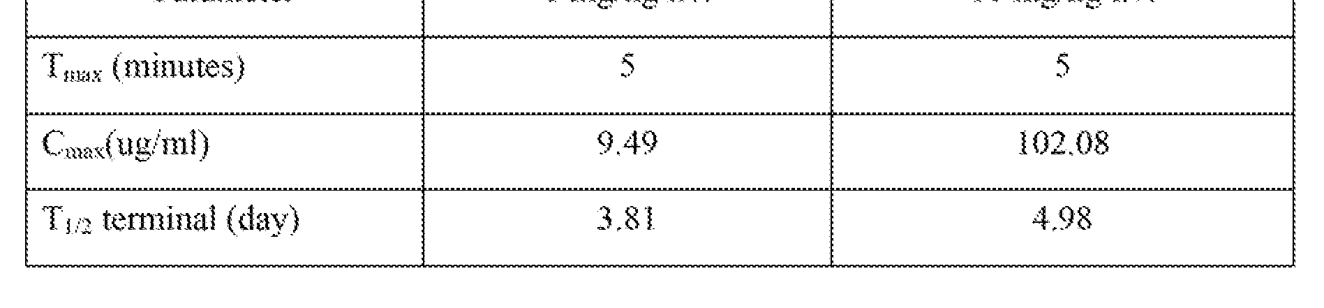


Claims
Priority Applications (20)
| Application Number | Priority Date | Filing Date | Title |
|---|---|---|---|
| US12/299,013 US8163497B2 (en) | 2008-09-07 | 2008-09-07 | Anti-extended type I glycosphingolipid antibody, derivatives thereof and use |
| CA2735433A CA2735433C (en) | 2008-09-07 | 2008-09-07 | Anti-extended type i glycosphingolipid antibody, derivatives thereof and use |
| DK08799283.0T DK2324060T3 (en) | 2008-09-07 | 2008-09-07 | ANTI-EXTENDED TYPE I glycosphingolipid ANTIBODY, DERIVATIVES AND USE |
| KR1020147011352A KR101432474B1 (en) | 2008-09-07 | 2008-09-07 | Anti-extended type i glycosphingolipid antibody, derivatives thereof and use |
| JP2011526023A JP5611210B2 (en) | 2008-09-07 | 2008-09-07 | Anti-expanded type I glycosphingolipid antibodies, derivatives and uses thereof |
| KR1020117007613A KR101442323B1 (en) | 2008-09-07 | 2008-09-07 | Anti-extended type i glycosphingolipid antibody, derivatives thereof and use |
| NZ591488A NZ591488A (en) | 2008-09-07 | 2008-09-07 | Anti-extended type i glycosphingolipid antibody, derivatives thereof and use |
| RU2011108579/10A RU2478648C2 (en) | 2008-09-07 | 2008-09-07 | Anti-extended type i glycosphingolipid antibody, its derivatives and use thereof |
| AU2008361352A AU2008361352B2 (en) | 2008-09-07 | 2008-09-07 | Anti-extended Type I glycosphingolipid antibody, derivatives thereof and use |
| CN200880131006.5A CN102282172B (en) | 2008-09-07 | 2008-09-07 | Anti-extended type i glycosphingolipid antibody, derivatives thereof and use |
| EP08799283.0A EP2324060B1 (en) | 2008-09-07 | 2008-09-07 | Anti-extended type i glycosphingolipid antibody, derivatives thereof and use |
| ES08799283.0T ES2549877T3 (en) | 2008-09-07 | 2008-09-07 | Antiglucosphingolipid antibody of extended type I, derivatives thereof and use |
| BRPI0823049-8A BRPI0823049A2 (en) | 2008-09-07 | 2008-09-07 | Anti-extended type 1 glycosphingolipid antibodies, derivatives thereof and use. |
| MX2011002478A MX2011002478A (en) | 2008-09-07 | 2008-09-07 | Anti-extended type i glycosphingolipid antibody, derivatives thereof and use. |
| PCT/US2008/075533 WO2010027364A1 (en) | 2008-09-07 | 2008-09-07 | Anti-extended type i glycosphingolipid antibody, derivatives thereof and use |
| TW098126571A TWI433684B (en) | 2008-09-07 | 2009-08-06 | Anti-extended type i glycosphingolipid antibody, derivatives thereof and use |
| ZA2011/01547A ZA201101547B (en) | 2008-09-07 | 2011-02-28 | Anti-extended type i glycosphingolipid antibody,derivatives thereof and use |
| IL211561A IL211561A (en) | 2008-09-07 | 2011-03-03 | Anti-extended type 1 glycosphingolipid antibody and compositions and articles of manufacture comprising same |
| HK11111704.1A HK1157363A1 (en) | 2008-09-07 | 2011-10-31 | Anti-extended type i glycosphingolipid antibody, derivatives thereof and use |
| US13/433,881 US8703129B2 (en) | 2008-09-07 | 2012-03-29 | Antibodies against extended type 1 chain antigens, derivatives thereof and use |
Applications Claiming Priority (1)
| Application Number | Priority Date | Filing Date | Title |
|---|---|---|---|
| PCT/US2008/075533 WO2010027364A1 (en) | 2008-09-07 | 2008-09-07 | Anti-extended type i glycosphingolipid antibody, derivatives thereof and use |
Related Child Applications (2)
| Application Number | Title | Priority Date | Filing Date |
|---|---|---|---|
| US12/299,013 A-371-Of-International US8163497B2 (en) | 2008-09-07 | 2008-09-07 | Anti-extended type I glycosphingolipid antibody, derivatives thereof and use |
| US13/433,881 Continuation-In-Part US8703129B2 (en) | 2008-09-07 | 2012-03-29 | Antibodies against extended type 1 chain antigens, derivatives thereof and use |
Publications (1)
| Publication Number | Publication Date |
|---|---|
| WO2010027364A1 true WO2010027364A1 (en) | 2010-03-11 |
Family
ID=41797355
Family Applications (1)
| Application Number | Title | Priority Date | Filing Date |
|---|---|---|---|
| PCT/US2008/075533 WO2010027364A1 (en) | 2008-09-07 | 2008-09-07 | Anti-extended type i glycosphingolipid antibody, derivatives thereof and use |
Country Status (18)
| Country | Link |
|---|---|
| US (1) | US8163497B2 (en) |
| EP (1) | EP2324060B1 (en) |
| JP (1) | JP5611210B2 (en) |
| KR (2) | KR101442323B1 (en) |
| CN (1) | CN102282172B (en) |
| AU (1) | AU2008361352B2 (en) |
| BR (1) | BRPI0823049A2 (en) |
| CA (1) | CA2735433C (en) |
| DK (1) | DK2324060T3 (en) |
| ES (1) | ES2549877T3 (en) |
| HK (1) | HK1157363A1 (en) |
| IL (1) | IL211561A (en) |
| MX (1) | MX2011002478A (en) |
| NZ (1) | NZ591488A (en) |
| RU (1) | RU2478648C2 (en) |
| TW (1) | TWI433684B (en) |
| WO (1) | WO2010027364A1 (en) |
| ZA (1) | ZA201101547B (en) |
Cited By (3)
| Publication number | Priority date | Publication date | Assignee | Title |
|---|---|---|---|---|
| US20120184717A1 (en) * | 2008-09-07 | 2012-07-19 | Glyconex Inc. | Antibodies against extended type 1 chain antigens, derivatives thereof and use |
| JP2014522402A (en) * | 2011-06-03 | 2014-09-04 | エルジー ライフ サイエンシーズ リミテッド | Stable liquid formulation of etanercept |
| WO2015063500A1 (en) * | 2013-11-01 | 2015-05-07 | The University Of Nottingham | Glycans as functional cancer targets and antibodies thereto |
Citations (115)
| Publication number | Priority date | Publication date | Assignee | Title |
|---|---|---|---|---|
| US3773919A (en) | 1969-10-23 | 1973-11-20 | Du Pont | Polylactide-drug mixtures |
| US4179337A (en) | 1973-07-20 | 1979-12-18 | Davis Frank F | Non-immunogenic polypeptides |
| US4256746A (en) | 1978-11-14 | 1981-03-17 | Takeda Chemical Industries | Dechloromaytansinoids, their pharmaceutical compositions and method of use |
| US4275149A (en) | 1978-11-24 | 1981-06-23 | Syva Company | Macromolecular environment control in specific receptor assays |
| US4294757A (en) | 1979-01-31 | 1981-10-13 | Takeda Chemical Industries, Ltd | 20-O-Acylmaytansinoids |
| US4301144A (en) | 1979-07-11 | 1981-11-17 | Ajinomoto Company, Incorporated | Blood substitute containing modified hemoglobin |
| US4307016A (en) | 1978-03-24 | 1981-12-22 | Takeda Chemical Industries, Ltd. | Demethyl maytansinoids |
| US4313946A (en) | 1981-01-27 | 1982-02-02 | The United States Of America As Represented By The Secretary Of Agriculture | Chemotherapeutically active maytansinoids from Trewia nudiflora |
| US4315929A (en) | 1981-01-27 | 1982-02-16 | The United States Of America As Represented By The Secretary Of Agriculture | Method of controlling the European corn borer with trewiasine |
| US4318980A (en) | 1978-04-10 | 1982-03-09 | Miles Laboratories, Inc. | Heterogenous specific binding assay employing a cycling reactant as label |
| US4322348A (en) | 1979-06-05 | 1982-03-30 | Takeda Chemical Industries, Ltd. | Maytansinoids |
| US4331598A (en) | 1979-09-19 | 1982-05-25 | Takeda Chemical Industries, Ltd. | Maytansinoids |
| US4362663A (en) | 1979-09-21 | 1982-12-07 | Takeda Chemical Industries, Ltd. | Maytansinoid compound |
| US4364866A (en) | 1979-09-21 | 1982-12-21 | Takeda Chemical Industries, Ltd. | Maytansinoids |
| US4371533A (en) | 1980-10-08 | 1983-02-01 | Takeda Chemical Industries, Ltd. | 4,5-Deoxymaytansinoids, their use and pharmaceutical compositions thereof |
| US4376110A (en) | 1980-08-04 | 1983-03-08 | Hybritech, Incorporated | Immunometric assays using monoclonal antibodies |
| US4424219A (en) | 1981-05-20 | 1984-01-03 | Takeda Chemical Industries, Ltd. | 9-Thiomaytansinoids and their pharmaceutical compositions and use |
| US4444887A (en) | 1979-12-10 | 1984-04-24 | Sloan-Kettering Institute | Process for making human antibody producing B-lymphocytes |
| US4450254A (en) | 1980-11-03 | 1984-05-22 | Standard Oil Company | Impact improvement of high nitrile resins |
| US4496689A (en) | 1983-12-27 | 1985-01-29 | Miles Laboratories, Inc. | Covalently attached complex of alpha-1-proteinase inhibitor with a water soluble polymer |
| US4560655A (en) | 1982-12-16 | 1985-12-24 | Immunex Corporation | Serum-free cell culture medium and process for making same |
| WO1986005807A1 (en) | 1985-04-01 | 1986-10-09 | Celltech Limited | Transformed myeloma cell-line and a process for the expression of a gene coding for a eukaryotic polypeptide employing same |
| US4640835A (en) | 1981-10-30 | 1987-02-03 | Nippon Chemiphar Company, Ltd. | Plasminogen activator derivatives |
| US4657866A (en) | 1982-12-21 | 1987-04-14 | Sudhir Kumar | Serum-free, synthetic, completely chemically defined tissue culture media |
| US4670417A (en) | 1985-06-19 | 1987-06-02 | Ajinomoto Co., Inc. | Hemoglobin combined with a poly(alkylene oxide) |
| US4676980A (en) | 1985-09-23 | 1987-06-30 | The United States Of America As Represented By The Secretary Of The Department Of Health And Human Services | Target specific cross-linked heteroantibodies |
| WO1987005330A1 (en) | 1986-03-07 | 1987-09-11 | Michel Louis Eugene Bergh | Method for enhancing glycoprotein stability |
| EP0239400A2 (en) | 1986-03-27 | 1987-09-30 | Medical Research Council | Recombinant antibodies and methods for their production |
| US4716111A (en) | 1982-08-11 | 1987-12-29 | Trustees Of Boston University | Process for producing human antibodies |
| US4737456A (en) | 1985-05-09 | 1988-04-12 | Syntex (U.S.A.) Inc. | Reducing interference in ligand-receptor binding assays |
| US4767704A (en) | 1983-10-07 | 1988-08-30 | Columbia University In The City Of New York | Protein-free culture medium |
| US4791192A (en) | 1986-06-26 | 1988-12-13 | Takeda Chemical Industries, Ltd. | Chemically modified protein with polyethyleneglycol |
| WO1989001036A1 (en) | 1987-07-23 | 1989-02-09 | Celltech Limited | Recombinant dna expression vectors |
| US4816397A (en) | 1983-03-25 | 1989-03-28 | Celltech, Limited | Multichain polypeptides or proteins and processes for their production |
| EP0332424A2 (en) | 1988-03-09 | 1989-09-13 | Hybritech Incorporated | Chimeric antibodies directed against human carcinoembryonic antigen |
| WO1989009622A1 (en) | 1988-04-15 | 1989-10-19 | Protein Design Labs, Inc. | Il-2 receptor-specific chimeric antibodies |
| EP0338745A1 (en) | 1988-04-16 | 1989-10-25 | Celltech Limited | Method for producing recombinant DNA proteins |
| WO1989012624A2 (en) | 1988-06-14 | 1989-12-28 | Cetus Corporation | Coupling agents and sterically hindered disulfide linked conjugates prepared therefrom |
| US4946778A (en) | 1987-09-21 | 1990-08-07 | Genex Corporation | Single polypeptide chain binding molecules |
| EP0396387A2 (en) | 1989-05-05 | 1990-11-07 | Research Development Foundation | A novel antibody delivery system for biological response modifiers |
| US4980286A (en) | 1985-07-05 | 1990-12-25 | Whitehead Institute For Biomedical Research | In vivo introduction and expression of foreign genetic material in epithelial cells |
| EP0413622A1 (en) | 1989-08-03 | 1991-02-20 | Rhone-Poulenc Sante | Albumin derivatives with therapeutic functions |
| US5011920A (en) | 1986-01-30 | 1991-04-30 | Fred Hutchinson Cancer Research Center | Disialofucoganglioside immunogen and fucoganglioside monosialosyl Lea II |
| WO1991009967A1 (en) | 1989-12-21 | 1991-07-11 | Celltech Limited | Humanised antibodies |
| WO1991010741A1 (en) | 1990-01-12 | 1991-07-25 | Cell Genesys, Inc. | Generation of xenogeneic antibodies |
| EP0439095A2 (en) | 1990-01-22 | 1991-07-31 | Bristol-Myers Squibb Company | Therapeutic antibody based fusion proteins |
| WO1991014438A1 (en) | 1990-03-20 | 1991-10-03 | The Trustees Of Columbia University In The City Of New York | Chimeric antibodies with receptor binding ligands in place of their constant region |
| WO1992001047A1 (en) | 1990-07-10 | 1992-01-23 | Cambridge Antibody Technology Limited | Methods for producing members of specific binding pairs |
| WO1992006180A1 (en) | 1990-10-01 | 1992-04-16 | University Of Connecticut | Targeting viruses and cells for selective internalization by cells |
| WO1992008495A1 (en) | 1990-11-09 | 1992-05-29 | Abbott Biotech, Inc. | Cytokine immunoconjugates |
| US5122469A (en) | 1990-10-03 | 1992-06-16 | Genentech, Inc. | Method for culturing Chinese hamster ovary cells to improve production of recombinant proteins |
| US5122464A (en) | 1986-01-23 | 1992-06-16 | Celltech Limited, A British Company | Method for dominant selection in eucaryotic cells |
| WO1992020316A2 (en) | 1991-05-14 | 1992-11-26 | University Of Connecticut | Targeted delivery of genes encoding immunogenic proteins |
| US5168043A (en) | 1986-01-10 | 1992-12-01 | Sloan-Kettering Institute For Cancer Research | Blood group antigen panel |
| EP0519596A1 (en) | 1991-05-17 | 1992-12-23 | Merck & Co. Inc. | A method for reducing the immunogenicity of antibody variable domains |
| WO1992022635A1 (en) | 1991-06-05 | 1992-12-23 | University Of Connecticut | Targeted delivery of genes encoding secretory proteins |
| WO1993008829A1 (en) | 1991-11-04 | 1993-05-13 | The Regents Of The University Of California | Compositions that mediate killing of hiv-infected cells |
| US5223409A (en) | 1988-09-02 | 1993-06-29 | Protein Engineering Corp. | Directed evolution of novel binding proteins |
| US5225539A (en) | 1986-03-27 | 1993-07-06 | Medical Research Council | Recombinant altered antibodies and methods of making altered antibodies |
| WO1993014188A1 (en) | 1992-01-17 | 1993-07-22 | The Regents Of The University Of Michigan | Targeted virus |
| WO1993015199A1 (en) | 1992-01-31 | 1993-08-05 | Rhone-Poulenc Rorer S.A. | Novel biologically active polypeptides, preparation thereof and pharmaceutical composition containing said polypeptides |
| WO1993015200A1 (en) | 1992-01-31 | 1993-08-05 | Rhone-Poulenc Rorer S.A. | Antithrombotic polypeptides as antagonists of the binding of vwf to platelets or to subendothelium |
| WO1993016185A2 (en) | 1992-02-06 | 1993-08-19 | Creative Biomolecules, Inc. | Biosynthetic binding protein for cancer marker |
| US5240833A (en) | 1989-01-30 | 1993-08-31 | The Biomembrane Institute | Method for the production of monoclonal antibodies directed to tumor-associated gangliosides and fucogangliosides |
| WO1993020221A1 (en) | 1992-04-03 | 1993-10-14 | Young Alexander T | Gene therapy using targeted viral vectors |
| WO1993021319A1 (en) | 1992-04-08 | 1993-10-28 | Cetus Oncology Corporation | HUMANIZED C-erbB-2 SPECIFIC ANTIBODIES |
| WO1993021232A1 (en) | 1992-04-10 | 1993-10-28 | Research Development Foundation | IMMUNOTOXINS DIRECTED AGAINST c-erbB-2 (HER-2/neu) RELATED SURFACE ANTIGENS |
| EP0592106A1 (en) | 1992-09-09 | 1994-04-13 | Immunogen Inc | Resurfacing of rodent antibodies |
| WO1994008598A1 (en) | 1992-10-09 | 1994-04-28 | Advanced Tissue Sciences, Inc. | Liver reserve cells |
| EP0598877A1 (en) | 1992-06-09 | 1994-06-01 | Hoppe Ag | Latch and lockset system |
| WO1994012649A2 (en) | 1992-12-03 | 1994-06-09 | Genzyme Corporation | Gene therapy for cystic fibrosis |
| WO1994025591A1 (en) | 1993-04-29 | 1994-11-10 | Unilever N.V. | PRODUCTION OF ANTIBODIES OR (FUNCTIONALIZED) FRAGMENTS THEREOF DERIVED FROM HEAVY CHAIN IMMUNOGLOBULINS OF $i(CAMELIDAE) |
| US5413923A (en) | 1989-07-25 | 1995-05-09 | Cell Genesys, Inc. | Homologous recombination for universal donor cells and chimeric mammalian hosts |
| US5436146A (en) | 1989-09-07 | 1995-07-25 | The Trustees Of Princeton University | Helper-free stocks of recombinant adeno-associated virus vectors |
| US5474981A (en) | 1992-08-26 | 1995-12-12 | President And Fellows Of Harvard College | Use of the cytokine IP-10 as an anti-tumor agent |
| US5475092A (en) | 1992-03-25 | 1995-12-12 | Immunogen Inc. | Cell binding agent conjugates of analogues and derivatives of CC-1065 |
| US5530101A (en) | 1988-12-28 | 1996-06-25 | Protein Design Labs, Inc. | Humanized immunoglobulins |
| US5534617A (en) | 1988-10-28 | 1996-07-09 | Genentech, Inc. | Human growth hormone variants having greater affinity for human growth hormone receptor at site 1 |
| US5545806A (en) | 1990-08-29 | 1996-08-13 | Genpharm International, Inc. | Ransgenic non-human animals for producing heterologous antibodies |
| WO1996025436A1 (en) | 1995-02-14 | 1996-08-22 | Sloan-Kettering Institute For Cancer Research | Recombinant human anti-lewis b antibodies |
| US5565332A (en) | 1991-09-23 | 1996-10-15 | Medical Research Council | Production of chimeric antibodies - a combinatorial approach |
| US5569825A (en) | 1990-08-29 | 1996-10-29 | Genpharm International | Transgenic non-human animals capable of producing heterologous antibodies of various isotypes |
| WO1996033735A1 (en) | 1995-04-27 | 1996-10-31 | Abgenix, Inc. | Human antibodies derived from immunized xenomice |
| WO1996034096A1 (en) | 1995-04-28 | 1996-10-31 | Abgenix, Inc. | Human antibodies derived from immunized xenomice |
| US5625126A (en) | 1990-08-29 | 1997-04-29 | Genpharm International, Inc. | Transgenic non-human animals for producing heterologous antibodies |
| US5633425A (en) | 1990-08-29 | 1997-05-27 | Genpharm International, Inc. | Transgenic non-human animals capable of producing heterologous antibodies |
| US5661016A (en) | 1990-08-29 | 1997-08-26 | Genpharm International Inc. | Transgenic non-human animals capable of producing heterologous antibodies of various isotypes |
| WO1997033899A1 (en) | 1996-03-14 | 1997-09-18 | Human Genome Sciences, Inc. | Apoptosis inducing molecule i |
| WO1997034631A1 (en) | 1996-03-18 | 1997-09-25 | Board Of Regents, The University Of Texas System | Immunoglobin-like domains with increased half lives |
| WO1997034911A1 (en) | 1996-03-22 | 1997-09-25 | Human Genome Sciences, Inc. | Apoptosis inducing molecule ii |
| US5698417A (en) | 1985-11-01 | 1997-12-16 | Xoma Corporation | Modular assembly of antibody genes, antibodies prepared thereby and use |
| US5712163A (en) | 1989-06-05 | 1998-01-27 | Organogenesis, Inc. | Chemically defined cell culture media and system and methods for use, particularly for culturing epithelial cells |
| WO1998016654A1 (en) | 1996-10-11 | 1998-04-23 | Japan Tobacco, Inc. | Production of a multimeric protein by cell fusion method |
| WO1998023289A1 (en) | 1996-11-27 | 1998-06-04 | The General Hospital Corporation | MODULATION OF IgG BINDING TO FcRn |
| WO1998024893A2 (en) | 1996-12-03 | 1998-06-11 | Abgenix, Inc. | TRANSGENIC MAMMALS HAVING HUMAN IG LOCI INCLUDING PLURAL VH AND Vλ REGIONS AND ANTIBODIES PRODUCED THEREFROM |
| US5807715A (en) | 1984-08-27 | 1998-09-15 | The Board Of Trustees Of The Leland Stanford Junior University | Methods and transformed mammalian lymphocyte cells for producing functional antigen-binding protein including chimeric immunoglobulin |
| US5814318A (en) | 1990-08-29 | 1998-09-29 | Genpharm International Inc. | Transgenic non-human animals for producing heterologous antibodies |
| WO1998046645A2 (en) | 1997-04-14 | 1998-10-22 | Micromet Gesellschaft Für Biomedizinische Forschung Mbh | Method for the production of antihuman antigen receptors and uses thereof |
| WO1998050433A2 (en) | 1997-05-05 | 1998-11-12 | Abgenix, Inc. | Human monoclonal antibodies to epidermal growth factor receptor |
| US5869619A (en) | 1991-12-13 | 1999-02-09 | Xoma Corporation | Modified antibody variable domains |
| US5885793A (en) | 1991-12-02 | 1999-03-23 | Medical Research Council | Production of anti-self antibodies from antibody segment repertoires and displayed on phage |
| WO1999023105A1 (en) | 1997-11-03 | 1999-05-14 | Human Genome Sciences, Inc. | Vegi, an inhibitor of angiogenesis and tumor growth |
| US6048728A (en) | 1988-09-23 | 2000-04-11 | Chiron Corporation | Cell culture medium for enhanced cell growth, culture longevity, and product expression |
| US6083929A (en) | 1991-05-06 | 2000-07-04 | The Biomembrane Institute | Extended type 1 chain glycosphingolipids as tumor-associated antigens |
| US6277375B1 (en) | 1997-03-03 | 2001-08-21 | Board Of Regents, The University Of Texas System | Immunoglobulin-like domains with increased half-lives |
| WO2001077137A1 (en) | 2000-04-12 | 2001-10-18 | Human Genome Sciences, Inc. | Albumin fusion proteins |
| US6333410B1 (en) | 2000-08-18 | 2001-12-25 | Immunogen, Inc. | Process for the preparation and purification of thiol-containing maytansinoids |
| US6432402B1 (en) * | 1989-05-25 | 2002-08-13 | Sloan-Kettering Institute For Cancer Research | Anti-idiotypic antibody which induces an immune response against a glycosphingolipid and use thereof |
| US6514496B1 (en) | 1995-04-14 | 2003-02-04 | Inhale Therapeutic Systems, Inc. | Dispersible antibody compositions and methods for their preparation and use |
| US20030130496A1 (en) | 1988-11-11 | 2003-07-10 | Medical Research Council | Single domain ligands, receptors comprising said ligands, methods for their production, and use of said ligands and receptors |
| US6632670B1 (en) | 1995-09-08 | 2003-10-14 | Genzyme Corporation | AAV vectors for gene therapy |
| US6642051B1 (en) | 1997-10-21 | 2003-11-04 | Targeted Genetics Corporation | Amplifiable adeno-associated virus(AAV) packaging cassettes for the production of recombinant AAV vectors |
| WO2006042333A2 (en) | 2004-10-12 | 2006-04-20 | Xencor, Inc. | Prediction and assessment of immunogenicity |
| US7041870B2 (en) | 2000-11-30 | 2006-05-09 | Medarex, Inc. | Transgenic transchromosomal rodents for making human antibodies |
| US7115717B2 (en) | 2001-05-18 | 2006-10-03 | Kirin Beer Kabushiki Kaisha | Anti-TRAIL-R antibodies |
Family Cites Families (2)
| Publication number | Priority date | Publication date | Assignee | Title |
|---|---|---|---|---|
| EP0232706A3 (en) * | 1986-01-10 | 1988-11-30 | Sloan-Kettering Institute For Cancer Research | Blood group antigen panel |
| EP1307207A4 (en) * | 2000-07-03 | 2005-12-14 | Biomembrane Inst | Extended type 1 chain glycosphingolipids as tumor-associated antigens |
-
2008
- 2008-09-07 BR BRPI0823049-8A patent/BRPI0823049A2/en not_active IP Right Cessation
- 2008-09-07 US US12/299,013 patent/US8163497B2/en active Active
- 2008-09-07 MX MX2011002478A patent/MX2011002478A/en active IP Right Grant
- 2008-09-07 KR KR1020117007613A patent/KR101442323B1/en active IP Right Grant
- 2008-09-07 WO PCT/US2008/075533 patent/WO2010027364A1/en active Application Filing
- 2008-09-07 DK DK08799283.0T patent/DK2324060T3/en active
- 2008-09-07 NZ NZ591488A patent/NZ591488A/en not_active IP Right Cessation
- 2008-09-07 CN CN200880131006.5A patent/CN102282172B/en active Active
- 2008-09-07 JP JP2011526023A patent/JP5611210B2/en active Active
- 2008-09-07 AU AU2008361352A patent/AU2008361352B2/en active Active
- 2008-09-07 EP EP08799283.0A patent/EP2324060B1/en active Active
- 2008-09-07 KR KR1020147011352A patent/KR101432474B1/en active IP Right Grant
- 2008-09-07 CA CA2735433A patent/CA2735433C/en active Active
- 2008-09-07 ES ES08799283.0T patent/ES2549877T3/en active Active
- 2008-09-07 RU RU2011108579/10A patent/RU2478648C2/en active
-
2009
- 2009-08-06 TW TW098126571A patent/TWI433684B/en active
-
2011
- 2011-02-28 ZA ZA2011/01547A patent/ZA201101547B/en unknown
- 2011-03-03 IL IL211561A patent/IL211561A/en active IP Right Grant
- 2011-10-31 HK HK11111704.1A patent/HK1157363A1/en unknown
Patent Citations (126)
| Publication number | Priority date | Publication date | Assignee | Title |
|---|---|---|---|---|
| US3773919A (en) | 1969-10-23 | 1973-11-20 | Du Pont | Polylactide-drug mixtures |
| US4179337A (en) | 1973-07-20 | 1979-12-18 | Davis Frank F | Non-immunogenic polypeptides |
| US4361650A (en) | 1978-03-24 | 1982-11-30 | Takeda Chemical Industries, Ltd. | Fermentation process of preparing demethyl maytansinoids |
| US4307016A (en) | 1978-03-24 | 1981-12-22 | Takeda Chemical Industries, Ltd. | Demethyl maytansinoids |
| US4318980A (en) | 1978-04-10 | 1982-03-09 | Miles Laboratories, Inc. | Heterogenous specific binding assay employing a cycling reactant as label |
| US4256746A (en) | 1978-11-14 | 1981-03-17 | Takeda Chemical Industries | Dechloromaytansinoids, their pharmaceutical compositions and method of use |
| US4275149A (en) | 1978-11-24 | 1981-06-23 | Syva Company | Macromolecular environment control in specific receptor assays |
| US4294757A (en) | 1979-01-31 | 1981-10-13 | Takeda Chemical Industries, Ltd | 20-O-Acylmaytansinoids |
| US4322348A (en) | 1979-06-05 | 1982-03-30 | Takeda Chemical Industries, Ltd. | Maytansinoids |
| US4301144A (en) | 1979-07-11 | 1981-11-17 | Ajinomoto Company, Incorporated | Blood substitute containing modified hemoglobin |
| US4331598A (en) | 1979-09-19 | 1982-05-25 | Takeda Chemical Industries, Ltd. | Maytansinoids |
| US4362663A (en) | 1979-09-21 | 1982-12-07 | Takeda Chemical Industries, Ltd. | Maytansinoid compound |
| US4364866A (en) | 1979-09-21 | 1982-12-21 | Takeda Chemical Industries, Ltd. | Maytansinoids |
| US4444887A (en) | 1979-12-10 | 1984-04-24 | Sloan-Kettering Institute | Process for making human antibody producing B-lymphocytes |
| US4376110A (en) | 1980-08-04 | 1983-03-08 | Hybritech, Incorporated | Immunometric assays using monoclonal antibodies |
| US4371533A (en) | 1980-10-08 | 1983-02-01 | Takeda Chemical Industries, Ltd. | 4,5-Deoxymaytansinoids, their use and pharmaceutical compositions thereof |
| US4450254A (en) | 1980-11-03 | 1984-05-22 | Standard Oil Company | Impact improvement of high nitrile resins |
| US4315929A (en) | 1981-01-27 | 1982-02-16 | The United States Of America As Represented By The Secretary Of Agriculture | Method of controlling the European corn borer with trewiasine |
| US4313946A (en) | 1981-01-27 | 1982-02-02 | The United States Of America As Represented By The Secretary Of Agriculture | Chemotherapeutically active maytansinoids from Trewia nudiflora |
| US4424219A (en) | 1981-05-20 | 1984-01-03 | Takeda Chemical Industries, Ltd. | 9-Thiomaytansinoids and their pharmaceutical compositions and use |
| US4640835A (en) | 1981-10-30 | 1987-02-03 | Nippon Chemiphar Company, Ltd. | Plasminogen activator derivatives |
| US4716111A (en) | 1982-08-11 | 1987-12-29 | Trustees Of Boston University | Process for producing human antibodies |
| US4560655A (en) | 1982-12-16 | 1985-12-24 | Immunex Corporation | Serum-free cell culture medium and process for making same |
| US4657866A (en) | 1982-12-21 | 1987-04-14 | Sudhir Kumar | Serum-free, synthetic, completely chemically defined tissue culture media |
| US4816397A (en) | 1983-03-25 | 1989-03-28 | Celltech, Limited | Multichain polypeptides or proteins and processes for their production |
| US4767704A (en) | 1983-10-07 | 1988-08-30 | Columbia University In The City Of New York | Protein-free culture medium |
| US4496689A (en) | 1983-12-27 | 1985-01-29 | Miles Laboratories, Inc. | Covalently attached complex of alpha-1-proteinase inhibitor with a water soluble polymer |
| US5807715A (en) | 1984-08-27 | 1998-09-15 | The Board Of Trustees Of The Leland Stanford Junior University | Methods and transformed mammalian lymphocyte cells for producing functional antigen-binding protein including chimeric immunoglobulin |
| WO1986005807A1 (en) | 1985-04-01 | 1986-10-09 | Celltech Limited | Transformed myeloma cell-line and a process for the expression of a gene coding for a eukaryotic polypeptide employing same |
| US4737456A (en) | 1985-05-09 | 1988-04-12 | Syntex (U.S.A.) Inc. | Reducing interference in ligand-receptor binding assays |
| US4670417A (en) | 1985-06-19 | 1987-06-02 | Ajinomoto Co., Inc. | Hemoglobin combined with a poly(alkylene oxide) |
| US4980286A (en) | 1985-07-05 | 1990-12-25 | Whitehead Institute For Biomedical Research | In vivo introduction and expression of foreign genetic material in epithelial cells |
| US4676980A (en) | 1985-09-23 | 1987-06-30 | The United States Of America As Represented By The Secretary Of The Department Of Health And Human Services | Target specific cross-linked heteroantibodies |
| US6204023B1 (en) | 1985-11-01 | 2001-03-20 | Xoma Ltd. | Modular assembly of antibody genes, antibodies prepared thereby and use |
| US5698435A (en) | 1985-11-01 | 1997-12-16 | Xoma Corporation | Modular assembly of antibody genes, antibodies prepared thereby and use |
| US5698417A (en) | 1985-11-01 | 1997-12-16 | Xoma Corporation | Modular assembly of antibody genes, antibodies prepared thereby and use |
| US5168043A (en) | 1986-01-10 | 1992-12-01 | Sloan-Kettering Institute For Cancer Research | Blood group antigen panel |
| US5122464A (en) | 1986-01-23 | 1992-06-16 | Celltech Limited, A British Company | Method for dominant selection in eucaryotic cells |
| US5011920A (en) | 1986-01-30 | 1991-04-30 | Fred Hutchinson Cancer Research Center | Disialofucoganglioside immunogen and fucoganglioside monosialosyl Lea II |
| WO1987005330A1 (en) | 1986-03-07 | 1987-09-11 | Michel Louis Eugene Bergh | Method for enhancing glycoprotein stability |
| US5225539A (en) | 1986-03-27 | 1993-07-06 | Medical Research Council | Recombinant altered antibodies and methods of making altered antibodies |
| EP0239400A2 (en) | 1986-03-27 | 1987-09-30 | Medical Research Council | Recombinant antibodies and methods for their production |
| US4791192A (en) | 1986-06-26 | 1988-12-13 | Takeda Chemical Industries, Ltd. | Chemically modified protein with polyethyleneglycol |
| WO1989001036A1 (en) | 1987-07-23 | 1989-02-09 | Celltech Limited | Recombinant dna expression vectors |
| US4946778A (en) | 1987-09-21 | 1990-08-07 | Genex Corporation | Single polypeptide chain binding molecules |
| EP0332424A2 (en) | 1988-03-09 | 1989-09-13 | Hybritech Incorporated | Chimeric antibodies directed against human carcinoembryonic antigen |
| WO1989009622A1 (en) | 1988-04-15 | 1989-10-19 | Protein Design Labs, Inc. | Il-2 receptor-specific chimeric antibodies |
| EP0338745A1 (en) | 1988-04-16 | 1989-10-25 | Celltech Limited | Method for producing recombinant DNA proteins |
| WO1989012624A2 (en) | 1988-06-14 | 1989-12-28 | Cetus Corporation | Coupling agents and sterically hindered disulfide linked conjugates prepared therefrom |
| US5223409A (en) | 1988-09-02 | 1993-06-29 | Protein Engineering Corp. | Directed evolution of novel binding proteins |
| US6048728A (en) | 1988-09-23 | 2000-04-11 | Chiron Corporation | Cell culture medium for enhanced cell growth, culture longevity, and product expression |
| US5534617A (en) | 1988-10-28 | 1996-07-09 | Genentech, Inc. | Human growth hormone variants having greater affinity for human growth hormone receptor at site 1 |
| US20030130496A1 (en) | 1988-11-11 | 2003-07-10 | Medical Research Council | Single domain ligands, receptors comprising said ligands, methods for their production, and use of said ligands and receptors |
| US5585089A (en) | 1988-12-28 | 1996-12-17 | Protein Design Labs, Inc. | Humanized immunoglobulins |
| US5530101A (en) | 1988-12-28 | 1996-06-25 | Protein Design Labs, Inc. | Humanized immunoglobulins |
| US5240833A (en) | 1989-01-30 | 1993-08-31 | The Biomembrane Institute | Method for the production of monoclonal antibodies directed to tumor-associated gangliosides and fucogangliosides |
| EP0396387A2 (en) | 1989-05-05 | 1990-11-07 | Research Development Foundation | A novel antibody delivery system for biological response modifiers |
| US6432402B1 (en) * | 1989-05-25 | 2002-08-13 | Sloan-Kettering Institute For Cancer Research | Anti-idiotypic antibody which induces an immune response against a glycosphingolipid and use thereof |
| US5712163A (en) | 1989-06-05 | 1998-01-27 | Organogenesis, Inc. | Chemically defined cell culture media and system and methods for use, particularly for culturing epithelial cells |
| US5413923A (en) | 1989-07-25 | 1995-05-09 | Cell Genesys, Inc. | Homologous recombination for universal donor cells and chimeric mammalian hosts |
| EP0413622A1 (en) | 1989-08-03 | 1991-02-20 | Rhone-Poulenc Sante | Albumin derivatives with therapeutic functions |
| US5436146A (en) | 1989-09-07 | 1995-07-25 | The Trustees Of Princeton University | Helper-free stocks of recombinant adeno-associated virus vectors |
| WO1991009967A1 (en) | 1989-12-21 | 1991-07-11 | Celltech Limited | Humanised antibodies |
| US5939598A (en) | 1990-01-12 | 1999-08-17 | Abgenix, Inc. | Method of making transgenic mice lacking endogenous heavy chains |
| WO1991010741A1 (en) | 1990-01-12 | 1991-07-25 | Cell Genesys, Inc. | Generation of xenogeneic antibodies |
| EP0439095A2 (en) | 1990-01-22 | 1991-07-31 | Bristol-Myers Squibb Company | Therapeutic antibody based fusion proteins |
| US5314995A (en) | 1990-01-22 | 1994-05-24 | Oncogen | Therapeutic interleukin-2-antibody based fusion proteins |
| WO1991014438A1 (en) | 1990-03-20 | 1991-10-03 | The Trustees Of Columbia University In The City Of New York | Chimeric antibodies with receptor binding ligands in place of their constant region |
| WO1992001047A1 (en) | 1990-07-10 | 1992-01-23 | Cambridge Antibody Technology Limited | Methods for producing members of specific binding pairs |
| US5633425A (en) | 1990-08-29 | 1997-05-27 | Genpharm International, Inc. | Transgenic non-human animals capable of producing heterologous antibodies |
| US5661016A (en) | 1990-08-29 | 1997-08-26 | Genpharm International Inc. | Transgenic non-human animals capable of producing heterologous antibodies of various isotypes |
| US5569825A (en) | 1990-08-29 | 1996-10-29 | Genpharm International | Transgenic non-human animals capable of producing heterologous antibodies of various isotypes |
| US5545806A (en) | 1990-08-29 | 1996-08-13 | Genpharm International, Inc. | Ransgenic non-human animals for producing heterologous antibodies |
| US5625126A (en) | 1990-08-29 | 1997-04-29 | Genpharm International, Inc. | Transgenic non-human animals for producing heterologous antibodies |
| US5814318A (en) | 1990-08-29 | 1998-09-29 | Genpharm International Inc. | Transgenic non-human animals for producing heterologous antibodies |
| WO1992006180A1 (en) | 1990-10-01 | 1992-04-16 | University Of Connecticut | Targeting viruses and cells for selective internalization by cells |
| US5122469A (en) | 1990-10-03 | 1992-06-16 | Genentech, Inc. | Method for culturing Chinese hamster ovary cells to improve production of recombinant proteins |
| WO1992008495A1 (en) | 1990-11-09 | 1992-05-29 | Abbott Biotech, Inc. | Cytokine immunoconjugates |
| US6083929A (en) | 1991-05-06 | 2000-07-04 | The Biomembrane Institute | Extended type 1 chain glycosphingolipids as tumor-associated antigens |
| US6294523B1 (en) | 1991-05-06 | 2001-09-25 | The Biomembrane Institute | Extended type 1 chain glycosphingolipids as tumor-associated antigens |
| WO1992020316A2 (en) | 1991-05-14 | 1992-11-26 | University Of Connecticut | Targeted delivery of genes encoding immunogenic proteins |
| EP0519596A1 (en) | 1991-05-17 | 1992-12-23 | Merck & Co. Inc. | A method for reducing the immunogenicity of antibody variable domains |
| WO1992022635A1 (en) | 1991-06-05 | 1992-12-23 | University Of Connecticut | Targeted delivery of genes encoding secretory proteins |
| US5565332A (en) | 1991-09-23 | 1996-10-15 | Medical Research Council | Production of chimeric antibodies - a combinatorial approach |
| WO1993008829A1 (en) | 1991-11-04 | 1993-05-13 | The Regents Of The University Of California | Compositions that mediate killing of hiv-infected cells |
| US5885793A (en) | 1991-12-02 | 1999-03-23 | Medical Research Council | Production of anti-self antibodies from antibody segment repertoires and displayed on phage |
| US5869619A (en) | 1991-12-13 | 1999-02-09 | Xoma Corporation | Modified antibody variable domains |
| WO1993014188A1 (en) | 1992-01-17 | 1993-07-22 | The Regents Of The University Of Michigan | Targeted virus |
| WO1993015200A1 (en) | 1992-01-31 | 1993-08-05 | Rhone-Poulenc Rorer S.A. | Antithrombotic polypeptides as antagonists of the binding of vwf to platelets or to subendothelium |
| WO1993015199A1 (en) | 1992-01-31 | 1993-08-05 | Rhone-Poulenc Rorer S.A. | Novel biologically active polypeptides, preparation thereof and pharmaceutical composition containing said polypeptides |
| WO1993016185A2 (en) | 1992-02-06 | 1993-08-19 | Creative Biomolecules, Inc. | Biosynthetic binding protein for cancer marker |
| US5585499A (en) | 1992-03-25 | 1996-12-17 | Immunogen Inc. | Cyclopropylbenzindole-containing cytotoxic drugs |
| US5846545A (en) | 1992-03-25 | 1998-12-08 | Immunogen, Inc. | Targeted delivery of cyclopropylbenzindole-containing cytotoxic drugs |
| US5475092A (en) | 1992-03-25 | 1995-12-12 | Immunogen Inc. | Cell binding agent conjugates of analogues and derivatives of CC-1065 |
| WO1993020221A1 (en) | 1992-04-03 | 1993-10-14 | Young Alexander T | Gene therapy using targeted viral vectors |
| WO1993021319A1 (en) | 1992-04-08 | 1993-10-28 | Cetus Oncology Corporation | HUMANIZED C-erbB-2 SPECIFIC ANTIBODIES |
| WO1993021232A1 (en) | 1992-04-10 | 1993-10-28 | Research Development Foundation | IMMUNOTOXINS DIRECTED AGAINST c-erbB-2 (HER-2/neu) RELATED SURFACE ANTIGENS |
| EP0598877A1 (en) | 1992-06-09 | 1994-06-01 | Hoppe Ag | Latch and lockset system |
| US5474981A (en) | 1992-08-26 | 1995-12-12 | President And Fellows Of Harvard College | Use of the cytokine IP-10 as an anti-tumor agent |
| EP0592106A1 (en) | 1992-09-09 | 1994-04-13 | Immunogen Inc | Resurfacing of rodent antibodies |
| US5639641A (en) | 1992-09-09 | 1997-06-17 | Immunogen Inc. | Resurfacing of rodent antibodies |
| WO1994008598A1 (en) | 1992-10-09 | 1994-04-28 | Advanced Tissue Sciences, Inc. | Liver reserve cells |
| WO1994012649A2 (en) | 1992-12-03 | 1994-06-09 | Genzyme Corporation | Gene therapy for cystic fibrosis |
| WO1994025591A1 (en) | 1993-04-29 | 1994-11-10 | Unilever N.V. | PRODUCTION OF ANTIBODIES OR (FUNCTIONALIZED) FRAGMENTS THEREOF DERIVED FROM HEAVY CHAIN IMMUNOGLOBULINS OF $i(CAMELIDAE) |
| WO1996025436A1 (en) | 1995-02-14 | 1996-08-22 | Sloan-Kettering Institute For Cancer Research | Recombinant human anti-lewis b antibodies |
| US6514496B1 (en) | 1995-04-14 | 2003-02-04 | Inhale Therapeutic Systems, Inc. | Dispersible antibody compositions and methods for their preparation and use |
| WO1996033735A1 (en) | 1995-04-27 | 1996-10-31 | Abgenix, Inc. | Human antibodies derived from immunized xenomice |
| WO1996034096A1 (en) | 1995-04-28 | 1996-10-31 | Abgenix, Inc. | Human antibodies derived from immunized xenomice |
| US6632670B1 (en) | 1995-09-08 | 2003-10-14 | Genzyme Corporation | AAV vectors for gene therapy |
| WO1997033899A1 (en) | 1996-03-14 | 1997-09-18 | Human Genome Sciences, Inc. | Apoptosis inducing molecule i |
| WO1997034631A1 (en) | 1996-03-18 | 1997-09-25 | Board Of Regents, The University Of Texas System | Immunoglobin-like domains with increased half lives |
| WO1997034911A1 (en) | 1996-03-22 | 1997-09-25 | Human Genome Sciences, Inc. | Apoptosis inducing molecule ii |
| WO1998016654A1 (en) | 1996-10-11 | 1998-04-23 | Japan Tobacco, Inc. | Production of a multimeric protein by cell fusion method |
| US5916771A (en) | 1996-10-11 | 1999-06-29 | Abgenix, Inc. | Production of a multimeric protein by cell fusion method |
| WO1998023289A1 (en) | 1996-11-27 | 1998-06-04 | The General Hospital Corporation | MODULATION OF IgG BINDING TO FcRn |
| WO1998024893A2 (en) | 1996-12-03 | 1998-06-11 | Abgenix, Inc. | TRANSGENIC MAMMALS HAVING HUMAN IG LOCI INCLUDING PLURAL VH AND Vλ REGIONS AND ANTIBODIES PRODUCED THEREFROM |
| US6277375B1 (en) | 1997-03-03 | 2001-08-21 | Board Of Regents, The University Of Texas System | Immunoglobulin-like domains with increased half-lives |
| WO1998046645A2 (en) | 1997-04-14 | 1998-10-22 | Micromet Gesellschaft Für Biomedizinische Forschung Mbh | Method for the production of antihuman antigen receptors and uses thereof |
| WO1998050433A2 (en) | 1997-05-05 | 1998-11-12 | Abgenix, Inc. | Human monoclonal antibodies to epidermal growth factor receptor |
| US6642051B1 (en) | 1997-10-21 | 2003-11-04 | Targeted Genetics Corporation | Amplifiable adeno-associated virus(AAV) packaging cassettes for the production of recombinant AAV vectors |
| WO1999023105A1 (en) | 1997-11-03 | 1999-05-14 | Human Genome Sciences, Inc. | Vegi, an inhibitor of angiogenesis and tumor growth |
| WO2001077137A1 (en) | 2000-04-12 | 2001-10-18 | Human Genome Sciences, Inc. | Albumin fusion proteins |
| US6333410B1 (en) | 2000-08-18 | 2001-12-25 | Immunogen, Inc. | Process for the preparation and purification of thiol-containing maytansinoids |
| US7041870B2 (en) | 2000-11-30 | 2006-05-09 | Medarex, Inc. | Transgenic transchromosomal rodents for making human antibodies |
| US7115717B2 (en) | 2001-05-18 | 2006-10-03 | Kirin Beer Kabushiki Kaisha | Anti-TRAIL-R antibodies |
| WO2006042333A2 (en) | 2004-10-12 | 2006-04-20 | Xencor, Inc. | Prediction and assessment of immunogenicity |
Non-Patent Citations (218)
| Title |
|---|
| "Expression Mechanism of Human Uterine Endometrial Cancer-Specific Fucosylated Carbohydrate Chain", INTERNATIONAL JOURNAL OF ONCOLOGY, vol. 6, 1995, pages 93 - 97 |
| "Extended Type I Chain Glycosphingolipid Antigens Isolation and Characterization of Trifucosyl-Leb Antigen", EUR. J. BIOCHEM, vol. 203, 1992, pages 577 - 586 |
| "High Expression of Lewis y/b Antigen is Associated with Decreased Survival in Lymph Node Negative Breast Carcinomas", BREAST CANCER RESEARCH |
| "Immunochemical and Immunohistological Expression of Lewis Hosto-blood Group Antigens in Small Intestine Including Individuals of the Le(a+b+) and Le(a-b-) Nonsecretor Phenotypes", GLYCOCONJUGATE JOURNAL, 1994, pages 600 - 607 |
| A. OSAL,: "Remington's Pharmaceutical Sciences, 16th edition,", 1980 |
| ADEY ET AL.: "Phage Display of Peptides and Proteins", 1996, ACADEMIC PRESS, pages: 277 - 291 |
| ADV DRUG DEL REV, vol. 58, 2006, pages 671 |
| AMIT ET AL., SCIENCE, vol. 233, 1986, pages 747 - 753 |
| APLIN; WRISTON, CRC CRIT REV BIOCHEM, 1981, pages 259 - 306 |
| ARNON ET AL.: "Monoclonal Antibodies and Cancer Therapy", 1985, ALAN R. LISS |
| AUSUBEL ET AL.,: "Current Protocols in Molecular Biology", 1998, JOHN WILEY & SONS |
| AUSUBEL ET AL.: "Current Protocols in Molecular Biology", 1993, JOHN WILEY & SONS |
| BALDWIN ET AL.: "Monoclonal Antibodies For Cancer Detection and Therapy", 1985, ACADEMIC PRESS |
| BARBAS ET AL., PROC NATL ACAD SCI USA, vol. 88, 1991, pages 7978 |
| BARBAS ET AL., PROC NATL ACAD SCI USA, vol. 91, 1994, pages 3809 |
| BARNES ET AL., ANAL BIOCHEM, vol. 102, 1980, pages 255 |
| BASS ET AL., PROTEINS, vol. 8, 1990, pages 309 |
| BEBBINGTON ET AL.: "DNA Cloning", vol. 3, 1987, ACADEMIC PRESS |
| BIRD, SCIENCE, vol. 242, 1988, pages 423 |
| BODER ET AL., PROC NATL ACAD SCI USA, vol. 97, 2000, pages 10701 - 10705 |
| BOERNER ET AL., J IMMUNOL, vol. 147, 1991, pages 86 |
| BOUT ET AL., HUMAN GENE THERAPY, vol. 5, 1994, pages 3 |
| BRANDEN & TOOZE: "Introduction to Protein Structure", 1991, GARLAND PUBLISHING |
| BRENNAN ET AL., SCIENCE, vol. 229, 1985, pages 81 |
| BRODEUR ET AL.: "Monoclonal Antibody Production Techniques and Applications", 1987, MARCEL DEKKER, INC, pages: 51 - 63 |
| BRUGGERMANN ET AL., YEAR IN IMMUNOL, vol. 7, 1993, pages 33 |
| BUCHWALD ET AL., SURGERY, vol. 88, 1980, pages 507 |
| CANCER RES., vol. 3, 1997, pages 1433 - 1442 |
| CARON ET AL., J EXP MED, vol. 176, 1992, pages 1191 - 1195 |
| CARTER ET AL., PROC NATL ACAD SCI USA, vol. 89, 1992, pages 4285 |
| CHANG ET AL., NATURE, vol. 275, 1978, pages 615 |
| CHOTHIA ET AL., J MOL BIOL, vol. 196, 1987, pages 901 |
| CHOTHIA ET AL., J. MOL. BIOL., vol. 196, 1987, pages 901 - 917 |
| CLACKSON ET AL., NATURE, vol. 352, 1991, pages 624 - 628 |
| CLARKSON ET AL., NATURE, vol. 352, 1991, pages 624 |
| CLIN. CANCER RES., vol. 11, no. 8, 2005, pages 3126 - 3135 |
| CLINE, PHARM THER, vol. 29, 1985, pages 69 |
| CLOWES ET AL., J CLIN INVEST, vol. 93, 1994, pages 644 |
| COCKETT ET AL., BIO/TECHNOLOGY, vol. 8, 1990, pages 2 |
| COHEN ET AL., METH ENZYMOL, vol. 217, 1993, pages 618 |
| COLBERRE-GARAPIN ET AL., J MOL BIOL, vol. 150, 1981, pages 1 |
| COLE ET AL., PROC NATL ACAD SCI USA, vol. 80, 1983, pages 2026 |
| COLE ET AL.: "Monoclonal Antibodies and Cancer Therapy", 1985, ALAN R. LISS |
| COLE ET AL.: "Monoclonal Antibodies and Cancer Therapy", 1985, ALAN R. LISS, pages: 77 - 96 |
| COLIGEN ET AL.,: "Current Protocols in Immunology", vol. 12, 1991, WILEY-INTERSCIENCE |
| CREIGHTON,: "Proteins, Structures and Molecular Principles", 1984, W. H. FREEMAN AND COMPANY |
| CREIGHTON: "Proteins: Structure and Molecular Properties", 1983, W.H. FREEMAN & CO., pages: 79 - 86 |
| CROUSE ET AL., MOL CELL BIOL, vol. 3, 1983, pages 257 |
| CUNNINGHAM; WELLS, PROC NAT. ACAD SCI USA, vol. 84, 1991, pages 6434 - 6437 |
| CUNNINGHAM; WELLS, SCIENCE, vol. 244, 1989, pages 1081 - 1085 |
| CWIRLA ET AL., PROC NATL ACAD SCI USA, vol. 8, 1990, pages 309 |
| DAVIES; RIECHMANN, IMMUNOTECH, vol. 2, 1996, pages 169 - 179 |
| DEVLIN ET AL., SCIENCE, vol. 249, 1990, pages 404 |
| DRACOPOLI ET AL.,: "Current Protocols in Human Genetics", 1994, JOHN WILEY & SONS |
| DUCHOSAL ET AL., NATURE, vol. 355, 1992, pages 258 |
| DURING ET AL., ANN NEUROL, vol. 25, 1989, pages 351 |
| EDGE ET AL., ANAL BIOCHEM, vol. 118, 1981, pages 131 |
| FELL ET AL., J IMMUNOL, vol. 146, 1991, pages 2446 |
| FLEER ET AL., GENE, vol. 107, 1991, pages 285 |
| FOECKING ET AL., GENE, vol. 45, 1986, pages 101 |
| FURUKAWA ET AL., J BIOL CHEM, vol. 276, 2001, pages 27622 - 27628 |
| GARRARD ET AL., BIOTECHNOL, vol. 9, 1991, pages 1373 |
| GENTZ ET AL., PROC NATL ACAD SCI USA, vol. 86, 1989, pages 821 |
| GILLIES ET AL., J IMMUNOL METHODS, vol. 125, 1989, pages 191 |
| GILLIES ET AL., J IMMUNOL METHODS, vol. 125, 1989, pages 191 - 202 |
| GILLIES ET AL., PROC NATL ACAD SCI USA, vol. 89, 1992, pages 1428 |
| GODING: "Monoclonal Antibodies: Principles and Practice", 1986, ACADEMIC PRESS, pages: 59 - 103 |
| GOEDDEL ET AL., NATURE, vol. 281, 1979, pages 544 |
| GOEDDEL ET AL., NUCL ACIDS RES, vol. 8, 1980, pages 4057 |
| GOLDSPIEL ET AL., CLINICAL PHARMACY, vol. 12, 1993, pages 488 |
| GRAM ET AL., PROC NATL ACAD SCI USA, vol. 89, 1992, pages 3576 - 3580 |
| GREENSPAN ET AL., FASEB J, vol. 7, 1989, pages 437 |
| GROSSMAN ET AL., CURR OPIN GEN AND DEV, vol. 3, 1993, pages 110 |
| GUSS ET AL., EMBO J, vol. 5, 1986, pages 1567 |
| HAKIMUDDIN ET AL., ARCH BIOCHEM BIOPHYS, vol. 259, 1987, pages 52 |
| HAKOMORI, PNAS, vol. 99, 2002, pages 10231 - 10233 |
| HAM ET AL., METH ENZYMOL, vol. 58, 1979, pages 44 |
| HAMMERLING ET AL.: "Monoclonal Antibodies and T-Cell Hybridomas", 1981, ELSEVIER |
| HARLOW ET AL.: "Antibodies: a Laboratory Manual, 2nd ed.", 1988, COLD SPRING HARBOR LABORATORY PRESS |
| HAWKINS ET AL., J MOL BIOL, vol. 254, 1992, pages 889 - 896 |
| HELLSTROM ET AL.: "Controlled Drug Delivery, 2nd ed.,", 1987, MARCEL DEKKER |
| HINNEN ET AL., PROC NATL ACAD SCI, vol. 75, 1978, pages 1929 |
| HITZEMAN ET AL., J BIOL CHEM, vol. 255, 1980, pages 2073 |
| HOLLAND ET AL., BIOCHEM, vol. 17, 1978, pages 4900 |
| HOWARD ET AL., J NEUROSURG, vol. 71, 1989, pages 105 |
| HUDSON, CURR OPIN IMM, vol. 11, 1999, pages 548 - 557 |
| HUSTON ET AL., PROC NATL ACAD SCI USA, vol. 85, 1988, pages 5879 |
| IMAKIRE ET AL., INT. J. CANCER, vol. 108, 2004, pages 564 - 570 |
| INNIS ET AL.: "PCR Protocols. A Guide to Methods and Applications", 1990, ACADEMIC |
| ITO ET AL., CANCER RES., vol. 52, 1992, pages 3739 - 3745 |
| JAKOBOVITIS ET AL., NATURE, vol. 362, 1993, pages 255 |
| JAKOBOVITIS ET AL., PROC NATL ACAD SCI USA, vol. 90, 1993, pages 2551 |
| JESPERS ET AL., BIO/TECHNOLOGY, vol. 12, 1988, pages 899 |
| KABAT ET AL.: "Sequences of Proteins of Immunological Interest", 1987, NATIONAL INSTITUTE OF HEALTH |
| KIEM ET AL., BLOOD, vol. 83, 1994, pages 1467 |
| KOHLER ET AL., NATURE, vol. 256, 1975, pages 495 |
| KOHLER, PROC NATL ACAD SCI USA, vol. 77, 1980, pages 2197 |
| KOLLER ET AL., PROC NATL ACAD SCI USA, vol. 86, 1989, pages 8932 |
| KOSBOR ET AL., IMMUNOLOGY TODAY, vol. 4, 1983, pages 72 |
| KOZARSKY ET AL., CURR OPIN GEN DEV, vol. 3, 1993, pages 499 |
| KOZBOR, J IMMUNOL, vol. 133, 1984, pages 3001 |
| KRIEGLER: "Gene Transfer and Expression, A Laboratory Manual", 1990, STOCKTON PRESS |
| KRUSE ET AL.,: "Tissue Culture", 1973, ACADEMIC PRESS |
| KUFER ET AL., TRENDS BIOTECH, vol. 22, 2004, pages 238 - 244 |
| KUTMEIER ET AL., BIO/TECHNIQUES, vol. 17, 1994, pages 242 |
| LANGER ET AL.,: "Medical Applications of Controlled Release", 1974, CRC PRESS |
| LANGER, SCIENCE, vol. 249, 1990, pages 1527 |
| LEVY ET AL., SCIENCE, vol. 228, 1985, pages 190 |
| LINDMARK ET AL., J IMMUNOL METH, vol. 62, 1983, pages 1 |
| LOEFFLER ET AL., METH ENZYMOL, vol. 217, 1993, pages 599 |
| LONBERG ET AL., INT REV IMMUNOL, vol. 13, 1995, pages 65 - 93 |
| LOWMAN ET AL., BIOCHEMISTRY, vol. 30, no. 45, 1991, pages 10832 - 10838 |
| LOWMAN; WELLS, J MOL BIOL, vol. 234, 1993, pages 564 - 578 |
| LOWY ET AL., CELL, vol. 22, 1980, pages 817 |
| LUCKOW ET AL., BIO/TECHNOLOGY, vol. 6, 1988, pages 47 |
| MAEDA ET AL., NATURE, vol. 315, 1985, pages 592 |
| MARKS ET AL., BIO/TECHNOLOGY, vol. 10, 1992, pages 779 |
| MARKS ET AL., J MOL BIOL, vol. 222, 1991, pages 581 |
| MASTRANGELI ET AL., J CLIN INVEST, vol. 91, 1993, pages 225 |
| MAY, TIBTECH, vol. 11, 1993, pages 155 |
| MCCAFFERTY ET AL., NATURE, vol. 348, 1990, pages 552 |
| MCCAFFERTY ET AL., NATURE, vol. 348, 1990, pages 552 - 554 |
| MEIJER ET AL., CANE RES, vol. 66, 2006, pages 9576 - 9582 |
| MILLER ET AL.: "Genetic Engineering", vol. 8, 1986, PLENUM PUBLISHING, pages: 277 - 9 |
| MILSTEIN ET AL., NATURE, vol. 305, 1983, pages 537 |
| MOL IMM, vol. 44, 2007, pages 1986 - 1988 |
| MORGAN ET AL., ANN REV BIOCHEM, vol. 62, 1993, pages 191 |
| MORIMOTO ET AL., J BIOCHEM BIOPHYS METHODS, vol. 24, 1992, pages 107 |
| MORRISON ET AL., PROC NATL ACAD SCI USA, vol. 81, 1984, pages 6851 |
| MORRISON, SCIENCE, vol. 229, 1985, pages 1202 |
| MULLIGAN ET AL., PROC NATL ACAD SCI USA, vol. 78, 1981, pages 2072 |
| MULLIGAN, SCIENCE, vol. 260, 1993, pages 926 |
| MUNSON ET AL., ANAL BIOCHEM, vol. 107, 1980, pages 220 |
| NARAMURA ET AL., IMMUNOL LETT, vol. 39, 1994, pages 91 |
| NISONOFF ET AL., ARCH. BIOCHEM. BIOPHYS., vol. 89, 1960, pages 230 - 244 |
| NISSINOFF, J IMMUNOL, vol. 147, 1991, pages 2429 |
| NOZAWA ET AL., CLIN CANCER RES., vol. 10, 2004, pages 7071 - 7078 |
| O'HARE ET AL., PROC NATL ACAD SCI USA, vol. 78, 1981, pages 1527 |
| OI ET AL., BIOTECHNIQUES, vol. 4, 1986, pages 214 |
| O'SULLIVAN ET AL.: "Meth Enz", vol. 73, 1981, ACADEMIC PRESS |
| PADLAN, MOLEC IMM, vol. 28, no. 4/5, 1991, pages 489 - 498 |
| PADLAN, MOLECULAR IMMUNOLOGY, vol. 28, 1991, pages 489 |
| PARHAM, J. IMMUNOL., vol. 131, 1983, pages 2895 - 2902 |
| PEARSON; LIPMAN, PROC NATL ACAD SCI USA, vol. 85, 1988, pages 2444 - 2448 |
| PITTELKOW ET AL., MAYO CLINIC PROC, vol. 61, 1986, pages 771 |
| PRESTA ET AL., J IMMUNOL, vol. 151, 1993, pages 2623 |
| PROTEIN SCIENCE, vol. 14, 2005, pages 246 |
| PROUDFOOT, NATURE, vol. 322, 1986, pages 52 |
| QINPING ET AL., ONCOGENE, vol. 24, 2005, pages 573 - 584 |
| RADER ET AL., PROC NATL ACAD SCI USA, vol. 95, 1998, pages 8910 - 8915 |
| RANGER ET AL., J MACROMOL SCI REV MACROMOL CHEM, vol. 23, 1983, pages 61 |
| RHEINWALD, METH CELL BIO, vol. 21A, 1980, pages 229 |
| RIECHMANN ET AL., NATURE, vol. 332, 1988, pages 323 |
| ROGUSKA ET AL., PNAS, vol. 91, 1994, pages 969 - 973 |
| ROGUSKA ET AL., PROC NATL ACAD SCI USA, vol. 91, 1994, pages 969 |
| ROSENBERG ET AL., GENE, vol. 56, 1987, pages 125 |
| ROSENFELD ET AL., CELL, vol. 68, 1992, pages 143 |
| ROSENFELD ET AL., SCIENCE, vol. 252, 1991, pages 431 |
| SALMONS ET AL., HUMAN GENE THERAPY, vol. 4, 1993, pages 129 |
| SAMBROOK ET AL.: "Molecular Cloning, A Laboratory Manual 2nd ed.,", 1990, COLD SPRING HARBOR LABORATORY |
| SANGER ET AL., PROC NATL ACAD SCI, vol. 74, 1977, pages 5463 |
| SANTERRE ET AL., GENE, vol. 30, 1984, pages 147 |
| SAUDEK ET AL., N ENGL J MED, vol. 321, 1989, pages 574 |
| SCHOEPFER, GENE, vol. 124, 1993, pages 83 |
| SCOTT; SMITH, SCIENCE, vol. 249, 1990, pages 386 |
| See also references of EP2324060A4 |
| SEFTON, CRC CRIT REF BIOMED ENG, vol. 14, 1987, pages 201 |
| SEMPLE ET AL., CANCER RES, vol. 38, 1978, pages 1345 - 1355 |
| SHAPIRO: "Practical Flow Cytometry", 1985, ALAN R. LISS, INC. |
| SHOPES, IMMUNOL, vol. 148, 1993, pages 2918 - 2922 |
| SHORT ET AL., J BIOL CHEM, vol. 277, 2002, pages 16365 - 16370 |
| SIMS ET AL., J IMMUNOL, vol. 151, 1993, pages 2296 |
| SKERRA ET AL., SCIENCE, vol. 242, 1988, pages 1038 |
| SMITH, SCIENCE, vol. 228, 1985, pages 1315 |
| SMOLEN ET AL.,: "Controlled Drug Bioavailability, Drug Product Design and Performance", 1984, WILEY |
| SPRING ET AL., J. IMMUNOL., vol. 113, 1974, pages 470 - 478 |
| STEMPLE ET AL., CELL, vol. 71, 1992, pages 973 |
| STROUD ET AL., EUR. J. BIOCHEM., vol. 203, 1992, pages 577 - 586 |
| STROUD ET AL., JBC, vol. 266, 1991, pages 8439 - 8446 |
| STUDIER, J MOL BIOL, vol. 219, 1991, pages 37 |
| STUDNICKA ET AL., PROT ENG, vol. 7, no. 6, 1994, pages 805 - 814 |
| STUDNICKA ET AL., PROTEIN ENGINEERING, vol. 7, 1994, pages 805 |
| SURESH ET AL., METH ENZYM, vol. 121, 1986, pages 210 |
| SUZUKI ET AL., GYNECOL. ONCOL., vol. 95, 2004, pages 290 - 298 |
| SZYBALSKA ET AL., PROC NATL ACAD SCI USA, vol. 48, 1992, pages 202 |
| TAKAHASHI ET AL., INT IMMUNOL, vol. 6, 1994, pages 1567 |
| TAWARA ET AL., CANCER SCI., vol. 98, no. 6, 2007, pages 921 - 928 |
| THOMPSON ET AL., J MOL BIOL, vol. 256, 1996, pages 77 - 88 |
| THORNTON ET AL., NATURE, vol. 354, 1991, pages 105 |
| THORPE ET AL., IMMUNOL REV, vol. 62, 1982, pages 119 |
| THORPE ET AL.: "Monoclonal Antibodies '84: Biological And Clinical Applications", 1985 |
| THOTAKURA ET AL., METH ENZYMOL, vol. 138, 1987, pages 350 |
| TOLSTOSHEV, ANN REV PHARMACOL TOXICOL, vol. 32, 1993, pages 573 |
| TOMIZUKA ET AL., NAT. GENET, vol. 16, 1997, pages 133 - 143 |
| TRAUNECKER ET AL., EMBO J, vol. 10, 1991, pages 3655 |
| TREAT ET AL.: "Liposomes in the Therapy of Infectious Disease and Cancer", 1989, pages: 353 - 365 |
| URLAUB ET AL.: "Proc Natl Acad Sci USA", vol. 77, 1980, pages: 4216 |
| VAUGHAN ET AL., NAT BIOTECH, vol. 16, 1998, pages 535 - 539 |
| VAUGHAN ET AL., NATURE BIOTECHNOLOGY, vol. 16, 1998, pages 535 - 539 |
| VERHOEYEN ET AL., SCIENCE, vol. 239, 1988, pages 1534 |
| WALSH ET AL., PROC SOC EXP BIOL MED, vol. 204, 1993, pages 289 |
| WANG ET AL., GENE THERAPY, vol. 2, 1995, pages 775 |
| WARD ET AL., NATURE, vol. 334, 1989, pages 544 |
| WATANABE ET AL., CANCER RES., vol. 51, 1991, pages 2199 - 2204 |
| WATERHOUSE ET AL., NUCL ACIDS RES, vol. 21, 1993, pages 2265 |
| WELLS; LOWMAN, CURR OPIN STRUCT BIOL, vol. 2, 1992, pages 597 |
| WIGLER ET AL., CELL, vol. 11, 1977, pages 223 |
| WIGLER ET AL., PROC NATL ACAD SCI USA, vol. 77, 1980, pages 357 |
| WILSON ET AL., CELL, vol. 37, 1984, pages 767 |
| WINTER; JONES ET AL., NATURE, vol. 321, 1986, pages 522 |
| WINTER; MILSTEIN, NATURE, vol. 349, 1991, pages 293 - 299 |
| WU ET AL., BIOTHERAPY, vol. 3, 1991, pages 87 |
| WU ET AL., J BIOL CHEM, vol. 262, 1987, pages 4429 |
| YANG ET AL., J MOL BIOL, vol. 254, 1995, pages 392 |
| YANG ET AL., J MOL BIOL, vol. 254, 1995, pages 392 - 403 |
| YOUNG ET AL., J. EXP. MED., vol. 150, 1979, pages 1008 - 1019 |
| ZIJLSTRA ET AL., NATURE, vol. 342, 1989, pages 435 |
| ZURAWAKI ET AL.: "Monoclonal Antibodies", 1980, PLENUM PRESS, pages: 19 - 33 |
Cited By (7)
| Publication number | Priority date | Publication date | Assignee | Title |
|---|---|---|---|---|
| US20120184717A1 (en) * | 2008-09-07 | 2012-07-19 | Glyconex Inc. | Antibodies against extended type 1 chain antigens, derivatives thereof and use |
| US8703129B2 (en) * | 2008-09-07 | 2014-04-22 | GlycoNex, Inc. | Antibodies against extended type 1 chain antigens, derivatives thereof and use |
| JP2014522402A (en) * | 2011-06-03 | 2014-09-04 | エルジー ライフ サイエンシーズ リミテッド | Stable liquid formulation of etanercept |
| JP2016145192A (en) * | 2011-06-03 | 2016-08-12 | エルジー ライフ サイエンシーズ リミテッドLg Life Sciences Ltd. | Stable liquid formulation of etanercept |
| US10258689B2 (en) | 2011-06-03 | 2019-04-16 | Lg Chem, Ltd. | Stable liquid formulation of etanercept |
| WO2015063500A1 (en) * | 2013-11-01 | 2015-05-07 | The University Of Nottingham | Glycans as functional cancer targets and antibodies thereto |
| US10072073B2 (en) | 2013-11-01 | 2018-09-11 | The University Of Nottingham | Glycans as functional cancer targets and antibodies thereto |
Also Published As
| Publication number | Publication date |
|---|---|
| EP2324060A4 (en) | 2011-10-19 |
| TWI433684B (en) | 2014-04-11 |
| IL211561A (en) | 2016-04-21 |
| MX2011002478A (en) | 2011-04-05 |
| BRPI0823049A2 (en) | 2015-06-16 |
| DK2324060T3 (en) | 2015-10-26 |
| RU2011108579A (en) | 2012-09-10 |
| KR20110076912A (en) | 2011-07-06 |
| IL211561A0 (en) | 2011-05-31 |
| ZA201101547B (en) | 2012-05-30 |
| KR101432474B1 (en) | 2014-08-21 |
| NZ591488A (en) | 2012-11-30 |
| CA2735433C (en) | 2016-02-16 |
| TW201010724A (en) | 2010-03-16 |
| AU2008361352A1 (en) | 2010-03-11 |
| HK1157363A1 (en) | 2012-06-29 |
| ES2549877T3 (en) | 2015-11-02 |
| RU2478648C2 (en) | 2013-04-10 |
| EP2324060B1 (en) | 2015-07-22 |
| US20110142844A1 (en) | 2011-06-16 |
| EP2324060A1 (en) | 2011-05-25 |
| AU2008361352B2 (en) | 2013-05-09 |
| CN102282172B (en) | 2014-02-19 |
| CN102282172A (en) | 2011-12-14 |
| CA2735433A1 (en) | 2010-03-11 |
| US8163497B2 (en) | 2012-04-24 |
| JP2012502027A (en) | 2012-01-26 |
| JP5611210B2 (en) | 2014-10-22 |
| KR101442323B1 (en) | 2014-09-25 |
| KR20140066785A (en) | 2014-06-02 |
Similar Documents
| Publication | Publication Date | Title |
|---|---|---|
| JP6563446B2 (en) | Bound anti-CD38 antibody | |
| KR101599704B1 (en) | Humanized anti-CXCR5 antibodies, derivatives thereof and their uses | |
| US8163497B2 (en) | Anti-extended type I glycosphingolipid antibody, derivatives thereof and use | |
| US10227419B2 (en) | Antibodies targeting B-cell receptor complex membrane bound IgM and uses thereof | |
| AU2013201832B2 (en) | Anti-extended type I glycosphingolipid antibody, derivatives thereof and use | |
| US8703129B2 (en) | Antibodies against extended type 1 chain antigens, derivatives thereof and use | |
| AU2013203264A1 (en) | Humanized anti-CXCR5 antibodies, derivatives thereof and their uses |
Legal Events
| Date | Code | Title | Description |
|---|---|---|---|
| WWE | Wipo information: entry into national phase |
Ref document number: 200880131006.5 Country of ref document: CN |
|
| WWE | Wipo information: entry into national phase |
Ref document number: 12299013 Country of ref document: US |
|
| 121 | Ep: the epo has been informed by wipo that ep was designated in this application |
Ref document number: 08799283 Country of ref document: EP Kind code of ref document: A1 |
|
| DPE2 | Request for preliminary examination filed before expiration of 19th month from priority date (pct application filed from 20040101) | ||
| WWE | Wipo information: entry into national phase |
Ref document number: 2735433 Country of ref document: CA Ref document number: 376/MUMNP/2011 Country of ref document: IN Ref document number: 12011500417 Country of ref document: PH |
|
| WWE | Wipo information: entry into national phase |
Ref document number: 591488 Country of ref document: NZ Ref document number: 2008799283 Country of ref document: EP |
|
| ENP | Entry into the national phase |
Ref document number: 2011526023 Country of ref document: JP Kind code of ref document: A |
|
| WWE | Wipo information: entry into national phase |
Ref document number: 2011108579 Country of ref document: RU Ref document number: MX/A/2011/002478 Country of ref document: MX |
|
| WWE | Wipo information: entry into national phase |
Ref document number: 2008361352 Country of ref document: AU |
|
| NENP | Non-entry into the national phase |
Ref country code: DE |
|
| ENP | Entry into the national phase |
Ref document number: 2008361352 Country of ref document: AU Date of ref document: 20080907 Kind code of ref document: A |
|
| ENP | Entry into the national phase |
Ref document number: 20117007613 Country of ref document: KR Kind code of ref document: A |
|
| ENP | Entry into the national phase |
Ref document number: PI0823049 Country of ref document: BR Kind code of ref document: A2 Effective date: 20110303 |